



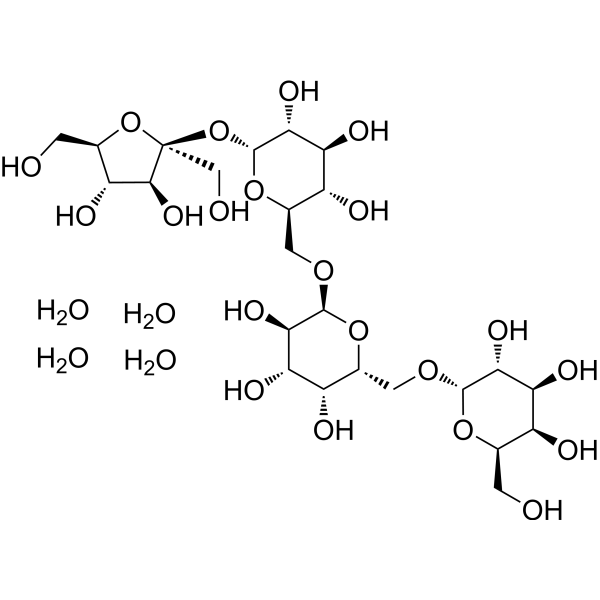
Stachyose tetrahydrate
Based on 1 publication(s) in Google Scholar
Stachyose tetrahydrate, a functional oligosaccharide, acts as a prebiotic. Stachyose tetrahydrate can prevent indirectly colon cancer cell growth by promoting the proliferation of probiotics or producing beneficial materials in the intestine.
For research use only. We do not sell to patients.
- Purity : 98.0%
- CAS No.: 10094-58-3
- Formula: C24H50O25
- Molecular Weight:738.64
-
Storage:
4°C, sealed storage, away from moisture and light
* In solvent : -80°C, 6 months; -20°C, 1 month (sealed storage, away from moisture and light)

To place orders, for customer services and technical support, please contact: MedChemExpress USA
Tel: 609-228-6898 E-mail: [email protected] [email protected]


-
 Biological Activity
-
Chemical Information
-
Solvent & Solubility
- Protocols
- Purity & Documentation
- References
-
 Help & FAQs


-
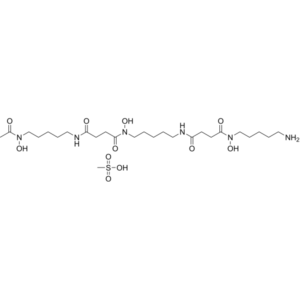
HY-B0988
Deferoxamine mesylateIron Chelator -
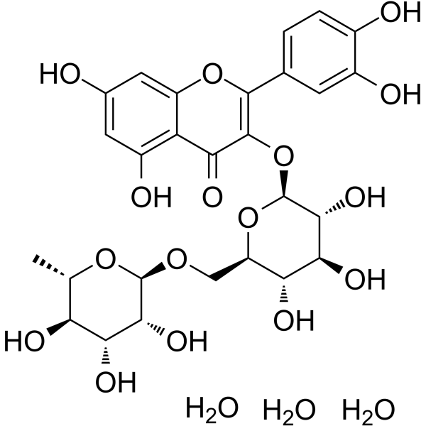
HY-W013075
Rutin trihydrateMultifunctional Natural Flavonoid Glycoside -
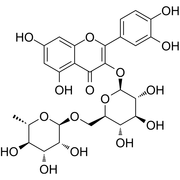
HY-N0148
RutinCBR1 Inhibitor -
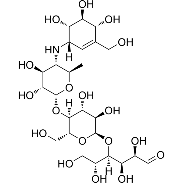
HY-B0089
AcarboseAlpha-Glucosidase Inhibitor -
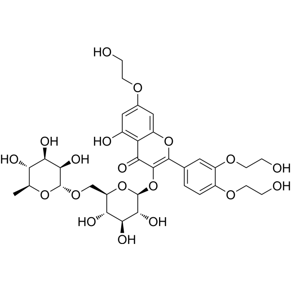
HY-N0139
TroxerutinNOD-like Receptor (NLR) Inhibitor -
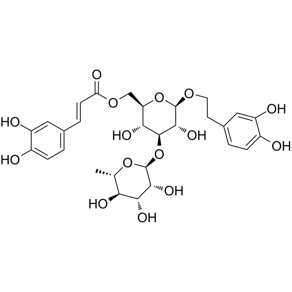
HY-N0022
IsoacteosideNatural Product -
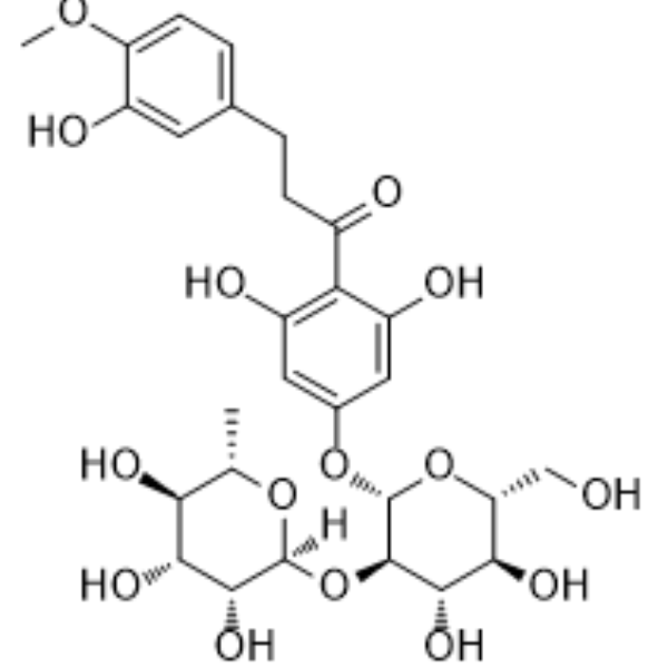
HY-N0154
Neohesperidin dihydrochalconeGlycoside Chalcone -
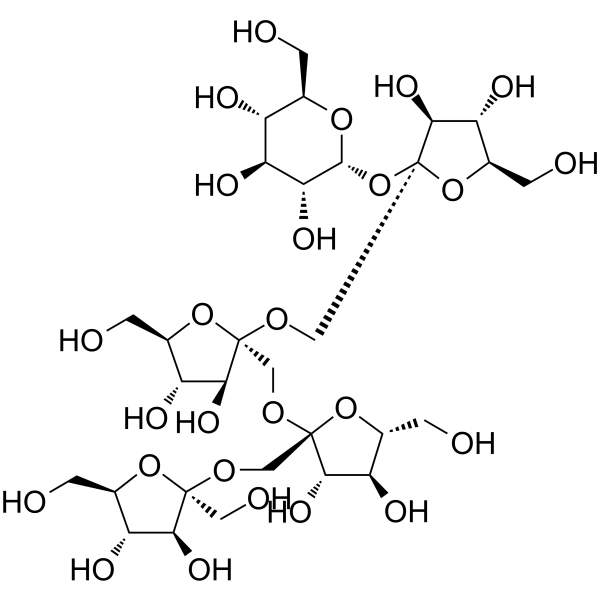
HY-N2577
1F-FructofuranosylnystoseFructo-oligose -
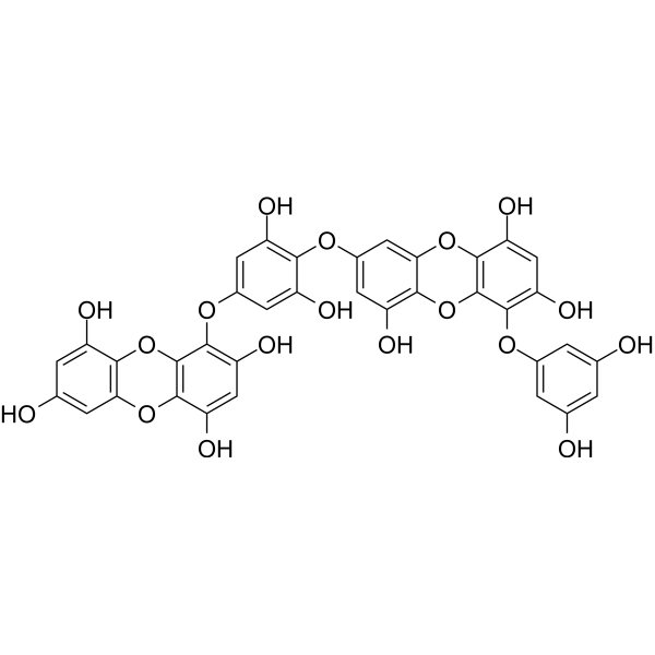
HY-147059
DieckolPhlorotannin Compound -
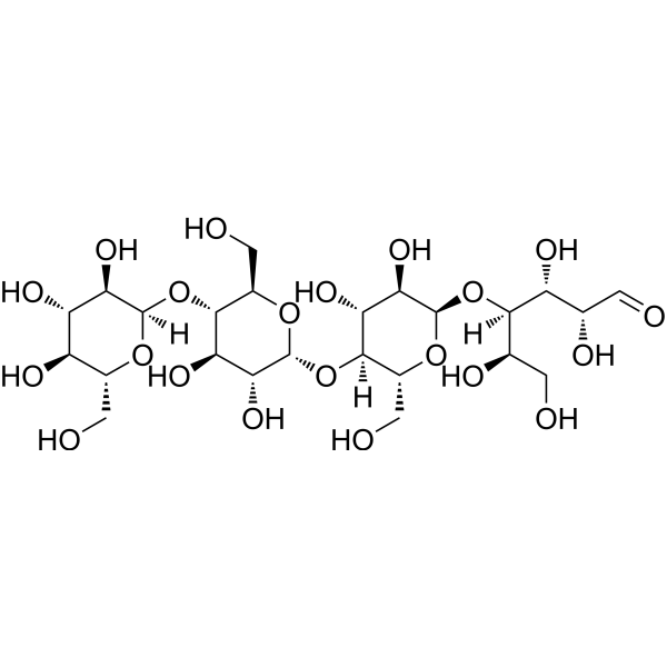
HY-N2464
MaltotetraoseAmylase Substrate -
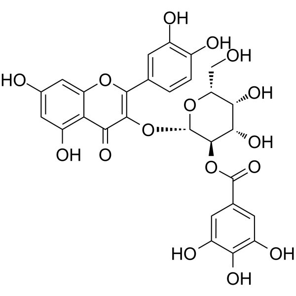
HY-N0526
2"-O-GalloylhyperinNatural Product -
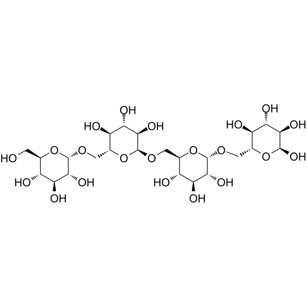
HY-N7948
IsomaltotetraosePrebiotic Oligosaccharide
Publications Citing Use of MedChemExpress (MCE) Stachyose tetrahydrate
More Customer Validation & Images

-
Cell Proliferation/Viability Assay

-
IF

-
RT-PCR

-
WB

-
Histological Imaging/Staining

All Endogenous Metabolite Isoforms
More

Biological Activity
Description
IC50 & Target
|
Human Endogenous Metabolite |
In Vitro
Stachyose tetrahydrate inhibits Caco-2 cell proliferation and induces apoptosis in a dose-dependent manner[1].
Stachyose highly promotes proliferation of lactic acid bacteria (LAB) by inducing LAB to produce more α-galactosidase to hydrolyze stachyose[2].
MedChemExpress (MCE) has not independently confirmed the accuracy of these methods. They are for reference only. Further protocols information, click here.


Chemical Information
-
CAS No. 10094-58-3
-
Appearance Solid
-
Molecular Weight 738.64
-
Formula C24H50O25
-
Color White to yellow
-
SMILES
OC[C@]1(O[C@H](CO)[C@@H](O)[C@@H]1O)O[C@H]2O[C@@H]([C@@H](O)[C@H](O)[C@H]2O)CO[C@H]3O[C@@H]([C@H](O)[C@H](O)[C@H]3O)CO[C@H]4O[C@@H]([C@H](O)[C@H](O)[C@H]4O)CO.O.O.O.O
-
Structure Classification
-
Initial Source
-
Shipping
Room temperature in continental US; may vary elsewhere.
-
Storage
4°C, sealed storage, away from moisture and light
* In solvent : -80°C, 6 months; -20°C, 1 month (sealed storage, away from moisture and light)


Publications (1)
-
Journal Impact Factor
-
Most Recent
-
Food Sci Nutr
Screening Based on Structural and Biological Verification of Stachyose as a PPARγ-Modulating Ligand for the Treatment of Non-Alcoholic Fatty Liver Disease. [Abstract]2025 Sep 22;13(9):e71009. PMID: 40994455
Stachyose tetrahydrate purchased from MedChemExpress. Usage Cited in: Food Sci Nutr. 2025 Sep 22;13(9):e71009. [Abstract]
Cytotoxicity of Stachyose (STA) (0, 0.4, 0.8, 1.6, 3.2, 6.4 mg/mL) on HepG2 cells.
Stachyose tetrahydrate purchased from MedChemExpress. Usage Cited in: Food Sci Nutr. 2025 Sep 22;13(9):e71009. [Abstract]
ORO staining images of HepG2 cells treated with Stachyose (STA) (0.8, 1.6 mg/mL).
Stachyose tetrahydrate purchased from MedChemExpress. Usage Cited in: Food Sci Nutr. 2025 Sep 22;13(9):e71009. [Abstract]
qRT‐PCR results of lipid metabolism‐related molecules (FASN, ACC, CPT2, ACO2), inflammation‐related molecules (TNF‐α, IL‐1β, IL6) and insulin resistance‐related molecules (GLUT4, IRS2, PTP1B, SOCS3) in HepG2 cells treated with Stachyose (STA) (0.8, 1.6 mg/mL).
Stachyose tetrahydrate purchased from MedChemExpress. Usage Cited in: Food Sci Nutr. 2025 Sep 22;13(9):e71009. [Abstract]
Protein expression of TNFα, IRS2, and ACC in HepG2 cells treated with Stachyose (STA) (0.8, 1.6 mg/mL).
Stachyose tetrahydrate purchased from MedChemExpress. Usage Cited in: Food Sci Nutr. 2025 Sep 22;13(9):e71009. [Abstract]
H&E staining images treated with Stachyose (STA) (0.3, 0.6 g/kg/day, i.g.).

Stachyose tetrahydrate purchased from MedChemExpress. Usage Cited in: Food Sci Nutr. 2025 Sep 22;13(9):e71009. [Abstract]
Co-IP analysis of PPARγ deacetylation in HepG2 cells treated with Stachyose (STA) (0.8, 1.6 mg/mL).


Solvent & Solubility
In Vitro:
DMSO : 250 mg/mL (338.46 mM; Need ultrasonic; Hygroscopic DMSO has a significant impact on the solubility of product, please use newly opened DMSO)
H2O : 100 mg/mL (135.38 mM; Need ultrasonic)
Please refer to the solubility information to select the appropriate solvent. Once prepared, please aliquot and store the solution to prevent product inactivation from repeated freeze-thaw cycles.
Storage method and period of stock solution: -80°C, 6 months; -20°C, 1 month (sealed storage, away from moisture and light). When stored at -80°C, please use it within 6 months. When stored at -20°C, please use it within 1 month.
* Note: If you choose water as the stock solution, please dilute it to the working solution, then filter and sterilize it with a 0.22 μm filter before use.
Please refer to the solubility information to select the appropriate solvent. Once prepared, please aliquot and store the solution to prevent product inactivation from repeated freeze-thaw cycles.
Storage method and period of stock solution: -80°C, 6 months; -20°C, 1 month (sealed storage, away from moisture and light). When stored at -80°C, please use it within 6 months. When stored at -20°C, please use it within 1 month.
* Note: If you choose water as the stock solution, please dilute it to the working solution, then filter and sterilize it with a 0.22 μm filter before use.
Concentration (start) × Volume (start) = Concentration (final) × Volume (final)
In Vivo:
Select the appropriate dissolution method based on your experimental animal and administration route.
- For the following dissolution methods, please ensure to first prepare a clear stock solution using an In Vitro approach and then sequentially add co-solvents:
- To ensure reliable experimental results, the clarified stock solution can be appropriately stored based on storage conditions. As for the working solution for In Vivo experiments, it is recommended to prepare freshly and use it on the same day.
- The percentages shown for the solvents indicate their volumetric ratio in the final prepared solution. If precipitation or phase separation occurs during preparation, heat and/or sonication can be used to aid dissolution.
Add each solvent one by one: 10% DMSO 40% PEG300 5% Tween-80 45% Saline
Solubility: ≥ 2.08 mg/mL (2.82 mM); Clear solution
This protocol yields a clear solution of ≥ 2.08 mg/mL (saturation unknown).
Taking 1 mL working solution as an example, add 100 μL DMSO stock solution (20.8 mg/mL) to 400 μL PEG300, and mix evenly; then add 50 μL Tween-80 and mix evenly; then add 450 μL Saline to adjust the volume to 1 mL.
Preparation of Saline: Dissolve 0.9 g sodium chloride in ddH₂O and dilute to 100 mL to obtain a clear Saline solution.
Add each solvent one by one: 10% DMSO 90% (20% SBE-β-CD in Saline)
Solubility: ≥ 2.08 mg/mL (2.82 mM); Clear solution
This protocol yields a clear solution of ≥ 2.08 mg/mL (saturation unknown).
Taking 1 mL working solution as an example, add 100 μL DMSO stock solution (20.8 mg/mL) to 900 μL 20% SBE-β-CD in Saline, and mix evenly.
Preparation of 20% SBE-β-CD in Saline (4°C, storage for one week): 2 g SBE-β-CD powder is dissolved in 10 mL Saline, completely dissolve until clear.
For the following dissolution methods, please prepare the working solution directly:
It is recommended to prepare fresh solutions and use them promptly within a short period of time.
The percentages shown for the solvents indicate their volumetric ratio in the final prepared solution. If precipitation or phase separation occurs during preparation, heat and/or sonication can be used to aid dissolution.
Add each solvent one by one: PBS
Solubility: 100 mg/mL (135.38 mM); Clear solution; Need ultrasonic
In Vivo Dissolution Calculator
Please enter the basic information of animal experiments:
-
-
-
-
Recommended: Prepare an additional quantity of animals to account for potential losses during experiments.
Working solution concentration: 0.22 mg/mL
This product has good water solubility, please refer to the measured solubility data in water/PBS/Saline for details.

Protocols
-
Protocol for Cell Counting and Cell Density Analysis
Cell counting and cell-density analysis estimate the number of cells in a known volume or field area. Manual hemocytometer counting uses a chamber of defined geometry to convert counted cells into cells/mL, while automated counters and image-analysis workflows detect cell objects from optical, brightfield, fluorescence, impedance, or digital-image features. Trypan blue viability counting is based on dye exclusion: viable cells with intact membranes exclude dye, while non-viable cells with compromised membranes stain blue. The readout is total cell density, viable-cell density, dead-cell density, and percent viability. Cell density can also be estimated from microscopy images by counting objects per image area, from flow cytometry using calibrated volume or reference particles, or from in situ microscopy in bioreactors after calibration against reference methods such as hemocytometer or flow cytometry.
-
Somatic Cell Culture
A method of simulating the in vivo environment in vitro to maintain the cell growth, differentation and main functions.
-
CCK-8/WST-8 Cell Proliferation Assay
The CCK-8/WST-8 assay is based on the reduction of the water-soluble tetrazolium salt WST-8 to a water-soluble formazan product by cellular dehydrogenases in metabolically active cells, where the generated formazan amount is proportional to the number of living cells and is quantified by measuring absorbance in the visible range, providing a colorimetric readout for cell viability and proliferation assessment. This class of tetrazolium-based assays improves upon earlier MTT-based systems by producing a water-soluble formazan, eliminating the need for organic solubilization steps and enabling direct spectrophotometric measurement in culture medium.
-
Cell Counting-Based Growth Curve Assay
Cell counting-based growth curve assays quantify cell proliferation by directly measuring changes in viable cell number over time using manual or automated counting methods such as hemocytometer-based counting or instrument-assisted cell enumeration, enabling construction of growth curves that reflect population expansion dynamics in response to culture conditions. A widely used approach is trypan blue exclusion with hemocytometer counting, where membrane-compromised (non-viable) cells take up the dye, allowing discrimination between viable and non-viable cells while simultaneously enabling total cell number quantification. Repeated sampling across time points allows estimation of proliferation rate, growth phases, and comparative growth kinetics between experimental conditions.
-
MTT Cell Proliferation Assay
The MTT assay is a colorimetric endpoint assay for estimating viable cell number, cell growth, cytotoxicity, or cell activation in cultured mammalian cells. Living cells reduce the yellow tetrazolium salt MTT into purple/blue formazan, while dead cells do not generate the same signal; the resulting color can be quantified with a multiwell spectrophotometer. MTT reduction is commonly interpreted as a readout of metabolic activity that often correlates with viable cell number, but it should not be treated as a direct cell-counting method unless the assay is optimized for the cell type and experimental condition. Studies show that MTT reduction can involve mitochondrial and non-mitochondrial reducing systems, and formazan may accumulate in intracellular lipid droplets rather than simply marking mitochondria.
-
Cell Viability Determination by MTT Colorimetric Assay
The following protocol uses the MTT colorimetric assay as a classic literature-established method for assessing cell viability/metabolic activity in cultured mammalian cells. MTT[3-(4,5-dimethylthiazol-2-yl)-2,5-diphenyltetrazolium bromide] is reduced by metabolically active cells to a colored formazan product; the amount of formazan is quantified spectrophotometrically and provides an indirect measure of metabolically active viable cells. Importantly, MTT reduction reflects cellular oxidoreductase/metabolic activity rather than an absolute direct count of living cells, so changes in cellular metabolism can alter the signal independently of cell number.


Purity & Documentation
-
Data Sheet (281 KB)
-
SDS (393 KB)
- English - EN (393 KB)
- Français - FR (393 KB)
- Deutsch - DE (393 KB)
- Norwegian - NO (393 KB)
- Español - ES (393 KB)
- Swedish - SV (393 KB)
- Italian - IT (393 KB)
- Korean - KR (393 KB)
- Portuguese - PT (393 KB)
-
Handling Instructions (2659 KB)
References
[1]. Huang G, et al. Stachyose-induced apoptosis of Caco-2 cells via the caspase-dependent mitochondrial pathway. Food Funct. 2015;6(3):765-771. [Content Brief]
[2]. Pan Q, et al. The Proliferation Mechanism of Lactobacillus plantarum RB1 Stimulated by Stachyose. Curr Microbiol. 2017 Jun;74(6):732-738. [Content Brief]


Complete Stock Solution Preparation Table
Please refer to the solubility information to select the appropriate solvent. Once prepared, please aliquot and store the solution to prevent product inactivation from repeated freeze-thaw cycles.
Storage method and period of stock solution: -80°C, 6 months; -20°C, 1 month (sealed storage, away from moisture and light). When stored at -80°C, please use it within 6 months. When stored at -20°C, please use it within 1 month.
| Optional Solvent | Concentration Solvent Mass | 1 mg | 5 mg | 10 mg | 25 mg |
|---|---|---|---|---|---|
| H2O / DMSO | 1 mM | 1.3538 mL | 6.7692 mL | 13.5384 mL | 33.8460 mL |
| 5 mM | 0.2708 mL | 1.3538 mL | 2.7077 mL | 6.7692 mL | |
| 10 mM | 0.1354 mL | 0.6769 mL | 1.3538 mL | 3.3846 mL | |
| 15 mM | 0.0903 mL | 0.4513 mL | 0.9026 mL | 2.2564 mL | |
| 20 mM | 0.0677 mL | 0.3385 mL | 0.6769 mL | 1.6923 mL | |
| 25 mM | 0.0542 mL | 0.2708 mL | 0.5415 mL | 1.3538 mL | |
| 30 mM | 0.0451 mL | 0.2256 mL | 0.4513 mL | 1.1282 mL | |
| 40 mM | 0.0338 mL | 0.1692 mL | 0.3385 mL | 0.8461 mL | |
| 50 mM | 0.0271 mL | 0.1354 mL | 0.2708 mL | 0.6769 mL | |
| 60 mM | 0.0226 mL | 0.1128 mL | 0.2256 mL | 0.5641 mL | |
| 80 mM | 0.0169 mL | 0.0846 mL | 0.1692 mL | 0.4231 mL | |
| 100 mM | 0.0135 mL | 0.0677 mL | 0.1354 mL | 0.3385 mL |
* Note: If you choose water as the stock solution, please dilute it to the working solution, then filter and sterilize it with a 0.22 μm filter before use.


Stachyose tetrahydrate Related Classifications
HY-113529 Related Classifications
 Powered by Bioz