



Acetylcysteine-d3

Based on 1 Customer Validation
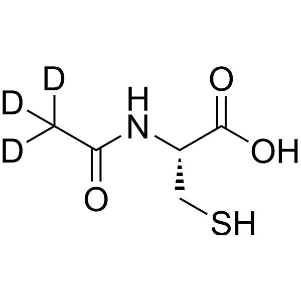
Acetylcysteine-d3 is the deuterium labeled Acetylcysteine. Acetylcysteine (N-Acetylcysteine) is a mucolytic agent which reduces the thickness of the mucus. Acetylcysteine is a ROS inhibitor. Acetylcysteine is a cysteine precursor, prevents hemin-induced ferroptosis by neutralizing toxic lipids generated by arachidonate-dependent activity of 5-lipoxygenases. Acetylcysteine induces cell apoptosis. Acetylcysteine also has anti-influenza virus activities. In addition, Acetylcysteine is the most stable form of cysteine during drug delivery and can be used in disulfidptosis studies.
Nur für Forschungszwecke. Wir verkaufen nicht an Patienten.
- Reinheit : 97.72%
- CAS. Nr.: 131685-11-5
- Formel: C5H6D3NO3S
- Molecular Weight:166.21
-
Speicherung:
4°C, protect from light, stored under nitrogen
* In solvent : -80°C, 6 months; -20°C, 1 month (protect from light, stored under nitrogen)

To place orders, for customer services and technical support, please contact: MedChemExpress USA
Tel: 609-228-6898 E-mail: [email protected] [email protected]


-
 Biologische Aktivität
-
Chemical Information
- Protokoll
- Reinheit & Dokumentation
- Verweise
-
 Help & FAQs


Alle Endogenous Metabolite Isoform-spezifische Produkte anzeigen
More

Biologische Aktivität
Beschreibung
In Vitro
Stable heavy isotopes of hydrogen, carbon, and other elements have been incorporated into drug molecules, largely as tracers for quantitation during the drug development process. Deuteration has gained attention because of its potential to affect the pharmacokinetic and metabolic profiles of drugs[1].
MedChemExpress (MCE) has not independently confirmed the accuracy of these methods. They are for reference only.
Anwendung
1. This compound can be used as a tracer
2. This compound can be used as an internal standard for quantitative analysis by NMR, GC-MS, or LC-MS.


Chemical Information
-
CAS. Nr. 131685-11-5
-
Unlabeled CAS 616-91-1
-
Appearance Solid
-
Molecular Weight 166.21
-
Formel C5H6D3NO3S
-
Color White to off-white
-
SMILES
[2H]C([2H])([2H])C(N[C@@H](CS)C(O)=O)=O
-
Synonyms
N-Acetylcysteine-d3; N-Acetyl-L-cysteine-d3; NAC-d3
-
Versand
Room temperature in continental US; may vary elsewhere.
-
Speicherung
4°C, protect from light, stored under nitrogen
* In solvent : -80°C, 6 months; -20°C, 1 month (protect from light, stored under nitrogen)


Reinheit & Dokumentation
-
Data Sheet (271 KB)
-
SDS (393 KB)
- English - EN (393 KB)
- Français - FR (393 KB)
- Deutsch - DE (393 KB)
- Norwegian - NO (393 KB)
- Español - ES (393 KB)
- Swedish - SV (393 KB)
- Italian - IT (393 KB)
- Korean - KR (393 KB)
- Portuguese - PT (393 KB)
-
Handling Instructions (2659 KB)
Verweise
[1]. Russak EM, et al. Impact of Deuterium Substitution on the Pharmacokinetics of Pharmaceuticals. Ann Pharmacother. 2019;53(2):211-216. [Content Brief]
[2]. Halasi M, et al. ROS inhibitor N-acetyl-L-cysteine antagonizes the activity of proteasome inhibitors. Biochem J. 2013 Sep 1;454(2):201-8. [Content Brief]
[3]. Ferrari G, et al. N-acetylcysteine (D- and L-stereoisomers) prevents apoptotic death of neuronal cells. J Neurosci. 1995 Apr;15(4):2857-66. [Content Brief]
[4]. Tsai JC, et al. Induction of apoptosis by pyrrolidinedithiocarbamate and N-acetylcysteine in vascular smooth muscle cells. J Biol Chem. 1996 Feb 16;271(7):3667-70. [Content Brief]
[5]. Yan CY, et al. Prevention of PC12 cell death by N-acetylcysteine requires activation of the Ras pathway. J Neurosci. 1998 Jun 1;18(11):4042-9. [Content Brief]
[6]. Farr SA, et al. The antioxidants alpha-lipoic acid and N-acetylcysteine reverse memory impairment and brain oxidative stress in aged SAMP8 mice. J Neurochem. 2003 Mar;84(5):1173-83. [Content Brief]
[7]. Kalimeris K, et al. N-acetylcysteine ameliorates liver injury in a rat model of intestinal ischemia reperfusion. J Surg Res. 2016 Dec;206(2):263-272. [Content Brief]
[8]. Garigliany MM, et al. N-acetylcysteine lacks universal inhibitory activity against influenza A viruses. J Negat Results Biomed. 2011 May 9;10:5. [Content Brief]
[9]. Gu Q, et al. Disulfidptosis, a novel cell death pathway: molecular landscape and therapeutic implications[J]. Aging Dis, 2024, 10. [Content Brief]

Calculators
Konzentration (Stammlösung) × Volumen (Stammlösung) = Konzentration (Ziellösung) × Volumen (Ziellösung)


Acetylcysteine-d3 Related Classifications
HY-B0215S Related Classifications
 Powered by Bioz