



AD-5584

Based on 1 Customer Validation
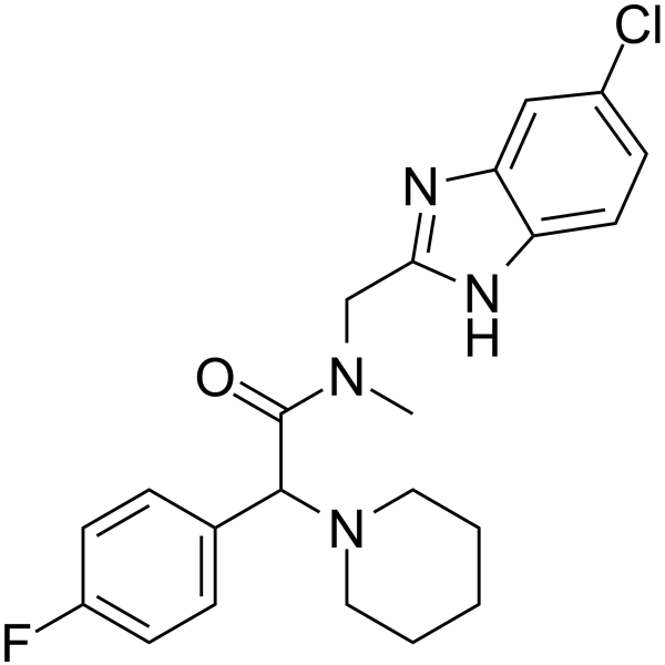
AD-5584 is a selective and blood-brain barrier-penetrant ACSS2 inhibitor (IC50 = 0.86 μM). AD-5584 binds to the nucleotide-binding pocket of ACSS2, blocking CoA binding and acetyl transfer through steric hindrance, thereby inhibiting the conversion of acetate to acetyl-CoA. AD-5584 decreases acetyl-CoA levels, lipid droplet content, FASN expression, E2F1, SLC7A11, and GPX4 levels, and reduces E2F1 occupancy at the SLC7A11 promoter. AD-5584 induces ferroptosis, lipid peroxidation, malondialdehyde accumulation, and cell death, and decreases clonogenic survival and proliferative capacity. AD-5584 can be used for research on breast cancer brain metastasis.
For research use only. We do not sell to patients.
- Purity : 99.86%
- CAS No.: 2306525-79-9
- Formula: C22H24ClFN4O
- Molecular Weight:414.90
-
Storage:Powder -20°C, 3 years ; In solvent -80°C, 6 months , -20°C, 1 month

To place orders, for customer services and technical support, please contact: MedChemExpress USA
Tel: 609-228-6898 E-mail: [email protected] [email protected]


-
 Biological Activity
-
Chemical Information
-
Solvent & Solubility
- Protocols
- Purity & Documentation
- References
-
 Help & FAQs




Biological Activity
Description
IC50 & Target
[1]|
ACSS2 0.86 μM (IC50) |
GPX4 |
In Vitro
AD-5584 binds selectively to human ACSS2 with low-micromolar affinity and shows no binding to hACSS1[1].
AD-5584 (100 μM; 48 h) significantly reduces clonogenic survival of MDA-MB-231BR breast cancer brain metastatic cells[1].
AD-5584 induces cell death in MDA-MB-231BR and 4T1BR breast cancer brain metastatic cells[1].
AD-5584 (100 μM; 48 h) significantly reduces acetyl-CoA levels in MDA-MB-231BR and 4T1BR breast cancer brain metastatic cells[1].
AD-5584 (100 μM; 48 h) significantly reduces lipid droplet content in MDA-MB-231BR breast cancer brain metastatic cells[1].
AD-5584 (100 μM; 24 h) reduces FASN protein levels in MDA-MB-231BR breast cancer brain metastatic cells[1].
AD-5584 has a metabolic half-life of 20 min in human liver microsomes, demonstrating moderate metabolic stability[1].
AD-5584 exhibits moderate permeability in the MDR1-MDCK blood-brain barrier assay[1].
AD-5584 (100 μM; 48 h) reduces E2F1, SLC7A11, and GPX4 protein levels in MDA-MB-231-BR cells[2].
AD-5584 (100 μM; 48 h) induces ferroptotic cell death in MDA-MB-231-BR cells, which is blocked by ferrostatin-1[2].
AD-5584 (100 μM; 48 h) induces lipid peroxidation in MDA-MB-231-BR cells, which is blocked by ferrostatin-1[2].
AD-5584 (100 μM; 48 h) E2F1 overexpression protects MDA-MB-231-BR cells from AD-5584-induced cell death and lipid peroxidation[2].
AD-5584 (100 μM; 48 h) reduces E2F1, SLC7A11, and GPX4 protein levels in 4T1-BR cells[2].
AD-5584 (100 μM; 48 h) induces ferroptotic cell death in 4T1-BR cells, which is blocked by Ferrostatin-1 (HY-100579) [2].
AD-5584 (100 μM; 48 h) induces lipid peroxidation in 4T1-BR cells, which is blocked by Ferrostatin-1 (HY-100579)[2].
AD-5584 induces ferroptosis-mediated tumor regression of MDA-MB-231-BR cells in ex vivo brain slices, with no observable toxicity to normal brain tissue[2].
AD-5584 induces ferroptosis-mediated tumor regression of 4T1-BR cells in ex vivo brain slices[2].
In MDA-MB-231-BR cells, AD-5584 (50 μM; 6 hours) reduces E2F1 occupancy at the SLC7A11 promoter[3].
AD-5584 (50 μM; 6 days) significantly reduces the growth of HCI-015 patient-derived TNBC brain metastasis organoids[3].
AD-5584 (24 hours) reduces proliferation equally in both MDA-MB-231 parental and MDA-MB-231-BR cells[3].
AD-5584 increases malondialdehyde levels, a marker of lipid peroxidation and ferroptosis, in breast cancer brain metastatic cells[3].
MedChemExpress (MCE) has not independently confirmed the accuracy of these methods. They are for reference only. Further protocols information, click here.
-
Cell Line:MDA-MB-231BR breast cancer brain metastatic cells
-
Concentration:100 μM
-
Incubation Time:48 h (treatment); 15 min (staining)
-
Result:Significantly reduced lipid droplet content compared to control.
-
Cell Line:MDA-MB-231BR breast cancer brain metastatic cells
-
Concentration:100 μM
-
Incubation Time:24 h
-
Result:Reduced protein levels of FASN compared to DMSO control.
-
Cell Line:MDA-MB-231-BR human breast cancer brain metastatic cells
-
Concentration:100 μM
-
Incubation Time:48 h
-
Result:Reduced E2F1, SLC7A11, and GPX4 protein levels compared to control-treated cells.
-
Cell Line:MDA-MB-231-BR human breast cancer brain metastatic cells
-
Concentration:100 μM
-
Incubation Time:48 h
-
Result:Significantly increased cell death compared to control-treated cells.
Reversed the increase in cell death by co-treatment with Ferrostatin-1.
-
Cell Line:4T1-BR mouse breast cancer brain metastatic cells
-
Concentration:100 μM
-
Incubation Time:48 h
-
Result:Reduced E2F1, SLC7A11, and GPX4 protein levels compared to control-treated cells.
-
Cell Line:4T1-BR mouse breast cancer brain metastatic cells
-
Concentration:100 μM
-
Incubation Time:48 h
-
Result:Significantly increased cell death compared to control-treated cells.
Reversed the increase in cell death by co-treatment with Ferrostatin-1.
In Vivo
AD-5584 (systemic; 14 days) inhibits breast cancer brain metastasis growth in immunocompromised mice bearing intracranial MDA-MB-231-BR tumors[2].
AD-5584 (50 mg/kg; i.p.; daily; 10 days) suppresses breast cancer brain metastatic growth and extends survival in mouse intracranial models[3].
AD-5584 (50-100 mg/kg; i.p.; single dose) penetrates the blood-brain barrier in healthy mice in vivo[1].
MedChemExpress (MCE) has not independently confirmed the accuracy of these methods. They are for reference only.
-
Animal Model:Nu/Nu athymic nude mice (4-6 weeks old; female for MFP model) injected with f MDA-
MB-231 or MDA-MB-231-BR cells[3] -
Dosage:50 mg/kg
-
Administration:i.p.; daily; 10 days
-
Result:Significantly blocked breast cancer brain metastatic growth of both 4T1-BR and MDA-MB-231-BR cells in vivo.
Significantly extended survival of treated mice compared to control.
No weight loss or observable behavioral changes were detected in treated animals.


Chemical Information
-
CAS No. 2306525-79-9
-
Appearance Solid
-
Molecular Weight 414.90
-
Formula C22H24ClFN4O
-
Color White to off-white
-
SMILES
FC1=CC=C(C(N2CCCCC2)C(N(C)CC3=NC4=CC(Cl)=CC=C4N3)=O)C=C1
-
Shipping
Room temperature in continental US; may vary elsewhere.
-
Storage
Powder -20°C 3 years In solvent -80°C 6 months -20°C 1 month


Solvent & Solubility
In Vitro:
DMSO : 12.5 mg/mL (30.13 mM; ultrasonic and warming and heat to 60°C; Hygroscopic DMSO has a significant impact on the solubility of product, please use newly opened DMSO)
Please refer to the solubility information to select the appropriate solvent. Once prepared, please aliquot and store the solution to prevent product inactivation from repeated freeze-thaw cycles.
Storage method and period of stock solution: -80°C, 6 months; -20°C, 1 month. When stored at -80°C, please use it within 6 months. When stored at -20°C, please use it within 1 month.
Please refer to the solubility information to select the appropriate solvent. Once prepared, please aliquot and store the solution to prevent product inactivation from repeated freeze-thaw cycles.
Storage method and period of stock solution: -80°C, 6 months; -20°C, 1 month. When stored at -80°C, please use it within 6 months. When stored at -20°C, please use it within 1 month.
Concentration (start) × Volume (start) = Concentration (final) × Volume (final)

Protocols
-
RNA extraction experimental
By lysing cells, releasing RNA, and removing impurities such as proteins and DNA, high-purity RNA products are finally obtained. The commonly used traditional method is the guanidine isothiocyanate/phenol/chloroform method (Trizol), which is suitable for a variety of animal materials including animal tissues, microorganisms, cultured cells, etc., and most plant materials.
-
Cell Cytotoxicity Assay
Cytotoxicity assays are usually based on the assessment of cell membrane damage, which can also be indirectly detected by measuring cell viability. Detection methods include MTT assay, CKK-8 assay, LDH assay and ATP assay, etc.
-
Mammalian live/dead viability and cytotoxicity staining
Live/dead viability and cytotoxicity staining assays are based on the simultaneous detection of intracellular esterase activity in metabolically active (viable) cells and membrane integrity loss in non-viable cells. In commonly used dual-staining approaches, membrane-permeant fluorogenic substrates are converted by intracellular esterases into fluorescent products in live cells, while impermeant DNA-binding dyes selectively enter cells with compromised plasma membranes and label nucleic acids in dead or dying cells, enabling discrimination between viable and non-viable populations by fluorescence microscopy or flow cytometry.
-
Apoptosis Solutions
Apoptosis is a regulated, generally non-lytic cell-death pathway that removes unwanted, damaged, infected, or abnormal cells through coordinated morphological changes, caspase activation, DNA fragmentation, and membrane remodeling. The intrinsic apoptosis pathway is controlled mainly by mitochondrial outer membrane permeabilization, BCL-2 family proteins, cytochrome c release, apoptosome formation, caspase-9 activation, and downstream executioner caspase-3/7 activation. The extrinsic apoptosis pathway is initiated by death receptors such as Fas, TNFR, and TRAIL receptors, which recruit adaptor proteins and activate caspase-8 before engaging executioner caspases or mitochondrial amplification through BID cleavage. Apoptosis is linked to many phenotypes, including cancer cell killing, tissue homeostasis, immune regulation, neurodegeneration, infection response, and treatment-induced cytotoxicity; unresolved questions include how apoptosis interacts with necroptosis, pyroptosis, ferroptos
-
Ferroptosis Solutions
Ferroptosis is an iron-dependent, non-apoptotic form of regulated cell death characterized by lethal lipid peroxidation and sensitivity to suppression by iron chelators or lipophilic radical-trapping antioxidants. The core pathway links cystine uptake through system Xc−, glutathione availability, GPX4-dependent detoxification of phospholipid hydroperoxides, iron-dependent oxidative reactions, and polyunsaturated-phospholipid metabolism into a cell-death program that is biochemically and morphologically distinct from apoptosis, necrosis, and autophagy. The ferroptosis pathway is experimentally linked to phenotype through chemical and genetic perturbation. Erastin induces ferroptosis by inhibiting cystine uptake through system Xc− and weakening antioxidant defenses, while GPX4 inhibition or depletion causes lipid peroxide accumulation and ferroptotic cancer-cell death. ACSL4 and oxidizable arachidonoyl- or adrenoyl-containing phosphatidylethanolamines shape ferroptosis sensitivity by con
-
Colony Formation (Clonogenic) Assay
The clonogenic (colony formation) assay measures the ability of a single cell to retain reproductive viability and form a macroscopic colony, typically defined as a cluster derived from one progenitor cell after a defined growth period. This assay is widely used to evaluate cell survival after exposure to ionizing radiation or cytotoxic treatments and is considered a standard method in radiation biology for generating dose-response relationships of reproductive cell death. Colony formation reflects long-term proliferative capacity rather than short-term metabolic activity, and survival is quantified by comparing treated versus untreated conditions based on colony number and derived survival fractions.
-
Breast Cancer Modeling
Breast cancer is a heterogeneous cancer, and it has been distinguished into four subtypes: luminal A, luminal B, HER2-positive and basal-like. Molecular mutations, epigenetic alterations, hormone exposure and immune microenvironment are related to the progression of breast cancer.
-
Orthotopic Cell-Line Xenograft
Orthotopic cell-line xenograft models involve implantation of human cancer cell lines into the anatomically corresponding organ of immunodeficient mice to reproduce tumor growth within a native microenvironment, enabling more clinically relevant tumor behavior compared with subcutaneous models. These models are widely used because orthotopic placement better recapitulates tumor progression, including invasion and metastatic spread, which are often underrepresented in heterotopic implantation systems. Compared with conventional xenografts, orthotopic implantation is described as more technically complex but provides improved simulation of tumor-microenvironment interactions and metastatic behavior, making it particularly valuable for translational oncology research. Surgical orthotopic implantation approaches have been emphasized as enabling faithful reproduction of clinical cancer features, including metastasis and disease progression patterns that align with the tumor’s organ of origi


Purity & Documentation
-
Data Sheet (299 KB)
-
SDS (251 KB)
- English - EN (251 KB)
- Français - FR (251 KB)
- Deutsch - DE (251 KB)
- Norwegian - NO (251 KB)
- Español - ES (251 KB)
- Swedish - SV (251 KB)
- Italian - IT (251 KB)
- Korean - KR (251 KB)
- Portuguese - PT (251 KB)
-
Handling Instructions (2659 KB)
References


Complete Stock Solution Preparation Table
Please refer to the solubility information to select the appropriate solvent. Once prepared, please aliquot and store the solution to prevent product inactivation from repeated freeze-thaw cycles.
Storage method and period of stock solution: -80°C, 6 months; -20°C, 1 month. When stored at -80°C, please use it within 6 months. When stored at -20°C, please use it within 1 month.
| Optional Solvent | Concentration Solvent Mass | 1 mg | 5 mg | 10 mg | 25 mg |
|---|---|---|---|---|---|
| DMSO | 1 mM | 2.4102 mL | 12.0511 mL | 24.1022 mL | 60.2555 mL |
| 5 mM | 0.4820 mL | 2.4102 mL | 4.8204 mL | 12.0511 mL | |
| 10 mM | 0.2410 mL | 1.2051 mL | 2.4102 mL | 6.0255 mL | |
| 15 mM | 0.1607 mL | 0.8034 mL | 1.6068 mL | 4.0170 mL | |
| 20 mM | 0.1205 mL | 0.6026 mL | 1.2051 mL | 3.0128 mL | |
| 25 mM | 0.0964 mL | 0.4820 mL | 0.9641 mL | 2.4102 mL | |
| 30 mM | 0.0803 mL | 0.4017 mL | 0.8034 mL | 2.0085 mL |


AD-5584 Related Classifications
HY-161116 Related Classifications
 Powered by Bioz