



Anti-Mouse CD317/BST2 Antibody (927)

Based on 1 Customer Validation
Anti-Mouse CD317/BST2 Antibody (927) is a rat-derived IgG2b κ type antibody inhibitor, targeting to mouse CD317/BST2. Anti-Mouse CD317/BST2 Antibody (927) can deplete plasmacytoid dendritic cells (pDC). Anti-Mouse CD317/BST2 Antibody (927) can be used for the researches of cancer, infection, inflammation and immunology, such as head and neck squamous cell carcinoma (HNSCC), murine cytomegalovirus (MCMV) infection and asthma.
For research use only. We do not sell to patients.
- Purity : ≥95.0%
- Molecular Weight:150 kDa
-
Storage:
Please store the product under the recommended conditions in the Certificate of Analysis.

To place orders, for customer services and technical support, please contact: MedChemExpress USA
Tel: 609-228-6898 E-mail: [email protected] [email protected]


-
 Biological Activity
-
Chemical Information
- Protocols
- Purity & Documentation
- References
-
 Help & FAQs




Biological Activity
Description
Isotype
Rat IgG2b kappa
Recommend Isotype Controls
Species Reactivity
Mouse
IC50 & Target
CD317/BST2
In Vitro
The antibody framework is stable, specific and adaptable, and has the ability to bind both antigens and endogenous immune receptors. Monoclonal antibodies have several derivatives, including bispecific antibodies, antibody-drug conjugates, and antibody fragments, and have significant effects in fields such as immunology and oncology. When designing inhibitory antibodies, considerations include identification of antigen-specific variable regions, choice of expression system, use of multispecific formats, and antibody derivatives based on fragmentation, oligomerization, or conjugation with other functional moieties[1].
MedChemExpress (MCE) has not independently confirmed the accuracy of these methods. They are for reference only. Further protocols information, click here.
In Vivo
Anti-Mouse CD317/BST2 Antibody (927) (15 mg/kg, i.p., every other day) effectively depletes plasmacytoid dendritic cells (pDC) and inhibits tumor growth in head and neck squamous cell carcinoma (HNSCC) mice models[2].
Anti-Mouse CD317/BST2 Antibody (927) (50 μg, i.v., twice a week for 5 weeks from 1 h before HDM) reduces plasmacytoid dendritic cells (pDC) and promotes inflammation responses in house dust mite (HDM)-induced asthma mice models[3].
Anti-Mouse CD317/BST2 Antibody (927) (250 μg, i.p., at days7 and 8) ameliorates EAE and shortens disease duration in experimental autoimmune encephalomyelitis (EAE) mice models[4].
MedChemExpress (MCE) has not independently confirmed the accuracy of these methods. They are for reference only.
-
Animal Model:Head and neck squamous cell carcinoma (HNSCC) mice models[2]
-
Dosage:15 mg/kg
-
Administration:Intraperitoneally injection, every other day
-
Result:Decreased progression rate of tumor and proportion of plasmacytoid DC.
Decreased the tumor cell proliferative capacity.
Reduced the size of spleen and lymph nodes.
Increased the CD4+ and CD8+ T cell populations.
Downregulated PD-1, LAG-3 and TIM-3 expression on T cell subsets.
Reduced the population of Tregs and monocytic MDSCs.
-
Animal Model:House dust mite (HDM)-induced asthma mice models[3]
-
Dosage:50 μg
-
Administration:Intravenously injection, twice a week for 5 weeks from 1 h before HDM
-
Result:Significantly reduced the percentages of plasmacytoid DC in the lungs.
Increased inflammatory cells, eosinophils, neutrophils, lymphocytes, and macrophages in the BAL fluid and lungs.
Increased the lung airway resistance and CD4+ cells, Th1, Th2, and Th17 cells.
Reduced Treg cells.
Gene ID
Accession
Conjugated
Unconjugated
Reconsititution
The product can be reconstituted/diluted with sterile PBS or saline.
Format
-
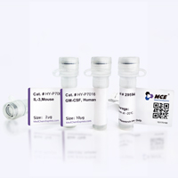
Product Image
Application
in vivo pDC depletion; Immunofluorescence; Flow cytometry


Chemical Information
-
Appearance Liquid
-
Molecular Weight 150 kDa
-
Color Colorless to light yellow
-
SMILES
[Anti-Mouse CD317/BST2 Antibody (927)]
-
Formulation
Please refer to the lot-specific COA for specific buffer information.
-
Storage
Please store the product under the recommended conditions in the Certificate of Analysis.

Protocols
-
Research Protocol for Infectious Diseases
Infectious-disease experiments test how pathogens interact with host barriers, innate immune receptors, inflammatory signaling, pathogen replication, and tissue injury; pattern-recognition receptors such as TLRs, RIG-I-like receptors, NOD-like receptors, and inflammasomes detect microbial molecules and activate NF-κB, interferon, and cytokine responses. The central hypothesis is that infection severity reflects the balance between pathogen burden and host response: protective inflammation restricts pathogen growth, whereas excessive or mislocalized inflammation contributes to tissue damage and disease phenotype. Unresolved questions include which host pathways are protective versus pathogenic, why some infection models fail to translate to human disease, and which combined readouts best predict clinically relevant infection outcomes.
-
Research Protocol for Inflammation-related Diseases
The NLRP3 inflammasome is a cytosolic innate immune signaling platform that integrates priming signals and danger-signal activation to promote caspase-1 activation, maturation of IL-1β and IL-18, and gasdermin D-mediated pyroptotic cell death. The core experimental logic is to determine whether inflammatory disease phenotypes are driven by increased NLRP3 expression, ASC-containing inflammasome assembly, caspase-1 cleavage, GSDMD cleavage, and extracellular release of IL-1β/IL-18 rather than by nonspecific cell injury alone. The pathway is strongly linked to inflammation-related disease phenotypes because monosodium urate crystals activate NALP3/NLRP3 inflammasome signaling in gout-like crystal inflammation, cholesterol crystals activate NLRP3 inflammasomes in atherogenesis models, and DSS-induced intestinal inflammation has been reported to involve NLRP3 inflammasome activity. However, experimental colitis studies also show context-dependent protective effects of NLRP3 inflammasome co
-
Monocyte-derived dendritic cell differentiation
Human monocyte-derived dendritic cells are generated by isolating PBMC-derived monocytes and culturing them with GM-CSF plus IL-4, which produces cells with dendritic-cell antigen-presenting properties, reduced monocyte phenotype, and increased dendritic-cell functional readouts such as antigen uptake, allogeneic T-cell stimulation, and expression of markers including HLA-DR, CD80, CD86, CD83, CD1a, or CD209 depending on protocol and maturation state. The main readout is phenotypic and functional differentiation: immature MoDCs are commonly evaluated by loss or reduction of CD14 with acquisition of dendritic-cell markers and antigen uptake capacity, whereas mature MoDCs are evaluated by increased CD83, CD80, CD86, HLA-DR, and T-cell stimulatory function after exposure to maturation stimuli such as TNF-α or a cytokine/PGE2 cocktail.


Purity & Documentation
-
Data Sheet (263 KB)
-
SDS (251 KB)
- English - EN (251 KB)
- Français - FR (251 KB)
- Deutsch - DE (251 KB)
- Norwegian - NO (251 KB)
- Español - ES (251 KB)
- Swedish - SV (251 KB)
- Italian - IT (251 KB)
- Korean - KR (251 KB)
- Portuguese - PT (251 KB)
-
Inhibitory Antibodies User Guide (603 KB)
References
[1]. Nash WT, et al. Murine Cytomegalovirus Disrupts Splenic Dendritic Cell Subsets via Type I Interferon-Dependent and -Independent Mechanisms. Front Immunol. 2017 Mar 9;8:251. [Content Brief]
[2]. Yang LL, et al. pDC depletion induced by CD317 blockade drives the antitumor immune response in head and neck squamous cell carcinoma. Oral Oncol. 2019 Sep;96:131-139. [Content Brief]
[3]. Park SY, et al. Peptidoglycan recognition protein 1 enhances experimental asthma by promoting Th2 and Th17 and limiting regulatory T cell and plasmacytoid dendritic cell responses. J Immunol. 2013 Apr 1;190(7):3480-92. [Content Brief]
[4]. Manouchehri N, et al. CD11c+CD88+CD317+ myeloid cells are critical mediators of persistent CNS autoimmunity. Proc Natl Acad Sci U S A. 2021 Apr 6;118(14):e2014492118. [Content Brief]

Calculators
Concentration (start) × Volume (start) = Concentration (final) × Volume (final)


Anti-Mouse CD317/BST2 Antibody (927) Related Classifications
HY-P990276 Related Classifications
 Powered by Bioz