



Anti-Mouse CD8a Antibody (53-6.7)
Based on 4 publication(s) in Google Scholar
Anti-Mouse CD8a Antibody (53-6.7) is an anti-mouse CD8a IgG2a antibody inhibitor derived from host Rat. Anti-Mouse CD8a Antibody (53-6.7) binds to CD8αβ stabilizes a conformation with a higher affinity for interaction with MHC class I. Anti-Mouse CD8a Antibody (53-6.7) induces the phosphorylation and activation of TCR proximal signaling pathway components Lck and ZAP70 in polyclonal memory T cells. Anti-Mouse CD8a Antibody (53-6.7) depletes CD8+ T cells and neutralizes cytokine in mice.
For research use only. We do not sell to patients.
- Purity : 98.18%
- Molecular Weight:154 kDa
-
Storage:
Please store the product under the recommended conditions in the Certificate of Analysis.

To place orders, for customer services and technical support, please contact: MedChemExpress USA
Tel: 609-228-6898 E-mail: [email protected] [email protected]


-
 Biological Activity
-
Chemical Information
- Protocols
- Purity & Documentation
- References
-
 Help & FAQs



Publications Citing Use of MedChemExpress (MCE) Anti-Mouse CD8a Antibody (53-6.7)
More

Biological Activity
Description
Isotype
Rat IgG2a kappa
Recommend Isotype Controls
Species Reactivity
Mouse
IC50 & Target
CD8a
In Vitro
Anti-Mouse CD8a Antibody (53-6.7) (4-10 μg) inhibits the binding to CD8αα expressing cells up to a maximum of 58% of wild type[1].
Anti-Mouse CD8a Antibody (53-6.7) (1 μg, 30 min) increases CD8αβ-MHC class I tetramer interaction in COS-7 cells[1].
Anti-Mouse CD8a Antibody (53-6.7) (10 μg/mL, 15 min) induces the phosphorylation and activation of TCR proximal signaling pathway components Lck and ZAP70 in polyclonal memory T cells[2].
MedChemExpress (MCE) has not independently confirmed the accuracy of these methods. They are for reference only. Further protocols information, click here.
In Vivo
Gene ID
Accession
Conjugated
Unconjugated
Reconsititution
The product can be reconstituted/diluted with sterile PBS or saline.
Format
-
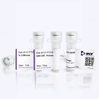
Product Image
Application
in vivo CD8+ T cell depletion; Immunofluorescence; Flow cytometry; Western blot


Chemical Information
-
Appearance Liquid
-
Molecular Weight 154 kDa
-
Color Colorless to light yellow
-
SMILES
[Anti-Mouse CD8a Antibody (53-6.7)]
-
Shipping
Shipping with dry ice.
-
Formulation
Please refer to the lot-specific COA for specific buffer information.
-
Storage
Please store the product under the recommended conditions in the Certificate of Analysis.


Publications (4)
-
Journal Impact Factor
-
Most Recent
-
J Clin Invest
Asparagine drives immune evasion in bladder cancer via RIG-I stability and type I IFN signaling. [Abstract]2025 Feb 18;135(8):e186648. PMID: 39964752 -
Adv Sci (Weinh)
Materials-Guided Gene-Ionizable Lipid Nanoparticles to Reverse Iron-Associated Immune Resistance in Renal Cancer. [Abstract]2026 Jun 9:e00078. PMID: 42261844 -
Proc Natl Acad Sci U S A
Bioresponsive immunomodulator nanocomplex for selective immunoengineering in metastatic lymph nodes. [Abstract]2026 Jan 27;123(4):e2519625123. PMID: 41557810 -
J Agric Food Chem
Combination of Ginsenoside Compound K and Anti-PD1 Inhibits Pancreatic Cancer by Activating CXCL13-CXCR5 Signaling Axis. [Abstract]2025 Jun 18;73(24):15125-15137. PMID: 40474585

Protocols
-
Kinase activity and phosphorylation assays
Kinase activity assays measure the ability of kinases to transfer phosphate groups from ATP to specific substrates, while phosphorylation assays detect the presence and levels of phosphorylated proteins. Common methods include radiolabeled ATP incorporation (e. g. ,), ADP release detection via bioluminescence (e. g. ,[3]), enzyme-linked immunosorbent assays (ELISA) for phospho-specific epitopes (e. g. ,[6]), and microtiter-based formats for high-throughput screening (e. g. ,[8]). The ADP-Glo assay quantifies kinase activity by measuring ADP produced during phosphorylation using a luciferase-based system. Radiometric assays involve autoradiography or scintillation counting after incorporation of 32P-labeled ATP into substrate proteins. ELISA-based approaches rely on phospho-specific antibodies to detect activated kinases in cell lysates or purified samples.
-
Western Blot
Western blotting (WB) is a commonly used experimental method in molecular biology, biochemistry, and immunogenetics for identifying and quantifying target proteins. It combines gel electrophoresis with immunoassay, enabling researchers to analyze protein expression, post-translational modifications, and molecular weight.
-
Protocol for Kinase activity and phosphorylation assays
Kinase activity assays measure transfer of phosphate from ATP to a protein or peptide substrate, generating phosphorylated substrate, ADP, or incorporated radiolabeled phosphate as the readout; phosphorylation assays measure site-specific phosphorylation in cells or tissues as a proxy for kinase-pathway activation, inhibition, or substrate regulation. Phosphorylation can be detected by phospho-specific Western blot, immunoprecipitation kinase assay, phospho-immunofluorescence, phospho-flow cytometry, luminescent ADP detection, radiolabeled ATP incorporation, or reporter-based pathway assays, and these readouts can be applied to cancer cells, primary neurons, mouse tumors, organoids, inflammatory macrophages, ferroptosis studies, and mitophagy studies when the kinase target is biologically relevant.
-
Research Protocol for Inflammation-related Diseases
The NLRP3 inflammasome is a cytosolic innate immune signaling platform that integrates priming signals and danger-signal activation to promote caspase-1 activation, maturation of IL-1β and IL-18, and gasdermin D-mediated pyroptotic cell death. The core experimental logic is to determine whether inflammatory disease phenotypes are driven by increased NLRP3 expression, ASC-containing inflammasome assembly, caspase-1 cleavage, GSDMD cleavage, and extracellular release of IL-1β/IL-18 rather than by nonspecific cell injury alone. The pathway is strongly linked to inflammation-related disease phenotypes because monosodium urate crystals activate NALP3/NLRP3 inflammasome signaling in gout-like crystal inflammation, cholesterol crystals activate NLRP3 inflammasomes in atherogenesis models, and DSS-induced intestinal inflammation has been reported to involve NLRP3 inflammasome activity. However, experimental colitis studies also show context-dependent protective effects of NLRP3 inflammasome co


Purity & Documentation
-
Data Sheet (262 KB)
-
SDS (251 KB)
- English - EN (251 KB)
- Français - FR (251 KB)
- Deutsch - DE (251 KB)
- Norwegian - NO (251 KB)
- Español - ES (251 KB)
- Swedish - SV (251 KB)
- Italian - IT (251 KB)
- Korean - KR (251 KB)
- Portuguese - PT (251 KB)
-
Inhibitory Antibodies User Guide (603 KB)
References
[1]. Devine L, et al. Location of the epitope for an anti-CD8alpha antibody 53.6.7 which enhances CD8alpha-MHC class I interaction indicates antibody stabilization of a higher affinity CD8 conformation. Immunol Lett. 2004 May 15;93(2-3):123-30. [Content Brief]
[2]. Madi A, et al. CD8 agonism functionally activates memory T cells and enhances antitumor immunity. Int J Cancer. 2022 Sep 1;151(5):797-808. [Content Brief]
[3]. Uddin MN, et al. TNF-α-dependent hematopoiesis following Bcl11b deletion in T cells restricts metastatic melanoma. J Immunol. 2014 Feb 15;192(4):1946-53. [Content Brief]

Calculators
Concentration (start) × Volume (start) = Concentration (final) × Volume (final)


Anti-Mouse CD8a Antibody (53-6.7) Related Classifications
HY-P99129 Related Classifications
 Powered by Bioz