



Anti-Mouse Vβ8 TCR Antibody (F23.1)
Anti-Mouse Vβ8 TCR Antibody (F23.1) is an anti-mouse Vβ8 TCR IgG2a monoclonal antibody. Anti-Mouse Vβ8 TCR Antibody (F23.1) can clear Vβ8+ T cells to alleviate immune conditions. Anti-Mouse Vβ8 TCR Antibody (F23.1) can be used for research on autoimmune conditions. Anti-Mouse Vβ8 TCR Antibody (F23.1) is often used in flow cytometry.
For research use only. We do not sell to patients.
- Molecular Weight:150 kDa
-
Storage:
Please store the product under the recommended conditions in the Certificate of Analysis.

To place orders, for customer services and technical support, please contact: MedChemExpress USA
Tel: 609-228-6898 E-mail: [email protected] [email protected]


-
 Biological Activity
-
Chemical Information
- Protocols
- Purity & Documentation
- References
-
 Help & FAQs




Biological Activity
Description
Isotype
Mouse IgG2a kappa
Recommend Isotype Controls
Species Reactivity
Mouse
In Vivo
MedChemExpress (MCE) has not independently confirmed the accuracy of these methods. They are for reference only.
-
Animal Model:Female MRL/Mp-lpr/lpr mice[1]
-
Dosage:100 μg
-
Administration:Intraperitoneal injection (i.p.), twice every 2 days, for 10-12 weeks
-
Result:Significantly prolonged the survival period.
Reduced proteinuria concentration, incidence of arthritis and skin ulcers.
Significantly reduced the accumulation of Vβ8+ and CD4-CD8- T cells.
Conjugated
Unconjugated
Reconsititution
The product can be reconstituted/diluted with sterile PBS or saline.
Format
-
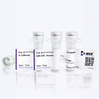
Product Image
Application
Flow cytometry


Chemical Information
-
Molecular Weight 150 kDa
-
SMILES
[Anti-Mouse Vβ8 TCR Antibody (F23.1)]
-
Formulation
Please refer to the lot-specific COA for specific buffer information.
-
Storage
Please store the product under the recommended conditions in the Certificate of Analysis.

Protocols
-
Flow Cytometry
Flow cytometry (FC) is a technique for high-speed, step-by-step quantitative analysis and sorting of single cells or other biological particles in a suspension by detecting labeled fluorescent signals.
-
Flow cytometric DNA-content cell-cycle staining
Flow cytometric DNA-content cell-cycle staining measures the fluorescence intensity of DNA-bound fluorochromes in single cells or nuclei to estimate DNA content distributions, allowing assignment of populations to G0/G1, S, and G2/M phases by DNA histogram deconvolution. Propidium iodide (PI) intercalates into DNA, and PI fluorescence is proportional to cellular DNA content when staining is performed under conditions that make DNA accessible and minimize non-DNA signal. Cells with G2/M DNA content are expected to show approximately twice the fluorescence intensity of G0/G1 cells, while S-phase cells occupy intermediate fluorescence values. PI-based DNA-content analysis can also detect cells with fractional DNA content, often reported as sub-G1, when DNA fragmentation and extraction during staining reduce retained DNA signal in apoptotic cells. DAPI is an alternative DNA fluorochrome for univariate DNA-content analysis, while bivariate approaches combining DNA content with proliferation
-
Protocol for Phospho-flow cytometry
Phospho-flow cytometry detects intracellular phosphorylated signaling proteins in single cells using phospho-specific antibodies after rapid fixation and permeabilization; the fluorescence intensity reflects phosphorylation state and therefore kinase-pathway activation, inhibition, or drug response in defined cell subsets. Unlike Western blot, phospho-flow preserves single-cell resolution and can measure signaling heterogeneity in cancer cells, primary immune cells, dissociated mouse tumors, macrophages, organoid-derived cells, and drug-screening samples when validated antibodies and fixation/permeabilization conditions are used.


Purity & Documentation
References
[1]. de Alborán IM, et al. Attenuation of autoimmune disease and lymphocyte accumulation in MRL/lpr mice by treatment with anti-V beta 8 antibodies. Eur J Immunol. 1992 Aug;22(8):2153-8. [Content Brief]
[2]. Yang Y, et al. Focused specificity of intestinal TH17 cells towards commensal bacterial antigens. Nature. 2014 Jun 5;510(7503):152-6. [Content Brief]

Calculators
Concentration (start) × Volume (start) = Concentration (final) × Volume (final)


Anti-Mouse Vβ8 TCR Antibody (F23.1) Related Classifications
HY-P990141 Related Classifications
 Powered by Bioz