



ABCG2 Antibody (YA648)

Based on 1 Customer Validation
ABCG2 Antibody (YA648) is a Rabbit-derived and non-conjugated IgG monoclonal antibody, targeting to ABCG2.
-
Host:
Rabbit
-
Isotype:
IgG
-
Application:
WB, ICC/IF, IHC-P, FC
-
Reactivity :
Human, Mouse
-
Formulation:
Supplied in 1*TBS (pH7.4), 0.05% BSA and 40% Glycerol. Preservative: 0.05% Sodium Azide.
-
Conjugation:
Non-conjugated




To place orders, for customer services and technical support, please contact: MedChemExpress USA
Tel: 609-228-6898 E-mail: [email protected] [email protected]





Applications
| Application |
WB

WB: Western Blot
|
ICC/IF
ICC/IF: Immunocytochemistry/
Immunofluorescence |
IHC-P
IHC-P: Immunohistochemistry-Paraffin
|
FC
FC: Flow Cytometry
|
|---|---|---|---|---|
| Dilution Ratio | 1:500-1:2000 | 1:50-1:200 | 1:50-1:200 | 1:50-1:100 |


Product Details
ABCG2 Antibody (YA648) is a Rabbit-derived and non-conjugated IgG monoclonal antibody, targeting to ABCG2.
-
Host Rabbit

-
Clonality Recombinant, Monoclonal

-
Species ReactivityHuman, Mouse
-
Observed Molecular WeightObserved band size: 72 kDa
Note: Due to possible protein modifications or aggregation, the molecular weight should be confirmed by actual measurement, and the predicted value is for reference only.

-
Calculated Molecular Weight Predicted band size: 72 kDa

Entrez Gene: 9429 Human ; 26357 Mouse ; 312382 Rat
SwissProt: Q9UNQ0 Human ; Q7TMS5 Mouse ; Q80W57 Rat
OMIM: 138900 Human
Synthetic peptide corresponding to Human ABCG2.AA range:140-180.
Endogenous
Protein A affinity purified.
Non-conjugated
Unmodified
IgG


Product Properties
-
Appearance
Liquid
-
Formulation
Supplied in 1*TBS (pH7.4), 0.05% BSA and 40% Glycerol. Preservative: 0.05% Sodium Azide.
-
Concentration
Batch-dependent, Please check the COA for the concentration of each lot. Check Lot Concentration
-
Storage & Stability
Stored at -20°C for 1 year. Avoid repeated freeze / thaw cycles.
-
Shipping
Shipping with blue ice.


Verification Images
-
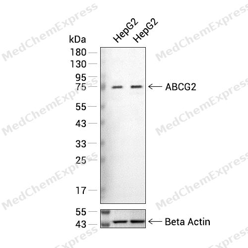 Western blot analysis of extracts from HepG2 (lane 2(20μg) and HepG2 (lane 3(40μg)using ABCG2 (HY-P80002) Rabbit mAb. Proteins were transferred to a PVDF membrane and blocked with 5% defatted milk powder in TBST for 2 hour at room temperature. The primary antibody (HY-P80002, 1/1000) and Loading control antibody (Beta Actin, HY-P80438, 1/3000) was used in 5% defatted milk powder in TBST at 4°C overnight. Goat Anti-Mouse/Rabbit IgG-HRP Secondary Antibody (HY-P8004/HY-P8001, 1/10,000) was used for 1 hour at room temperature.
 -
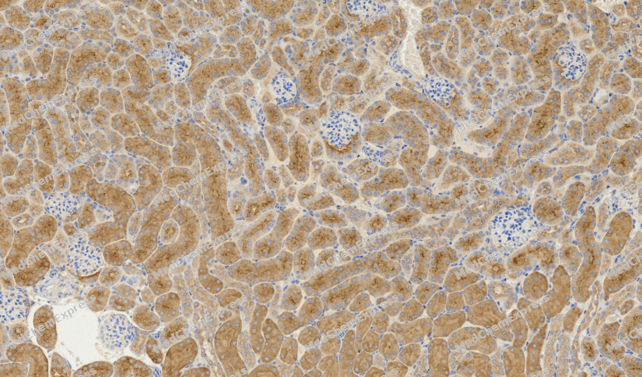 Immunohistochemical analysis of paraffin-embedded Mouse kidney tissue using ABCG2 Antibody. The section was pre-treated using heat mediated antigen retrieval with sodium citrate buffer (pH 6.0) for 8 minutes. The tissues were blocked in QuickBlock for 20 minutes at room temperature, washed with ddH2O and PBS, and then probed with the primary antibody (HY-P80002, 1/100) in 4℃ overnight. The detection was performed using an HRP conjugated compact polymer system. DAB was used as the chromogen. Tissues were counterstained with hematoxylin and mounted with DPX.
 -
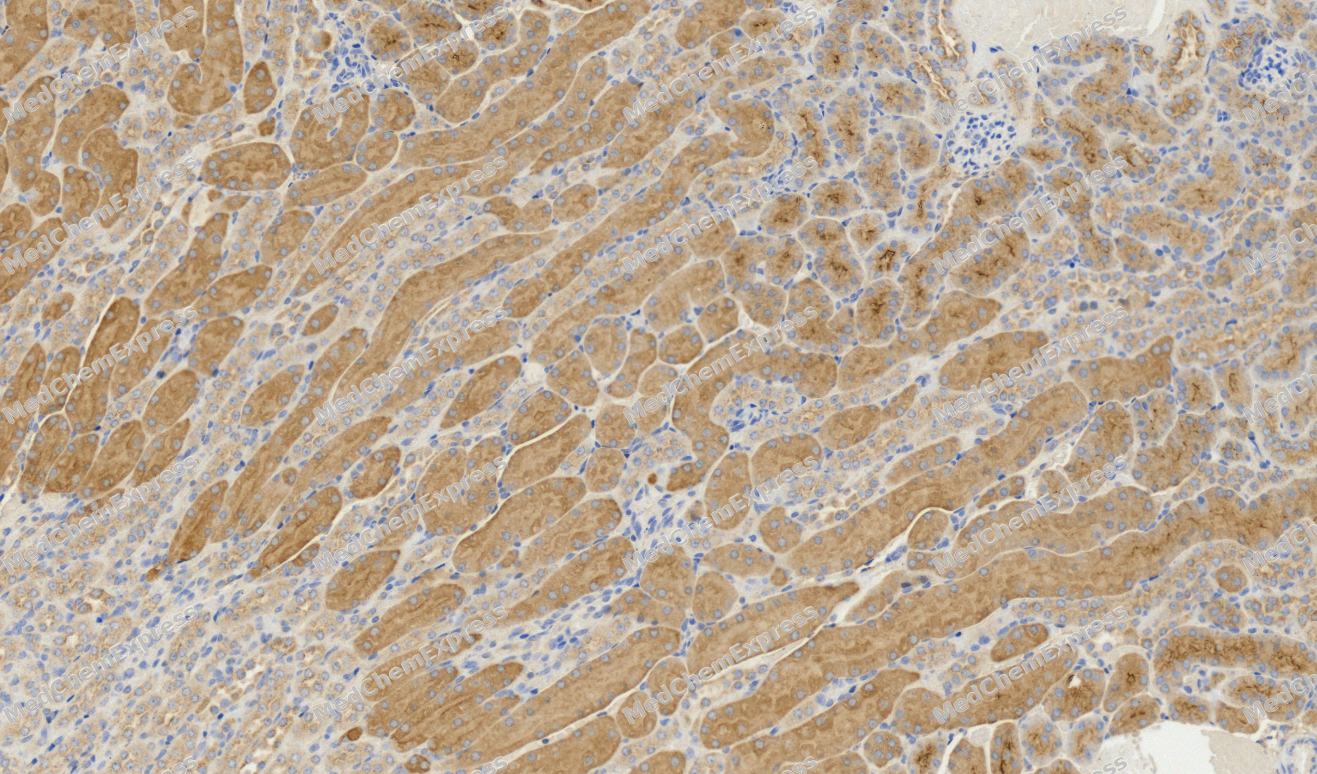
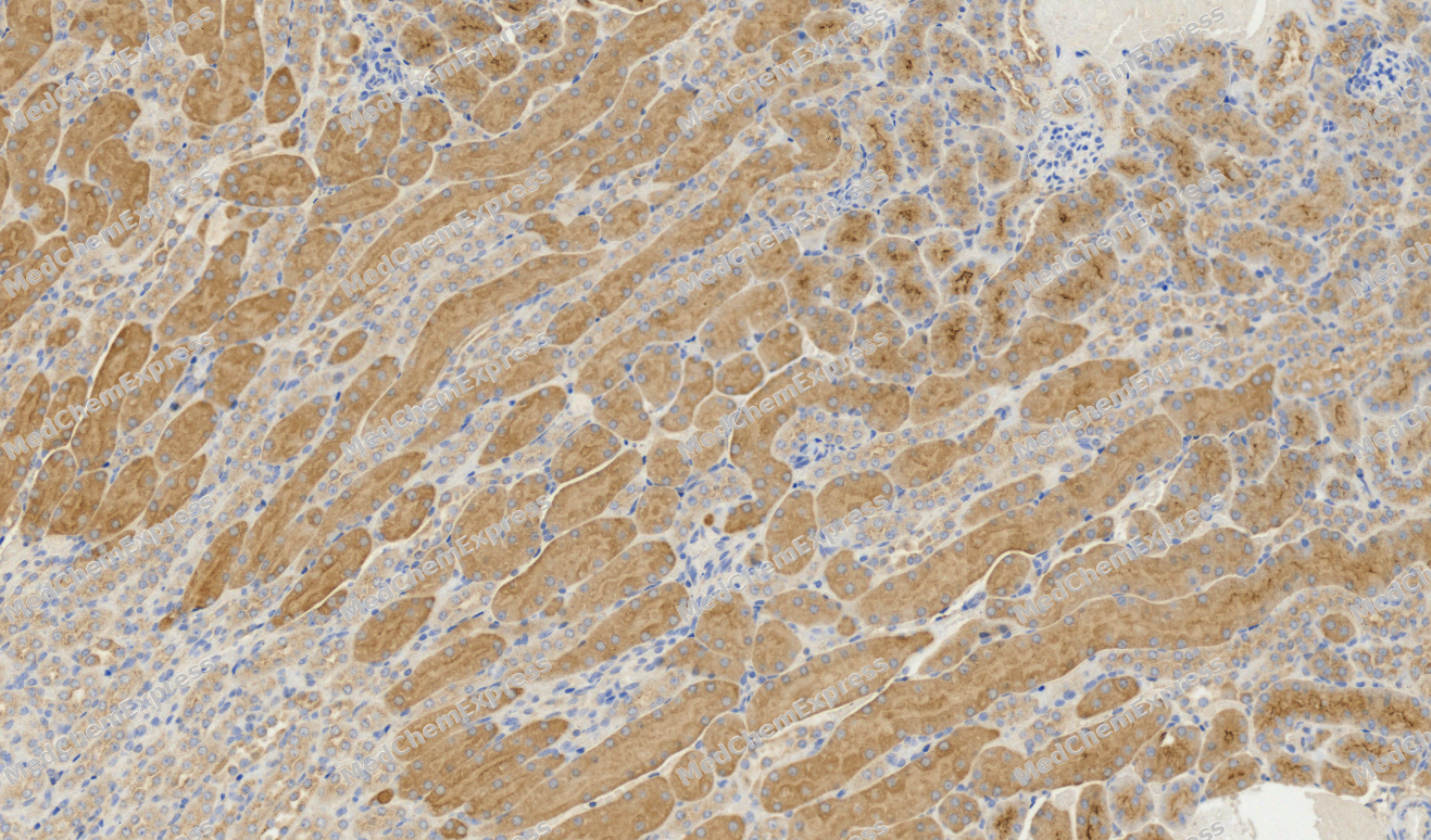 Immunohistochemical analysis of paraffin-embedded Mouse kidney tissue using ABCG2 Antibody. The section was pre-treated using heat mediated antigen retrieval with sodium citrate buffer (pH 6.0) for 8 minutes. The tissues were blocked in QuickBlock for 20 minutes at room temperature, washed with ddH2O and PBS, and then probed with the primary antibody (HY-P80002, 1/100) in 4℃ overnight. The detection was performed using an HRP conjugated compact polymer system. DAB was used as the chromogen. Tissues were counterstained with hematoxylin and mounted with DPX.
 -
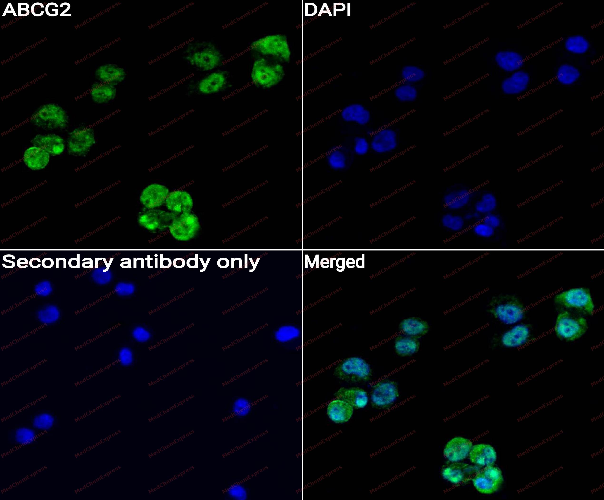 Immunocytochemistry analysis of Hela cells labeling ABCG2 with ABCG2 antibody (HY-P80002) at 1/50 dilution. Cells were fixed in 4% paraformaldehyde for 15 minutes at room temperature, permeabilized with 0.1% Triton X-100 for 10 minutes at room temperature, then blocked with QuickBlock™ Blocking Buffer for Immunol Staining for 10 min at room temperature. Cells were then incubated with ABCG2 antibody (HY-P80002) at 1/50 dilution in QuickBlock™ Blocking Buffer for Immunol Staining at 4 ℃. Alexa Fluor® 488-conjugated AffiniPure Goat Anti-Rabbit IgG H&L(HY-P8002, Green) was used as the secondary antibody at 1/1,000 dilution. PBS instead of the primary antibody was used as the secondary antibody only control. The Nuclear counterstain was DAPI (Blue).
 -
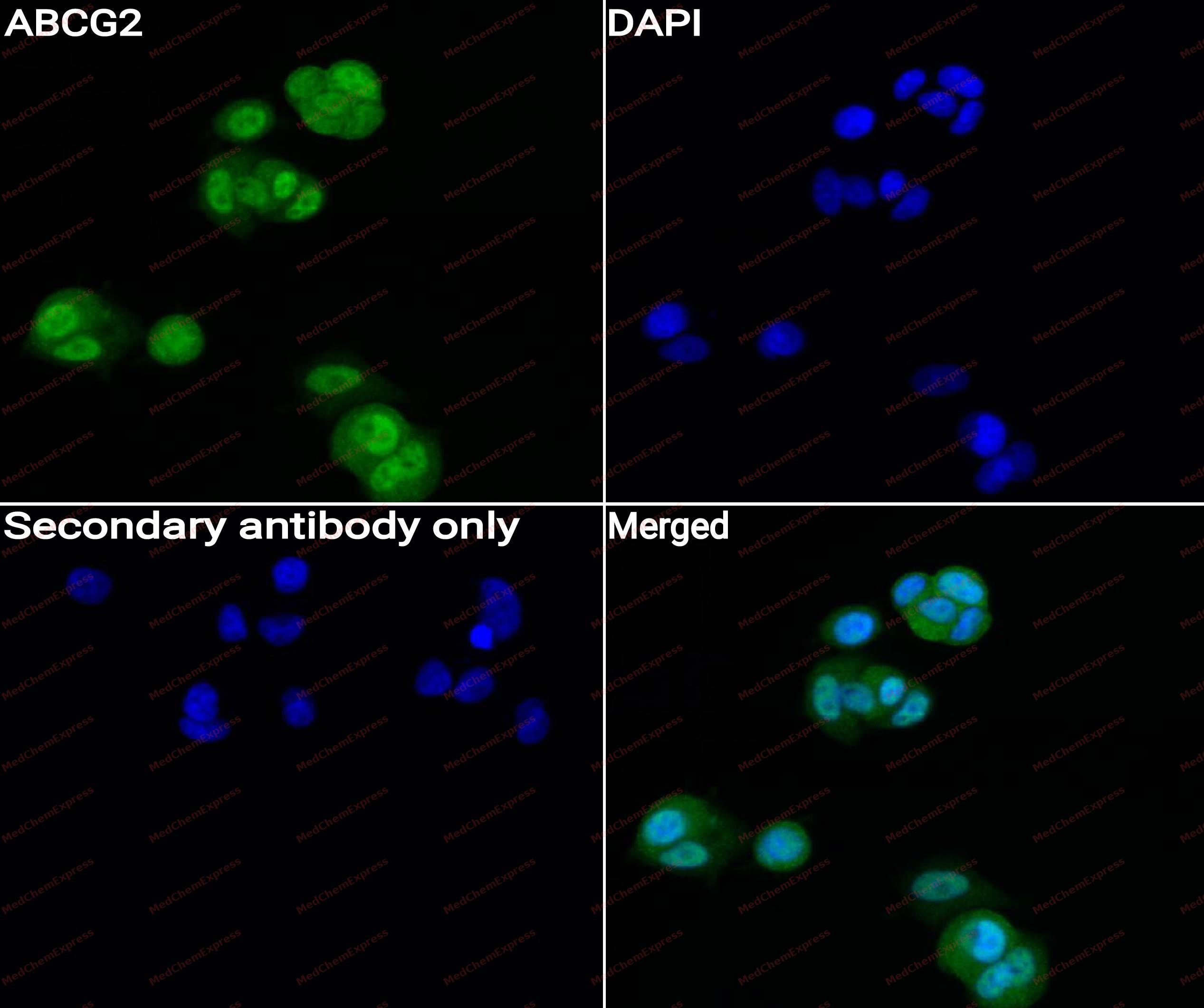 Immunocytochemistry analysis of MCF-7 cells labeling ABCG2 with ABCG2 antibody (HY-P80002) at 1/50 dilution. Cells were fixed in 4% paraformaldehyde for 15 minutes at room temperature, permeabilized with 0.1% Triton X-100 for 10 minutes at room temperature, then blocked with QuickBlock™ Blocking Buffer for Immunol Staining for 10 min at room temperature. Cells were then incubated with ABCG2 antibody (HY-P80002) at 1/50 dilution in QuickBlock™ Blocking Buffer for Immunol Staining at 4 ℃. Alexa Fluor® 488-conjugated AffiniPure Goat Anti-Rabbit IgG H&L(HY-P8002, Green) was used as the secondary antibody at 1/1,000 dilution. PBS instead of the primary antibody was used as the secondary antibody only control. The Nuclear counterstain was DAPI (Blue).
 -
 Flow cytometric analysis of 1X10⁶ A549 cells labeling ABCG2 Antibody(HY-P80002, red). Cells were stained with the primary antibody at 1/50 dilution for an hour at 4℃. AF488-conjugated Goat Anti-Rabbit IgG H&L (HY-P8002) was used as the secondary antibody at 1/1,000 dilution for 30 minutes at 4℃. Rabbit IgG Isotype Control (HY-P80879, blue) was used as the isotype control, cells without incubation with primary antibody were used as the unlabeled control (black).


Background
-
Function
ABCG2 is a Broad substrate specificity ATP-dependent transporter of the ATP-binding cassette (ABC) family that actively extrudes a wide variety of physiological compounds, dietary toxins and xenobiotics from cells. Involved in porphyrin homeostasis, mediating the export of protoporphyrin IX (PPIX) from both mitochondria to cytosol and cytosol to extracellular space, it also functions in the cellular export of heme. Also mediates the efflux of sphingosine-1-P from cells. Acts as a urate exporter functioning in both renal and extrarenal urate excretion. In kidney, it also functions as a physiological exporter of the uremic toxin indoxyl sulfate. Also involved in the excretion of steroids like estrone 3-sulfate/E1S, 3beta-sulfooxy-androst-5-en-17-one/DHEAS, and other sulfate conjugates. Mediates the secretion of the riboflavin and biotin vitamins into milk. Extrudes pheophorbide a, a phototoxic porphyrin catabolite of chlorophyll, reducing its bioavailability. Plays an important role in the exclusion of xenobiotics from the brain (Probable). It confers to cells a resistance to multiple drugs and other xenobiotics including mitoxantrone, pheophorbide, camptothecin, methotrexate, azidothymidine, and the anthracyclines daunorubicin and doxorubicin, through the control of their efflux. In placenta, it limits the penetration of drugs from the maternal plasma into the fetus. May play a role in early stem cell self-renewal by blocking differentiation. In inflammatory macrophages, exports itaconate from the cytosol to the extracellular compartment and limits the activation of TFEB-dependent lysosome biogenesis involved in antibacterial innate immune response[1][2][3][4][5][6][7][8][9][10][11][12][13][14][15][16][17].
-
Subcellular Localization
Cell membrane; Multi-pass membrane protein; Apical cell membrane; Multi-pass membrane protein; Mitochondrion membrane; Multi-pass membrane protein
-
Expression
Tissue_specificity:High expression in the placenta (PubMed: 9850061) . Low expression in the small intestine, liver, and colon (PubMed: 9861027) . Expression in brain tissue (protein level) (PubMed: 12958161) . -
Subunit
Homodimer; disulfide-linked (PubMed:15001581, PubMed:17686774, PubMed:18056989, PubMed:28554189). The minimal functional unit is a homodimer, but the major oligomeric form in plasma membrane is a homotetramer with possibility of higher order oligomerization up to homododecamers (PubMed:15001581)
-
SwissProt ID
-
Research Field
Stem Cells


Documentation
-
Data Sheet (235 KB)
-
SDS (252 KB)
- English - EN (252 KB)
- Français - FR (252 KB)
- Deutsch - DE (252 KB)
- Norwegian - NO (252 KB)
- Español - ES (252 KB)
- Swedish - SV (252 KB)
- Italian - IT (252 KB)
- Korean - KR (252 KB)
- Portuguese - PT (252 KB)
-
User Guide for Antibodies (1077 KB)

 Powered by Bioz
