



ADAM17 Antibody (YA1562)
(Synonyms: CD156b; ADAM17; CSVP; TACE)Based on 1 Customer Validation
ADAM17 Antibody (YA1562) is a Rabbit-derived and non-conjugated IgG monoclonal antibody, targeting to ADAM17.
-
Host:
Rabbit
-
Isotype:
IgG
-
Application:
WB, ICC/IF, IP, FC
-
Reactivity :
Human, Mouse, Rat
-
Formulation:
Supplied in 10mM PBS, pH 7.4, 150mM sodium chloride, 0.05% BSA, 0.02% sodium azide and 50% glycerol.
-
Conjugation:
Non-conjugated


To place orders, for customer services and technical support, please contact: MedChemExpress USA
Tel: 609-228-6898 E-mail: [email protected] [email protected]





Applications
| Application |
WB

WB: Western Blot
|
ICC/IF
ICC/IF: Immunocytochemistry/
Immunofluorescence |
IP
IP: Immunoprecipitation
|
FC
FC: Flow Cytometry
|
|---|---|---|---|---|
| Dilution Ratio | 1:500-1:1000 | 1:50-1:200 | 1:50 | 1:50-1:100 |


Product Details
ADAM17 Antibody (YA1562) is a Rabbit-derived and non-conjugated IgG monoclonal antibody, targeting to ADAM17.
-
Host Rabbit

-
Clonality Recombinant,Monoclonal

-
Species ReactivityHuman, Mouse, Rat
-
Observed Molecular WeightObserved band size: 100 kDa
Note: Due to possible protein modifications or aggregation, the molecular weight should be confirmed by actual measurement, and the predicted value is for reference only.

-
Calculated Molecular Weight Predicted band size: 93 kDa

A synthesized peptide derived from human ADAM17 aa750-800/824.
Endogenous
Affinity Chromatography
Non-conjugated
Unmodified
IgG


Product Properties
-
Appearance
Solution
-
Formulation
Supplied in 10mM PBS, pH 7.4, 150mM sodium chloride, 0.05% BSA, 0.02% sodium azide and 50% glycerol.
-
Concentration
Batch-dependent, Please check the COA for the concentration of each lot. Check Lot Concentration
-
Storage & Stability
Stored at -20°C for 1 year. Avoid repeated freeze / thaw cycles.
-
Shipping
Shipping with blue ice.


Verification Images
-
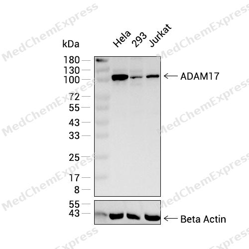 Western blot analysis was performed on extracts from Hela (lane 1, 20 μg), 293 (lane 2, 20 μg), and Jurkat (lane 3, 20 μg) using ADAM17 Rabbit mAb.Proteins were transferred to a PVDF membrane and blocked with 5% non - fat milk in TBST at 4°C overnight.The primary antibody (1:1000 dilution) and the loading control antibody (beta-Actin, HY-P83730, 1:5000 dilution) were incubated in 5% non-fat milk in TBST for 1 hour at 37°C.Goat Anti - Rabbit IgG - HRP Secondary Antibody (1:20000 dilution) was then applied for 40 minutes at 37°C.
 -
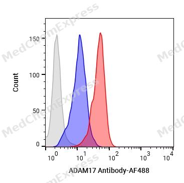 Flow cytometric analysis of 1X10⁶ HeLa cells labeling ADAM17 Antibody (HY-P81817, red). Cells were fixed with 4% paraformaldehyde and permeabilised with 90% methanol. Then stained with the primary antibody at 1/100 dilution for an hour at 4℃. AF488-conjugated Goat Anti-Rabbit IgG H&L (HY-P8002) was used as the secondary antibody at 1/1,000 dilution for 30 minutes at 4℃. Rabbit IgG Isotype Control (HY-P80879, blue) was used as the isotype control, cells without incubation with primary antibody were used as the unlabeled control (black).
 -
 Immunocytochemistry analysis of HepG2 cells labeling ADAM17 with ADAM17 Antibody (HY-P81817) at 1/100 dilution. Cells were fixed in 4% paraformaldehyde for 15 minutes at room temperature, permeabilized with 0.1% Triton X-100 in PBS for 15 minutes at room temperature, then blocked with quick block buffer for 10 minutes at room temperature. Cells were then incubated with ADAM17 Antibody (HY-P81817) at 1/100 dilution in quick block buffer overnight at 4 ℃. AF488-conjugated Goat Anti-Rabbit IgG H&L(HY-P8002, Green) was used as the secondary antibody at 1/1,000 dilution. PBS instead of the primary antibody was used as the secondary antibody only control. The Nuclear counterstain was DAPI (Blue).
 -
 Immunocytochemistry analysis of HepG2 cells labeling ADAM17 with ADAM17 Antibody (HY-P81817) at 1/200 dilution. Cells were fixed in 4% paraformaldehyde for 15 minutes at room temperature, permeabilized with 0.1% Triton X-100 in PBS for 15 minutes at room temperature, then blocked with quick block buffer for 10 minutes at room temperature. Cells were then incubated with ADAM17 Antibody (HY-P81817) at 1/200 dilution in quick block buffer overnight at 4 ℃. AF488-conjugated Goat Anti-Rabbit IgG H&L(HY-P8002, Green) was used as the secondary antibody at 1/1,000 dilution. PBS instead of the primary antibody was used as the secondary antibody only control. The Nuclear counterstain was DAPI (Blue).


Background
-
Function
ADAM17 (A Disintegrin and Metalloprotease 17), also known as TNF-α-converting enzyme (TACE), is a membrane-associated metalloprotease that regulates cell-cell communication through ectodomain shedding of cytokines, receptors, adhesion molecules, and growth factor ligands[1][2]. Mechanistically, ADAM17 is a major sheddase for tumor necrosis factor-α (TNF-α), interleukin-6 receptor (IL-6R), and multiple epidermal growth factor receptor (EGFR) ligands, thereby coordinating inflammatory, regenerative, and proliferative signaling pathways[2][3][4]. Through the generation of soluble TNF-α and soluble IL-6R, ADAM17 functions as a central signaling hub linking immune activation with systemic inflammatory responses[2][4]. In addition, ADAM17-dependent shedding of EGFR ligands regulates EGFR signaling in epithelial tissues and contributes to processes associated with tissue homeostasis, development, and tumor progression[3][5][6]. Disease studies further demonstrate that ADAM17 participates in inflammation, cancer, and other pathological conditions through its broad substrate repertoire and downstream signaling effects[1][2][6]. Compared with the closely related metalloprotease ADAM10, ADAM17 displays distinct substrate specificity and serves as the predominant sheddase for amphiregulin, transforming growth factor-α (TGF-α), epiregulin, and heparin-binding EGF-like growth factor (HB-EGF), whereas ADAM10 preferentially processes other EGFR ligands[7][8]. This functional distinction is important for experimental design because selective modulation of ADAM17 can differentially affect TNF-α, IL-6, and EGFR signaling networks[2][7]. For research applications, ADAM17 inhibitors and blocking antibodies are widely used to investigate cytokine release, inflammatory signaling, EGFR activation, and tumor-associated microenvironmental interactions in cellular and animal models[2][9].
-
Subcellular Localization
Cell membrane; Single-pass type I membrane protein
-
Expression
Tissue_specificity:Widely expressed. Highest expression levels are found in the human heart, placenta, skeletal muscle, pancreas, spleen, thymus, prostate, testes, ovaries, and small intestine; highest expression levels are also found in the fetal brain, lungs, liver, and kidneys. It is also expressed in natural killer cells (protein level) (PubMed:24337742) .
Induction:In arthritis-affected cartilage -
Isoforms & Post-Translational Modification
P78536 has 2 isomers: P78536-1: 93021 Da (predicted); P78536-2: 78543 Da (predicted).
The precursor is cleaved by a furin endopeptidase;Phosphorylated. Stimulation by growth factor or phorbol 12-myristate 13-acetate induces phosphorylation of Ser-819 but decreases phosphorylation of Ser-791. Phosphorylation at Thr-735 by MAPK14 is required for ADAM17-mediated ectodomain shedding -
Subunit
Interacts with MAD2L1, MAPK14 and MUC1 (PubMed:12441351, PubMed:20188673). Interacts with iRhom1/RHBDF1 and iRhom2/RHBDF2 (PubMed:29897333). Interacts with FRMD8 via its interaction with iRhom1/RHBDF1 and iRhom2/RHBDF2 (PubMed:29897333). Interacts with TSPAN8 (PubMed:36078095)
-
SwissProt ID
-
Synonyms
CD156b; ADAM17; CSVP; TACE
-
Research Field
Neuroscience


Documentation
-
Data Sheet (262 KB)
-
SDS (252 KB)
- English - EN (252 KB)
- Français - FR (252 KB)
- Deutsch - DE (252 KB)
- Norwegian - NO (252 KB)
- Español - ES (252 KB)
- Swedish - SV (252 KB)
- Italian - IT (252 KB)
- Korean - KR (252 KB)
- Portuguese - PT (252 KB)
-
User Guide for Antibodies (1077 KB)
References
[1]. Zunke F, et al. The shedding protease ADAM17: Physiology and pathophysiology. Biochim Biophys Acta Mol Cell Res. 2017 Nov;1864(11 Pt B):2059-2070. [Content Brief]
[2]. Schumacher N, et al. ADAM17 orchestrates Interleukin-6, TNFα and EGF-R signaling in inflammation and cancer. Biochim Biophys Acta Mol Cell Res. 2022 Jan;1869(1):119141. [Content Brief]
[3]. Rose-John S. ADAM17, et al. Pharmacol Res. 2013;71:19-22.
[4]. Liu L, et al. ADAM17 as a promising therapeutic target: from structural basis to inhibitor discovery in human diseases. Front Pharmacol. 2025 Sep 19;16:1640090. [Content Brief]
[5]. Lee DC, et al. TACE/ADAM17 processing of EGFR ligands indicates a role as a physiological convertase. Ann N Y Acad Sci. 2003 May;995:22-38. [Content Brief]
[6]. Gnosa SP, et al. ADAM17-mediated EGFR ligand shedding directs macrophage-promoted cancer cell invasion. JCI Insight. 2022 Sep 22;7(18):e155296. [Content Brief]
[7]. Sahin U, et al. Distinct roles for ADAM10 and ADAM17 in ectodomain shedding of six EGFR ligands. J Cell Biol. 2004 Mar 1;164(5):769-79. [Content Brief]
[8]. Hosur V, et al. ADAM17 is essential for ectodomain shedding of the EGF-receptor ligand amphiregulin. FEBS Open Bio. 2018 Mar 12;8(4):702-710. [Content Brief]
[9]. Moss ML, et al. Recent Advances in ADAM17 Research: A Promising Target for Cancer and Inflammation. Mediators Inflamm. 2017;2017:9673537. [Content Brief]

 Powered by Bioz
