



ATP Citrate Lyase Antibody (YA596)
(Synonyms: ATP-citrate synthase, ATP-citrate (pro-S-)-lyase, Citrate cleavage enzyme, ACL, ACLY)
Based on 1 Customer Validation
ATP Citrate Lyase Antibody (YA596) is a Rabbit-derived and non-conjugated IgG monoclonal antibody, targeting to ATP Citrate Lyase.
-
Host:
Rabbit
-
Isotype:
IgG
-
Application:
WB, ICC/IF, IHC-P, IP, FC
-
Reactivity :
Human, Mouse
-
Formulation:
Supplied in 1*TBS (pH7.4), 0.05% BSA and 40% Glycerol. Preservative: 0.05% Sodium Azide.
-
Conjugation:
Non-conjugated


To place orders, for customer services and technical support, please contact: MedChemExpress USA
Tel: 609-228-6898 E-mail: [email protected] [email protected]





Applications
| Application |
WB

WB: Western Blot
|
ICC/IF
ICC/IF: Immunocytochemistry/
Immunofluorescence |
IHC-P
IHC-P: Immunohistochemistry-Paraffin
|
FC
FC: Flow Cytometry
|
IP
IP: Immunoprecipitation
|
|---|---|---|---|---|---|
| Dilution Ratio | 1:500-1:2000 | 1:50-1:200 | 1:50-1:200 | 1:50-1:200 | Use at an assay dependent concentration. |


Product Details
ATP Citrate Lyase Antibody (YA596) is a Rabbit-derived and non-conjugated IgG monoclonal antibody, targeting to ATP Citrate Lyase.
-
Host Rabbit

-
Clonality Recombinant,Monoclonal

-
Species ReactivityHuman, Mouse
-
Observed Molecular WeightObserved band size: 121 kDa
Note: Due to possible protein modifications or aggregation, the molecular weight should be confirmed by actual measurement, and the predicted value is for reference only.

-
Calculated Molecular Weight Predicted band size: 121 kDa

Entrez Gene: 47 Human ; 104112 Mouse ; 24159 Rat
SwissProt: P53396 Human ; Q91V92 Mouse ; P16638 Rat
OMIM: 108728 Human
Synthetic peptide within C-terminal human ATP citrate lyase.
Endogenous
Protein A affinity purified.
Non-conjugated
Unmodified
IgG


Product Properties
-
Appearance
Solution
-
Formulation
Supplied in 1*TBS (pH7.4), 0.05% BSA and 40% Glycerol. Preservative: 0.05% Sodium Azide.
-
Concentration
Batch-dependent, Please check the COA for the concentration of each lot. Check Lot Concentration
-
Storage & Stability
Stored at -20°C for 1 year. Avoid repeated freeze / thaw cycles.
-
Shipping
Shipping with blue ice.


Verification Images
-
 Western blot analysis was performed on protein extracts (25 μg) from Hela (lane 2), NIH/3T3 (lane 3), A549 (lane 4), A431 (lane 5), Jurkat (lane 6) and Raji (lane 7) using ATP Citrate Lyase antibody. Proteins were transferred onto a 0.22 μm PVDF membrane using a wet transfer system at a voltage of 100 V for 90 mins. The membrane was then blocked with 5% nonfat milk in TBST (HY-K1025) for 1 h at room temperature. The primary antibody (1:1000) and loading control antibody GAPDH Antibody (HRP) (HY-P80954A) (1:5000) were diluted in 5% nonfat milk in TBST and incubated with the membrane overnight at 4°C. After washing, the membrane of primary antibody was incubated with HRP-conjugated goat anti-rabbit/mouse IgG secondary antibody (HY-P8001/HY-P8004) (1:5000) diluted in 5% nonfat milk in TBST for 1 h at room temperature. Protein bands were visualized using an Ultra High Sensitivity ECL detection kit (HY-K1005).
 -
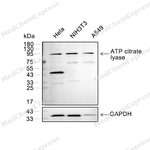 Western blot analysis of extracts from Hela(lane 2(20μg), NIH3T3(lane 3(20μg) and A549(lane 4(20μg) using ATP citrate lyase (HY-P80380) Rabbit mAb. Proteins were transferred to a PVDF membrane and blocked with 5% BSA in TBST for 2 hour at room temperature. The primary antibody (HY-P80380, 1/1000) and Loading control antibody (GAPDH, HY-P80954, 1/3000) was used in 5% BSA in TBST at 4°C overnight. Goat Anti-Mouse/Rabbit IgG-HRP Secondary Antibody (HY-P8004/HY-P8001, 1/10,000) was used for 1 hour at room temperature.
 -
 Flow cytometric analysis of 1X10⁶ NIH-3T3 cells labeling ATP Citrate Lyase Antibody(HY-P80380, red). Cells were fixed with 4% paraformaldehyde and permeabilised with 90% methanol. Then stained with the primary antibody at 1/100 dilution for an hour at 4℃. AF488-conjugated Goat Anti-Rabbit IgG H&L (HY-P8002) was used as the secondary antibody at 1/1,000 dilution for 30 minutes at 4℃. Rabbit IgG Isotype Control (HY-P80879, blue) was used as the isotype control, cells without incubation with primary antibody were used as the unlabeled control (black).
 -
 Immunocytochemistry analysis of Hela cells labeling ATP Citrate Lyase with ATP Citrate Lyase Antibody (HY-P80380) at 1/100 dilution. Cells were fixed in 4% paraformaldehyde for 15 minutes at room temperature, permeabilized with 0.1% Triton X-100 in PBS for 15 minutes at room temperature, then blocked with quick block buffer for 10 minutes at room temperature. Cells were then incubated with ATP Citrate Lyase Antibody (HY-P80380) at 1/100 dilution in quick block buffer overnight at 4 ℃. AF488-conjugated Goat Anti-Rabbit IgG H&L(HY-P8002, Green) was used as the secondary antibody at 1/1,000 dilution. PBS instead of the primary antibody was used as the secondary antibody only control. The Nuclear counterstain was DAPI (Blue).
 -
 Immunocytochemistry analysis of Hela cells labeling ATP Citrate Lyase with ATP Citrate Lyase Antibody (HY-P80380) at 1/200 dilution. Cells were fixed in 4% paraformaldehyde for 15 minutes at room temperature, permeabilized with 0.1% Triton X-100 in PBS for 15 minutes at room temperature, then blocked with quick block buffer for 10 minutes at room temperature. Cells were then incubated with ATP Citrate Lyase Antibody (HY-P80380) at 1/200 dilution in quick block buffer overnight at 4 ℃. AF488-conjugated Goat Anti-Rabbit IgG H&L(HY-P8002, Green) was used as the secondary antibody at 1/1,000 dilution. PBS instead of the primary antibody was used as the secondary antibody only control. The Nuclear counterstain was DAPI (Blue).


Background
-
Function
ATP-citrate lyase (ACLY) is a central metabolic enzyme and catalyses the ATP-dependent conversion of citrate and coenzyme A (CoA) to oxaloacetate and acetyl-CoA. The acetyl-CoA product is crucial for the metabolism of fatty acids, the biosynthesis of cholesterol, and the acetylation and prenylation of proteins. Acetyl CoA is also required for acetylation reactions that modify proteins, such as histone acetylation. ACLY is upregulated or activated in several types of cancers, and its inhibition is known to induce proliferation arrest in cancer cells both in vitro and in vivo.
ACLY links glucose and lipid metabolism by catalyzing the formation of acetyl-CoA and oxaloacetate from citrate produced by glycolysis in the presence of ATP and CoA. ACLY is aberrantly expressed in many immortalized cells and tumors, such as breast, liver, colon, lung and prostate cancers, and is correlated reversely with tumor stage and differentiation, serving as a negative prognostic marker. ACLY is an upstream enzyme of the long chain fatty acid synthesis, providing acetyl-CoA as an essential component of the fatty acid synthesis. Therefore, ACLY is a key enzyme of cellular lipogenesis and potent target for cancer therapy.
-
Subcellular Localization
Cytoplasm, cytosol
-
Subunit
Homotetramer
-
SwissProt ID
-
Synonyms
ATP-citrate synthase, ATP-citrate (pro-S-)-lyase, Citrate cleavage enzyme, ACL, ACLY
-
Research Field
Signal Transduction


Documentation
-
Data Sheet (234 KB)
-
SDS (251 KB)
- English - EN (251 KB)
- Français - FR (251 KB)
- Deutsch - DE (251 KB)
- Norwegian - NO (251 KB)
- Español - ES (251 KB)
- Swedish - SV (251 KB)
- Italian - IT (251 KB)
- Korean - KR (251 KB)
- Portuguese - PT (251 KB)
-
User Guide for Antibodies (1077 KB)

 Powered by Bioz
