



CD68 Antibody (YA798)
(Synonyms: CD 68 antibody; CD68 antibody; CD68 antigen antibody; CD68 molecule antibody; CD68_HUMAN antibody; DKFZp686M18236 antibody; gp11 antibody; Gp110 antibody; LAMP4 antibody; Macrophage antigen CD68 (microsialin) antibody; MACROPHAGE ANTIGEN CD68 antibody; Macrosialin antibody; SCARD1 antibody; Scavenger receptor class D member 1 antibody)
Based on 1 Customer Validation
CD68 Antibody (YA798) is a Mouse-derived and non-conjugated IgG1 monoclonal antibody, targeting to CD68.
-
Host:
Mouse
-
Isotype:
IgG
-
Application:
WB, IHC-P, FC
-
Reactivity :
Human
-
Formulation:
Supplied in 1*PBS (pH7.4), 0.2% BSA and 50% Glycerol. Preservative: 0.05% Sodium Azide.
-
Conjugation:
Non-conjugated


To place orders, for customer services and technical support, please contact: MedChemExpress USA
Tel: 609-228-6898 E-mail: [email protected] [email protected]





Applications
| Application |
WB

WB: Western Blot
|
IHC-P
IHC-P: Immunohistochemistry-Paraffin
|
FC
FC: Flow Cytometry
|
|---|---|---|---|
| Dilution Ratio | 1:1000-1:2000 | 1:50-1:200 | 1:50-1:100 |


Product Details
CD68 Antibody (YA798) is a Mouse-derived and non-conjugated IgG1 monoclonal antibody, targeting to CD68.
-
Host Mouse

-
Clonality Monoclonal

-
Species ReactivityHuman
-
Observed Molecular WeightObserved band size: 75-130 kDa
Note: Due to possible protein modifications or aggregation, the molecular weight should be confirmed by actual measurement, and the predicted value is for reference only.

-
Calculated Molecular Weight Predicted band size: 37 kDa;

Synthetic peptide corresponding to Human CD68.AA range:320-354.
Endogenous
Protein G affinity purified.
Non-conjugated
Unmodified
IgG


Product Properties
-
Appearance
Solution
-
Formulation
Supplied in 1*PBS (pH7.4), 0.2% BSA and 50% Glycerol. Preservative: 0.05% Sodium Azide.
-
Concentration
Batch-dependent, Please check the COA for the concentration of each lot. Check Lot Concentration
-
Storage & Stability
Stored at -20°C for 1 year. Avoid repeated freeze / thaw cycles.
-
Shipping
Shipping with blue ice.


Verification Images
-
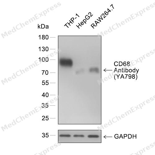 Western blot analysis of extracts from THP-1 (lane 1) and HepG2 (lane 2) and RAW264.7 (lane 3) using CD68 Antibody (YA798) antibody. Proteins were transferred to a PVDF membrane and blocked with 5% nonfat powdered milk in PBST for 2 hour at room temperature. The primary antibody (1/1000) and Loading control antibody (GAPDH, 1/3000) was diluted with 5% nonfat powdered milk in PBST at 4°C overnight. Goat Anti-Rabbit IgG-HRP Secondary Antibody (1/8,000) was incubated for 45min at room temperature.
 -
 Immunohistochemical analysis of paraffin-embedded human pancreatic cancer using CD68 antibody. The section was pre-treated using heat mediated antigen retrieval with EDTA (pH 9.0) for 14 minutes. The tissues were blocked in 5% BSA for 30 minutes at room temperature, washed with TBST, and then probed with the primary antibody (HY-P80066, 1/1000) for 30 minutes at room temperature. The detection was performed using Polymer HRP-conjugated Goat Anti-Mouse/Rabbit lgG(H&L) secondary antibody (HY-P83652). DAB was used as the chromogen. Tissues were counterstained with hematoxylin and mounted with DPX.
 -
 Immunohistochemical analysis of paraffin-embedded human non-hodgkin lymphoma using CD68 antibody. The section was pre-treated using heat mediated antigen retrieval with EDTA (pH 9.0) for 14 minutes. The tissues were blocked in 5% BSA for 30 minutes at room temperature, washed with TBST, and then probed with the primary antibody (HY-P80066, 1/1000) for 30 minutes at room temperature. The detection was performed using Polymer HRP-conjugated Goat Anti-Mouse/Rabbit lgG(H&L) secondary antibody (HY-P83652). DAB was used as the chromogen. Tissues were counterstained with hematoxylin and mounted with DPX.
 -
 Immunohistochemical analysis of paraffin-embedded human head and neck cancer using CD68 antibody. The section was pre-treated using heat mediated antigen retrieval with EDTA (pH 9.0) for 14 minutes. The tissues were blocked in 5% BSA for 30 minutes at room temperature, washed with TBST, and then probed with the primary antibody (HY-P80066, 1/1000) for 30 minutes at room temperature. The detection was performed using Polymer HRP-conjugated Goat Anti-Mouse/Rabbit lgG(H&L) secondary antibody (HY-P83652). DAB was used as the chromogen. Tissues were counterstained with hematoxylin and mounted with DPX.
 -
 Immunohistochemical analysis of paraffin-embedded human non-small cell lung cancer using CD68 antibody. The section was pre-treated using heat mediated antigen retrieval with EDTA (pH 9.0) for 14 minutes. The tissues were blocked in 5% BSA for 30 minutes at room temperature, washed with TBST, and then probed with the primary antibody (HY-P80066, 1/1000) for 30 minutes at room temperature. The detection was performed using Polymer HRP-conjugated Goat Anti-Mouse/Rabbit lgG(H&L) secondary antibody (HY-P83652). DAB was used as the chromogen. Tissues were counterstained with hematoxylin and mounted with DPX.
 -
 Flow cytometric analysis of 1X10⁶ THP-1 cells labeling CD68 Antibody (HY-P80066, red). Cells were stained with the primary antibody at 1/50 dilution for an hour at 4℃. AF488-conjugated Goat Anti-Mouse IgG H&L (HY-P8005) was used as the secondary antibody at 1/1,000 dilution for 30 minutes at 4℃. Mouse IgG Isotype Control (HY-P80757, blue) was used as the isotype control, cells without incubation with primary antibody were used as the unlabeled control (black).
 -
 Flow cytometric analysis of 1X10⁶ THP-1 cells labeling CD68 Antibody (HY-P80066, red). Cells were stained with the primary antibody at 1/50 dilution for an hour at 4℃. AF488-conjugated Goat Anti-Mouse IgG H&L (HY-P8005) was used as the secondary antibody at 1/1,000 dilution for 30 minutes at 4℃. Mouse IgG Isotype Control (HY-P80757, blue) was used as the isotype control, cells without incubation with primary antibody were used as the unlabeled control (black).


Background
-
Function
CD68: This gene encodes a 110-kD transmembrane glycoprotein that is highly expressed by human monocytes and tissue macrophages. It is a member of the lysosomal/endosomal-associated membrane glycoprotein (LAMP) family. The protein primarily localizes to lysosomes and endosomes with a smaller fraction circulating to the cell surface. It is a type I integral membrane protein with a heavily glycosylated extracellular domain and binds to tissue- and organ-specific lectins or selectins. The protein is also a member of the scavenger receptor family. Scavenger receptors typically function to clear cellular debris, promote phagocytosis, and mediate the recruitment and activation of macrophages. Alternative splicing results in multiple transcripts encoding different isoforms.
-
Subcellular Localization
Cell membrane; Single-pass type I membrane protein; Endosome membrane; Single-pass type I membrane protein; Lysosome membrane; Single-pass type I membrane protein
-
Expression
Tissue_specificity:It is highly expressed in blood monocytes and tissue macrophages. It is also expressed in lymphocytes, fibroblasts, and endothelial cells. It is expressed in many tumor cell lines, which may enable them to attach to selectins on vascular endothelium, thereby promoting their spread to secondary sites. -
Isoforms & Post-Translational Modification
CD68 has 3 isomers: P34810-1: 37408 Da (predicted); P34810-2: 31738 Da (predicted); P34810-3: 34654 Da (predicted).
N- and O-glycosylated -
SwissProt ID
-
Synonyms
CD 68 antibody; CD68 antibody; CD68 antigen antibody; CD68 molecule antibody; CD68_HUMAN antibody; DKFZp686M18236 antibody; gp11 antibody; Gp110 antibody; LAMP4 antibody; Macrophage antigen CD68 (microsialin) antibody; MACROPHAGE ANTIGEN CD68 antibody; Macrosialin antibody; SCARD1 antibody; Scavenger receptor class D member 1 antibody
-
Research Field
Immunology


Documentation
-
Data Sheet (234 KB)
-
SDS (315 KB)
- English - EN (315 KB)
- Français - FR (315 KB)
- Deutsch - DE (315 KB)
- Norwegian - NO (315 KB)
- Español - ES (315 KB)
- Swedish - SV (315 KB)
- Italian - IT (315 KB)
- Korean - KR (315 KB)
- Portuguese - PT (315 KB)
-
User Guide for Antibodies (1077 KB)

 Powered by Bioz
