



CDX2 Antibody (YA3498)
(Synonyms: Caudal type homeo box 2 antibody; Caudal type homeo box transcription factor 2 antibody; Caudal type homeobox 2 antibody; Caudal type homeobox protein 2 antibody; Caudal type homeobox transcription factor 2 antibody; Caudal-type homeobox protein 2 antibody; CDX 2 antibody; CDX 3 antibody; CDX-3 antibody; Cdx2 antibody; CDX2_HUMAN antibody; CDX3 antibody; Homeobox protein CDX 2 antibody; Homeobox protein CDX-2 antibody; )
Based on 1 Customer Validation
CDX2 Antibody (YA3498) is a Rabbit-derived and non-conjugated IgG monoclonal antibody, targeting to CDX2.
-
Host:
Rabbit
-
Isotype:
IgG
-
Application:
WB, IHC-P, ICC/IF, FC
-
Reactivity :
Human, Mouse, Rat
-
Formulation:
Supplied in 50mM Tris-Glycine(pH 7.4), 0.15M NaCl, 40%Glycerol, 0.01% sodium azide and 0.05% BSA.
-
Conjugation:
Non-conjugated


To place orders, for customer services and technical support, please contact: MedChemExpress USA
Tel: 609-228-6898 E-mail: [email protected] [email protected]





Applications
| Application |
WB

WB: Western Blot
|
IHC-P
IHC-P: Immunohistochemistry-Paraffin
|
ICC/IF
ICC/IF: Immunocytochemistry/
Immunofluorescence |
FC
FC: Flow Cytometry
|
|---|---|---|---|---|
| Dilution Ratio | 1:500-1:1000 | 1:50-1:100 | 1:50-1:200 | 1:50-1:100 |


Product Details
CDX2 Antibody (YA3498) is a Rabbit-derived and non-conjugated IgG monoclonal antibody, targeting to CDX2.
-
Host Rabbit

-
Clonality Recombinant,Monoclonal

-
Species ReactivityHuman, Mouse, Rat
-
Observed Molecular WeightObserved band size: 40 kDa
Note: Due to possible protein modifications or aggregation, the molecular weight should be confirmed by actual measurement, and the predicted value is for reference only.

-
Calculated Molecular Weight Predicted band size: 34 kDa

Entrez Gene: 1045 Human ; 12591 Mouse ; 66019 Rat
SwissProt: Q99626 Human ; P43241 Mouse ;
OMIM: 600297 Human
A synthesized peptide derived from human CDX2 aa11-76.
Endogenous
Non-conjugated
Unmodified
IgG


Product Properties
-
Appearance
Liquid
-
Formulation
Supplied in 50mM Tris-Glycine(pH 7.4), 0.15M NaCl, 40%Glycerol, 0.01% sodium azide and 0.05% BSA.
-
Concentration
Batch-dependent, Please check the COA for the concentration of each lot. Check Lot Concentration
-
Storage & Stability
Stored at -20°C for 1 year. Avoid repeated freeze / thaw cycles.
-
Shipping
Shipping with blue ice.


Verification Images
-
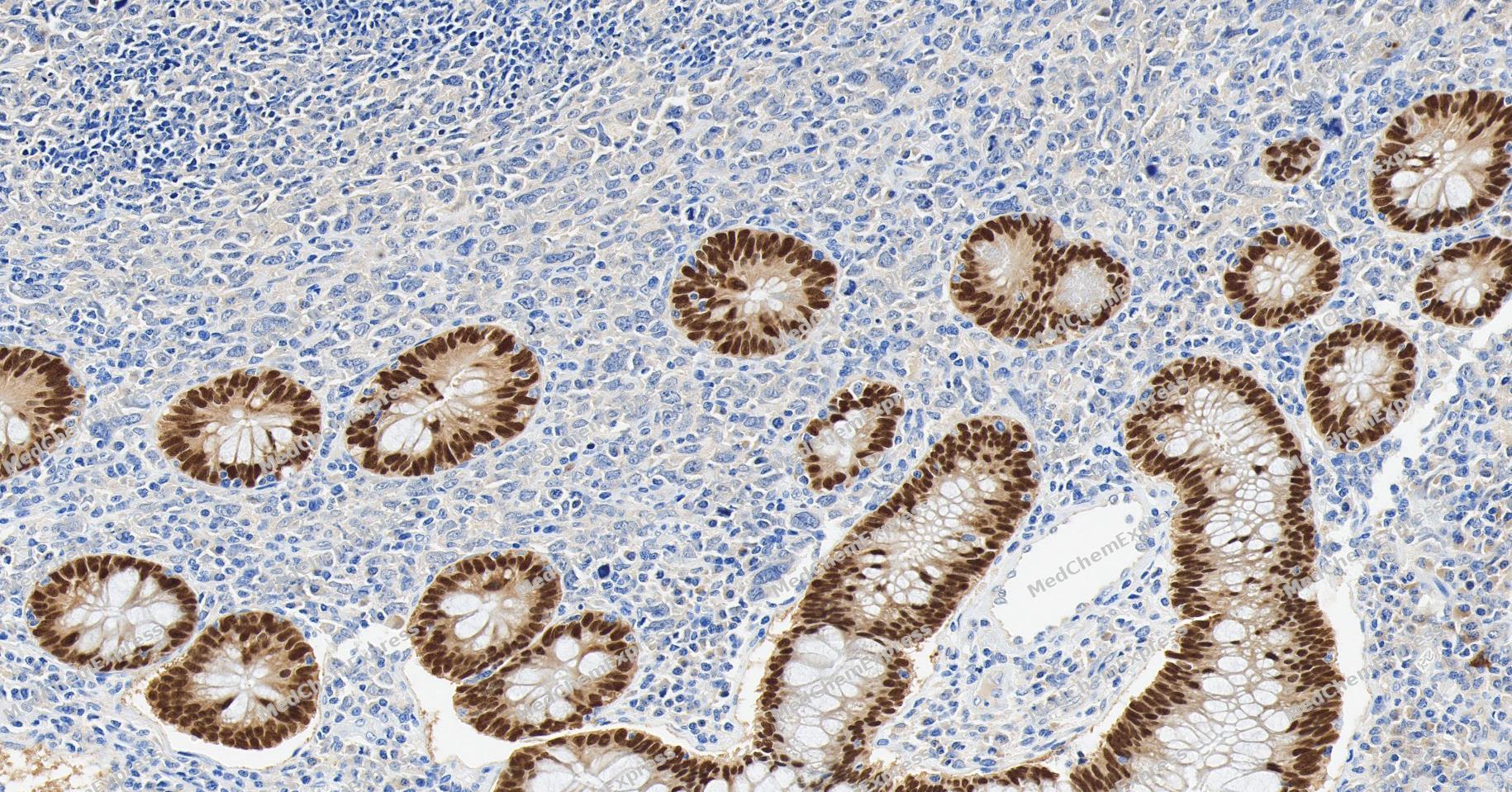 Immunohistochemical analysis of paraffin-embedded human colon cancer tissue using CDX2 Antibody (YA3498). The section was pre-treated using heat mediated antigen retrieval with Tris/EDTA buffer (pH 9.0) for 20 minutes. The tissues were probed with the primary antibody (HY-P80983,1/300) overnight at 4℃. The detection was performed using an HRP conjugated compact polymer system. DAB was used as the chromogen. Tissues were counterstained with hematoxylin and mounted with neutral balsam.
 -
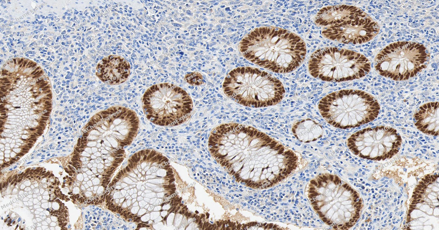 Immunohistochemical analysis of paraffin-embedded human colon cancer tissue using CDX2 Antibody (YA3498). The section was pre-treated using heat mediated antigen retrieval with Tris/EDTA buffer (pH 9.0) for 20 minutes. The tissues were probed with the primary antibody (HY-P80983,1/300) overnight at 4℃. The detection was performed using an HRP conjugated compact polymer system. DAB was used as the chromogen. Tissues were counterstained with hematoxylin and mounted with neutral balsam.
 -
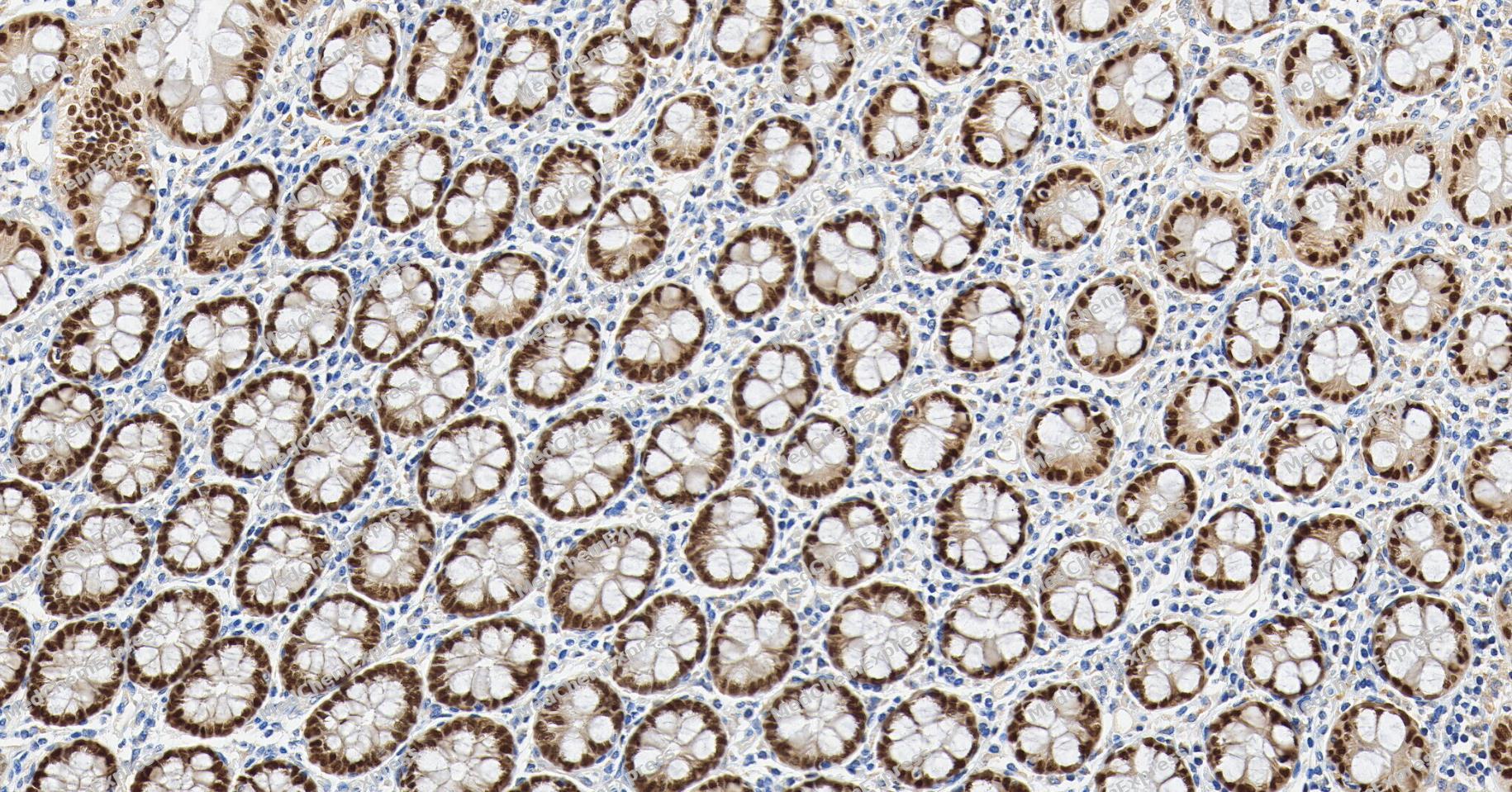 Immunohistochemical analysis of paraffin-embedded human small intestine tissue using CDX2 Antibody (YA3498). The section was pre-treated using heat mediated antigen retrieval with Tris/EDTA buffer (pH 9.0) for 20 minutes. The tissues were probed with the primary antibody (HY-P80983,1/300) overnight at 4℃. The detection was performed using an HRP conjugated compact polymer system. DAB was used as the chromogen. Tissues were counterstained with hematoxylin and mounted with neutral balsam.
 -
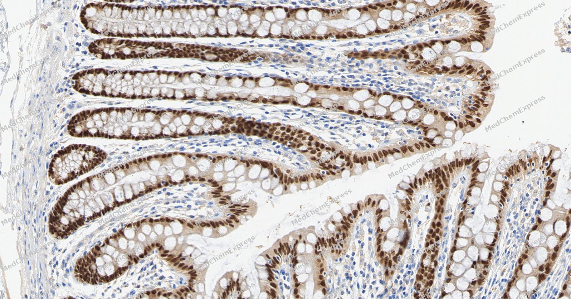 Immunohistochemical analysis of paraffin-embedded human small intestine tissue using CDX2 Antibody (YA3498). The section was pre-treated using heat mediated antigen retrieval with Tris/EDTA buffer (pH 9.0) for 20 minutes. The tissues were probed with the primary antibody (HY-P80983,1/300) overnight at 4℃. The detection was performed using an HRP conjugated compact polymer system. DAB was used as the chromogen. Tissues were counterstained with hematoxylin and mounted with neutral balsam.
 -
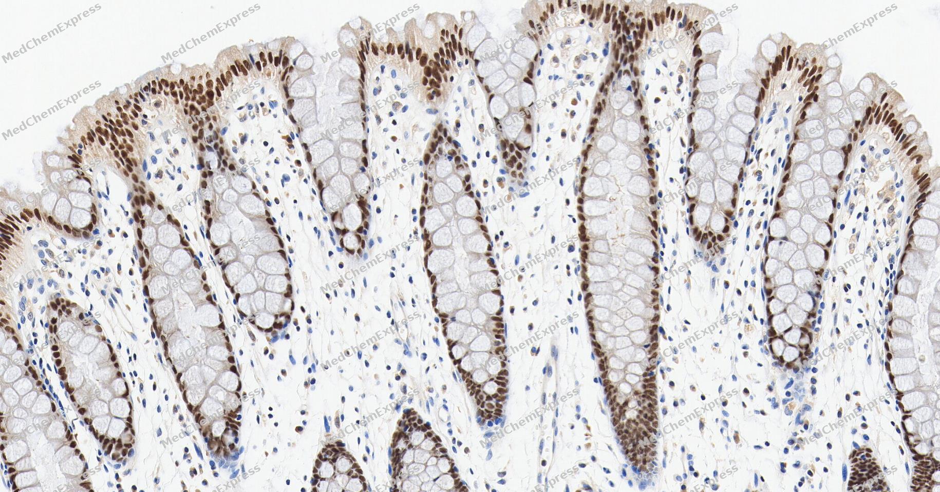 Immunohistochemical analysis of paraffin-embedded human colon tissue using CDX2 Antibody (YA3498). The section was pre-treated using heat mediated antigen retrieval with Tris/EDTA buffer (pH 9.0) for 20 minutes. The tissues were probed with the primary antibody (HY-P80983,1/300) overnight at 4℃. The detection was performed using an HRP conjugated compact polymer system. DAB was used as the chromogen. Tissues were counterstained with hematoxylin and mounted with neutral balsam.
 -
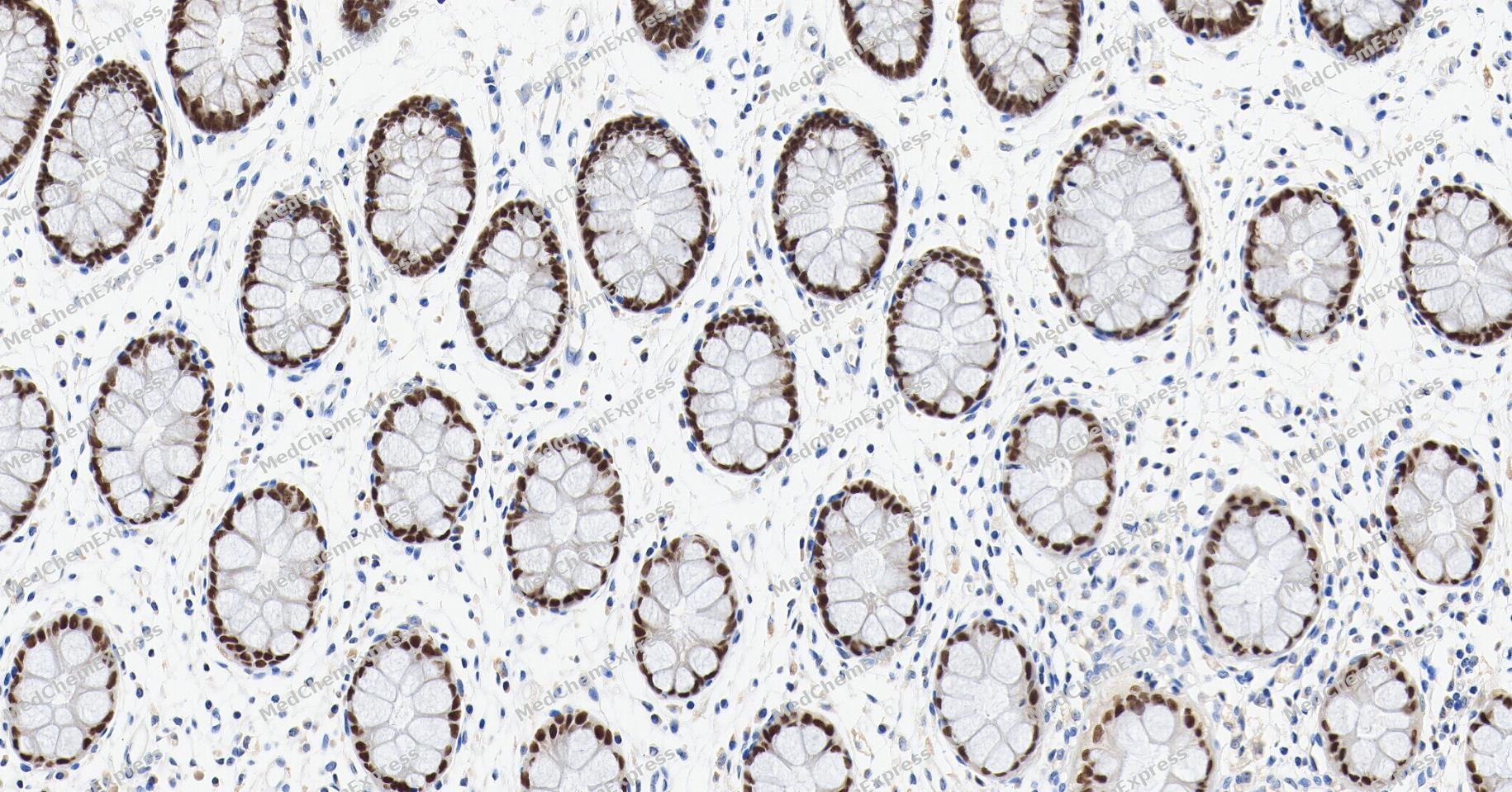 Immunohistochemical analysis of paraffin-embedded human colon tissue using CDX2 Antibody (YA3498). The section was pre-treated using heat mediated antigen retrieval with Tris/EDTA buffer (pH 9.0) for 20 minutes. The tissues were probed with the primary antibody (HY-P80983,1/300) overnight at 4℃. The detection was performed using an HRP conjugated compact polymer system. DAB was used as the chromogen. Tissues were counterstained with hematoxylin and mounted with neutral balsam.
 -
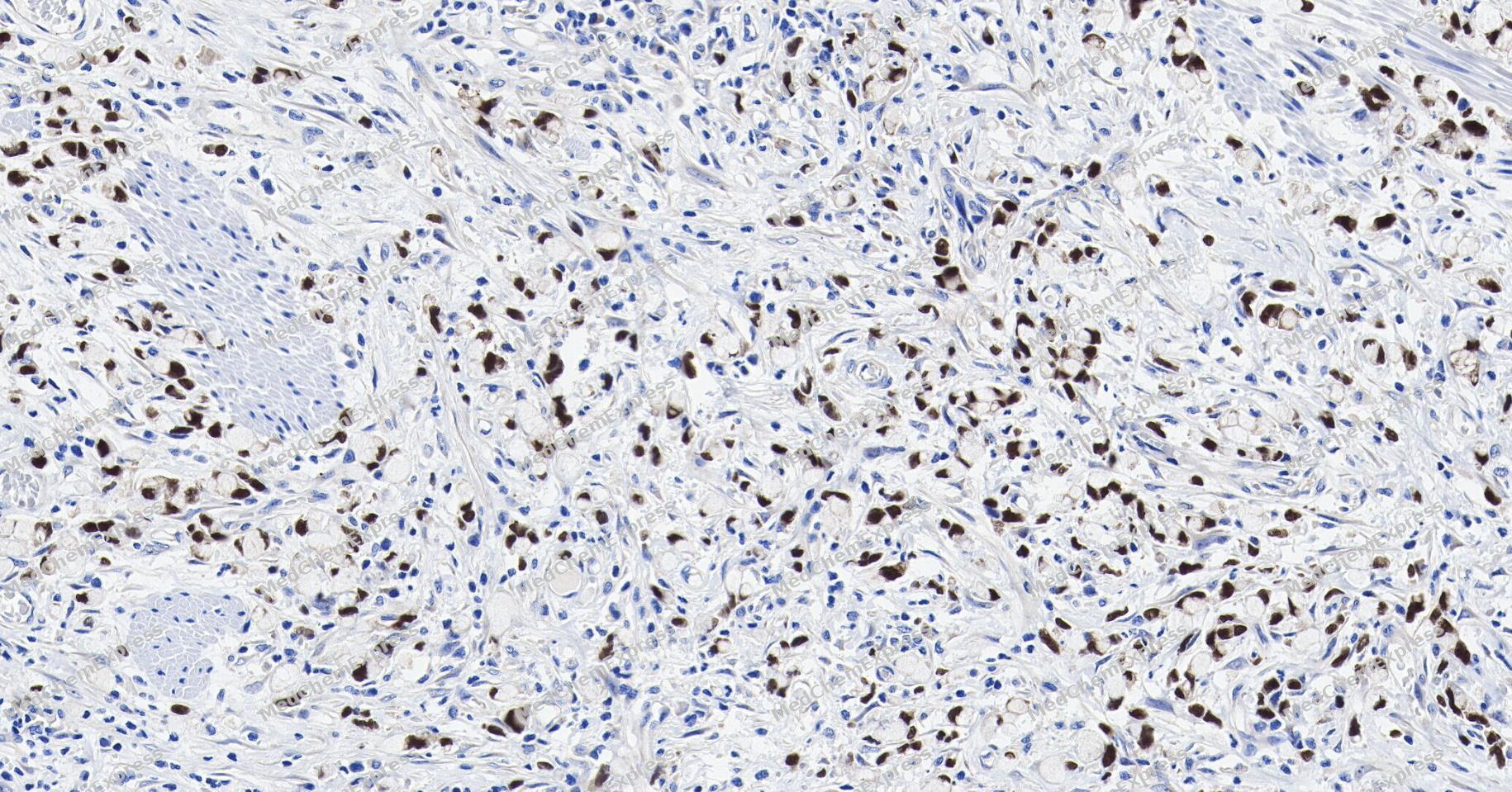 Immunohistochemical analysis of paraffin-embedded human gastric signet ring cell carcinoma tissue using CDX2 Antibody (YA3498). The section was pre-treated using heat mediated antigen retrieval with Tris/EDTA buffer (pH 9.0) for 20 minutes. The tissues were probed with the primary antibody (HY-P80983,1/300) overnight at 4℃. The detection was performed using an HRP conjugated compact polymer system. DAB was used as the chromogen. Tissues were counterstained with hematoxylin and mounted with neutral balsam.
 -
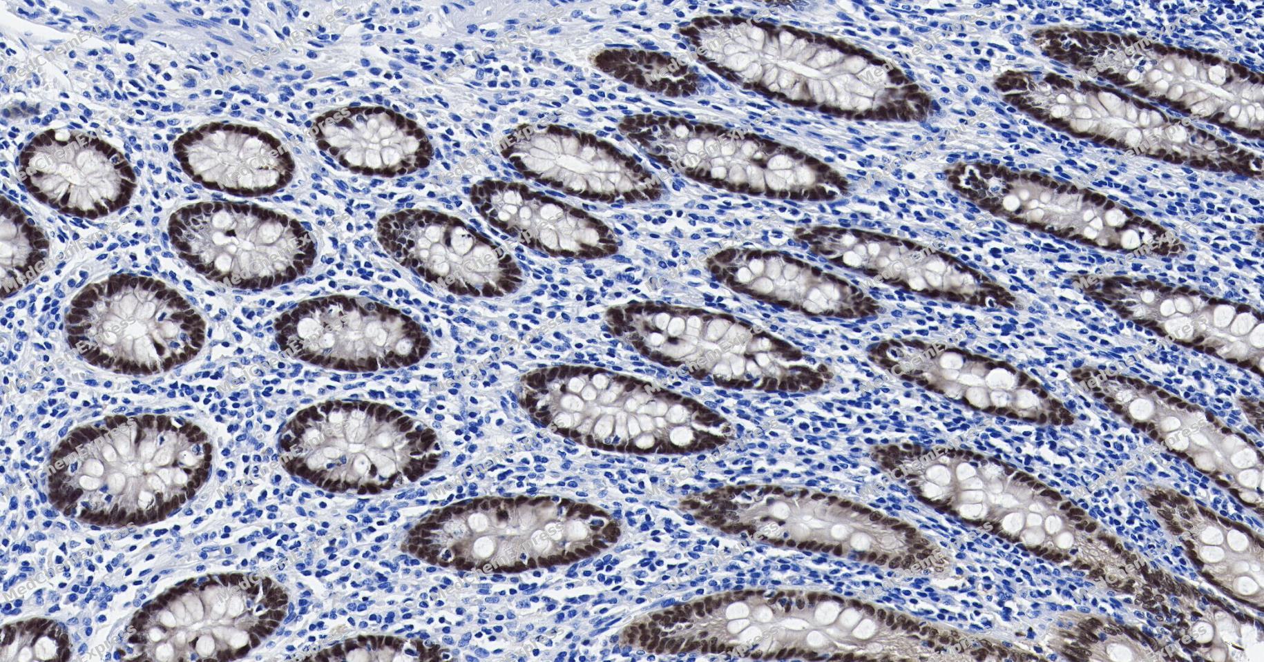 Immunohistochemical analysis of paraffin-embedded human appendix tissue using CDX2 Antibody (YA3498). The section was pre-treated using heat mediated antigen retrieval with Tris/EDTA buffer (pH 9.0) for 20 minutes. The tissues were probed with the primary antibody (HY-P80983,1/300) overnight at 4℃. The detection was performed using an HRP conjugated compact polymer system. DAB was used as the chromogen. Tissues were counterstained with hematoxylin and mounted with neutral balsam.
 -
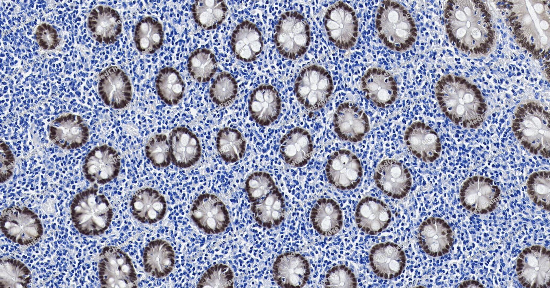 Immunohistochemical analysis of paraffin-embedded human appendix tissue using CDX2 Antibody (YA3498). The section was pre-treated using heat mediated antigen retrieval with Tris/EDTA buffer (pH 9.0) for 20 minutes. The tissues were probed with the primary antibody (HY-P80983,1/300) overnight at 4℃. The detection was performed using an HRP conjugated compact polymer system. DAB was used as the chromogen. Tissues were counterstained with hematoxylin and mounted with neutral balsam.
 -
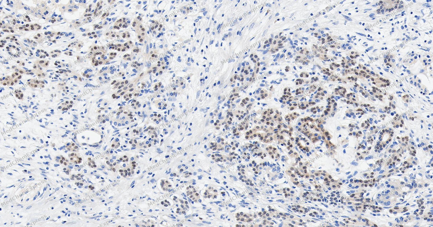 Immunohistochemical analysis of paraffin-embedded human pancreatic cancer tissue using CDX2 Antibody (YA3498). The section was pre-treated using heat mediated antigen retrieval with Tris/EDTA buffer (pH 9.0) for 20 minutes. The tissues were probed with the primary antibody (HY-P80983,1/300) overnight at 4℃. The detection was performed using an HRP conjugated compact polymer system. DAB was used as the chromogen. Tissues were counterstained with hematoxylin and mounted with neutral balsam.
 -
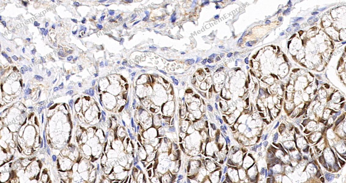 Immunohistochemical analysis of paraffin-embedded rat colon tissue using CDX2 Antibody. The section was pre-treated using heat mediated antigen retrieval with sodium citrate buffer (pH 6.0) for 8 minutes. The tissues were blocked in QuickBlock for 20 minutes at room temperature, washed with ddH2O and PBS, and then probed with the primary antibody at 1/100 dilution in 4℃ overnight. The detection was performed using an HRP conjugated compact polymer system. DAB was used as the chromogen. Tissues were counterstained with hematoxylin and mounted with DPX.
 -
Immunohistochemical analysis of paraffin-embedded rat colon tissue using CDX2 Antibody. The section was pre-treated using heat mediated antigen retrieval with sodium citrate buffer (pH 6.0) for 8 minutes. The tissues were blocked in QuickBlock for 20 minutes at room temperature, washed with ddH2O and PBS, and then probed with the primary antibody at 1/100 dilution in 4℃ overnight. The detection was performed using an HRP conjugated compact polymer system. DAB was used as the chromogen. Tissues were counterstained with hematoxylin and mounted with DPX.
-
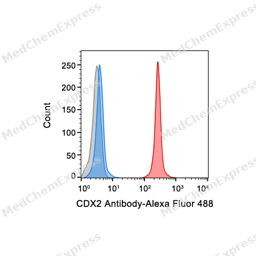 Flow cytometric analysis of 1X10^6 Hela cells labeling CDX2 Antibody (HY-P80983, red). Cells were fixed with 4% paraformaldehyde and permeabilised with intranuclear rupture fluid. Then stained with the primary antibody at 1/50 dilution for an hour at 4℃. Alexa Fluor® 488-conjugated AffiniPure Goat Anti-Rabbit IgG H&L (HY-P8002) was used as the secondary antibody at 1/1,000 dilution for 30 minutes at 4℃. Rabbit IgG Isotype Control (HY-P80879, blue) was used as the isotype control, cells without incubation with primary antibody were used as the unlabeled control (black).
 -
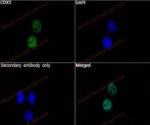 Immunocytochemistry analysis of HELA cells labeling CDX2 with CDX2 Antibody (HY-P80983) at 1/50 dilution. Cells were fixed in 4% paraformaldehyde for 15 minutes at room temperature, permeabilized with 0.1% Triton X-100 for 10 minutes at room temperature, then blocked with QuickBlock™ Blocking Buffer for Immunol Staining for 10 min at room temperature. Cells were then incubated with CDX2 Antibody (HY-P80983) at 1/50dilution in QuickBlock™ Blocking Buffer for Immunol Staining at 4 ℃. Alexa Fluor® 488-conjugated AffiniPure Goat Anti-Rabbit IgG H&L(HY-P8002, Green) was used as the secondary antibody at 1/1,000 dilution. PBS instead of the primary antibody was used as the secondary antibody only control. The Nuclear counterstain was DAPI (Blue).
 -
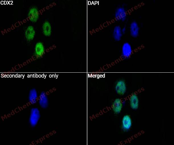 Immunocytochemistry analysis of HELA cells labelingalpha CDX2 with CDX2 Antibody (HY-P80983) at 1/100 dilution. Cells were fixed in 4% paraformaldehyde for 15 minutes at room temperature, permeabilized with 0.1% Triton X-100 for 10 minutes at room temperature, then blocked with QuickBlock™ Blocking Buffer for Immunol Staining for 10 min at room temperature. Cells were then incubated with CDX2 Antibody (HY-P80983) at 1/100 dilution in QuickBlock™ Blocking Buffer for Immunol Staining at 4 ℃. Alexa Fluor® 488-conjugated AffiniPure Goat Anti-Rabbit IgG H&L(HY-P8002, Green) was used as the secondary antibody at 1/1,000 dilution. PBS instead of the primary antibody was used as the secondary antibody only control. The Nuclear counterstain was DAPI (Blue).


Background
-
Function
CDX2 transcription factor which regulates the transcription of multiple genes expressed in the intestinal epithelium. Binds to the promoter of the intestinal sucrase-isomaltase SI and activates SI transcription. Binds to the DNA sequence 5'-ATAAAAACTTAT-3' in the promoter region of VDR and activates VDR transcription. Binds to and activates transcription of LPH. Activates transcription of CLDN2 and intestinal mucin MUC2. Binds to the 5'-AATTTTTTACAACACCT-3' DNA sequence in the promoter region of CA1 and activates CA1 transcription. Important in broad range of functions from early differentiation to maintenance of the intestinal epithelial lining of both the small and large intestine. Binds preferentially to methylated DNA[1].
-
Subcellular Localization
Nucleus
-
Expression
Tissue_specificity:It has been detected in the small intestine, colon, and pancreas.
Positive sample: Caco-2 cell lysate, LoVo cell lysate, AGS cell lysate, LOVO, AGS, human appendix tissue, Human stomach cancer tissue, human colon cancer -
Subunit
Can bind DNA as a monomer or homodimer
-
SwissProt ID
-
Synonyms
Caudal type homeo box 2 antibody; Caudal type homeo box transcription factor 2 antibody; Caudal type homeobox 2 antibody; Caudal type homeobox protein 2 antibody; Caudal type homeobox transcription factor 2 antibody; Caudal-type homeobox protein 2 antibody; CDX 2 antibody; CDX 3 antibody; CDX-3 antibody; Cdx2 antibody; CDX2_HUMAN antibody; CDX3 antibody; Homeobox protein CDX 2 antibody; Homeobox protein CDX-2 antibody;
-
Research Field
Stem Cells


Documentation
-
Data Sheet (258 KB)
-
SDS (253 KB)
- English - EN (253 KB)
- Français - FR (253 KB)
- Deutsch - DE (253 KB)
- Norwegian - NO (253 KB)
- Español - ES (253 KB)
- Swedish - SV (253 KB)
- Italian - IT (253 KB)
- Korean - KR (253 KB)
- Portuguese - PT (253 KB)
-
User Guide for Antibodies (1077 KB)

 Powered by Bioz
