



Claudin 18.2 Antibody
(Synonyms: CLDN 18.2,Claudin 18.2)
Based on 1 Customer Validation
Claudin 18.2 Antibody is a Rabbit-derived and non-conjugated IgG polyclonal antibody, targeting to Claudin 18.2.
-
Host:
Rabbit
-
Isotype:
IgG
-
Application:
WB, IHC-P, ICC/IF, FC
-
Reactivity :
Human, Mouse, Rat
-
Formulation:
Supplied in 1*PBS (pH7.4), 0.2% BSA, 50% Glycerol. Preservative: 0.05% Sodium Azide.
-
Conjugation:
Non-conjugated


To place orders, for customer services and technical support, please contact: MedChemExpress USA
Tel: 609-228-6898 E-mail: [email protected] [email protected]






Applications
| Application |
WB

WB: Western Blot
|
ICC/IF
ICC/IF: Immunocytochemistry/
Immunofluorescence |
IHC-P
IHC-P: Immunohistochemistry-Paraffin
|
FC
FC: Flow Cytometry
|
|---|---|---|---|---|
| Dilution Ratio | 1:500-1:2000 | 1:50-1:200 | 1:100-1:500 | 1:50-1:100 |


Product Details
Claudin 18.2 Antibody is a Rabbit-derived and non-conjugated IgG polyclonal antibody, targeting to Claudin 18.2.
-
Host Rabbit

-
Clonality Polyclonal

-
Species ReactivityHuman, Mouse, Rat
-
Observed Molecular WeightObserved band size: 35 kDa
Note: Due to possible protein modifications or aggregation, the molecular weight should be confirmed by actual measurement, and the predicted value is for reference only.

-
Calculated Molecular Weight Predicted band size: 28 kDa

Entrez Gene: 51208 Human ; 56492 Mouse ;
SwissProt: P56856 Human ; P56856-2 Human ; P56857 Mouse ;
OMIM: 609210 Human
Synthetic peptide corresponding to N terminal of Human CLDN18 (P56856).
Endogenous
affinity purified
Non-conjugated
Unmodified
IgG


Product Properties
-
Appearance
Liquid
-
Formulation
Supplied in 1*PBS (pH7.4), 0.2% BSA, 50% Glycerol. Preservative: 0.05% Sodium Azide.
-
Concentration
Batch-dependent, Please check the COA for the concentration of each lot. Check Lot Concentration
-
Storage & Stability
Stored at -20°C for 1 year. Avoid repeated freeze / thaw cycles.
-
Shipping
Shipping with blue ice.


Verification Images
-
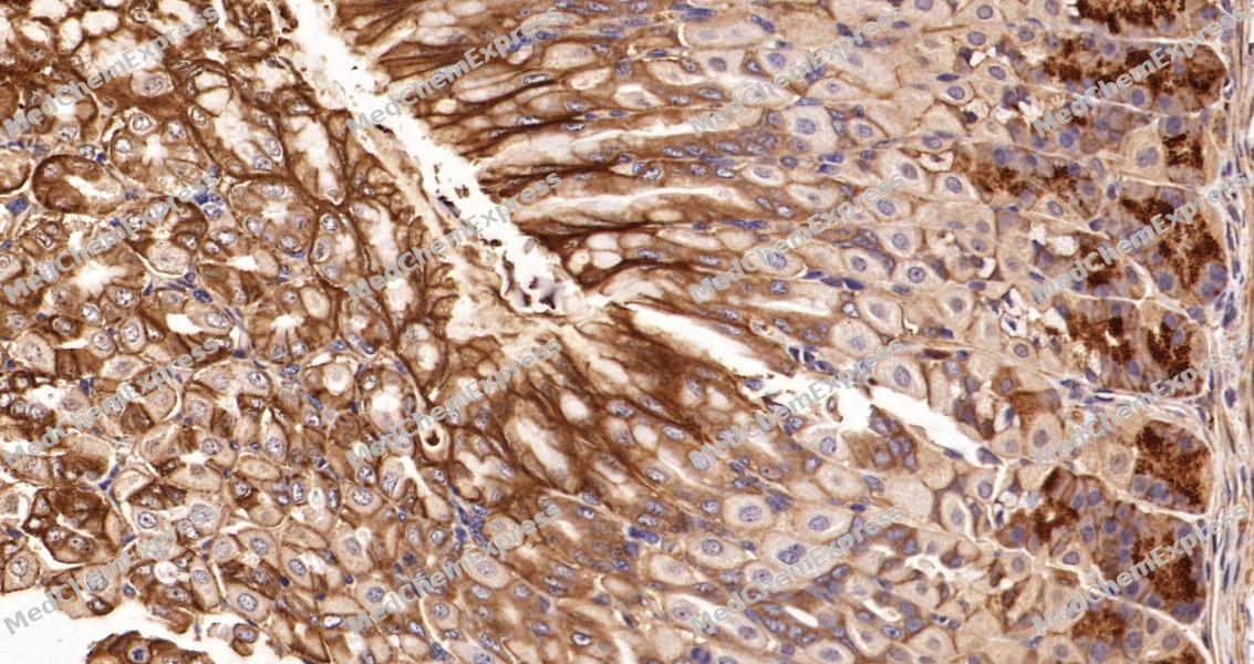 Immunohistochemical analysis of paraffin-embedded mouse stomach tissue using Claudin 18.2 Antibody. The section was pre-treated using heat mediated antigen retrieval with sodium citrate buffer (pH 6.0) for 8 minutes. The tissues were blocked in QuickBlock for 20 minutes at room temperature, washed with ddH2O and PBS, and then probed with the primary antibody at 1/100 dilution in 4℃ overnight. The detection was performed using an HRP conjugated compact polymer system. DAB was used as the chromogen. Tissues were counterstained with hematoxylin and mounted with DPX.
 -
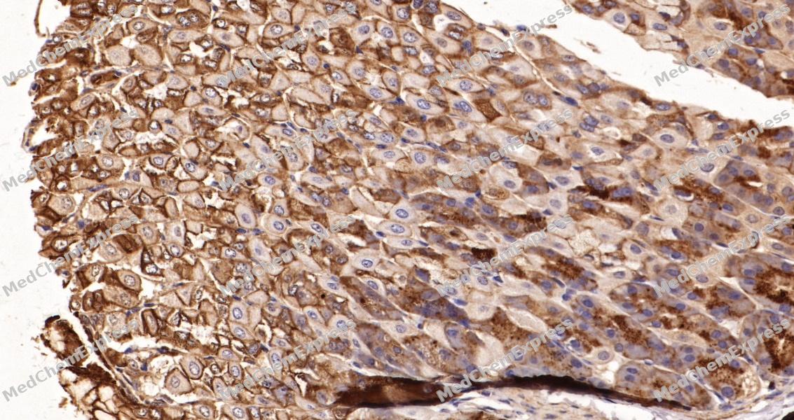 Immunohistochemical analysis of paraffin-embedded mouse stomach tissue using Claudin 18.2 Antibody. The section was pre-treated using heat mediated antigen retrieval with sodium citrate buffer (pH 6.0) for 8 minutes. The tissues were blocked in QuickBlock for 20 minutes at room temperature, washed with ddH2O and PBS, and then probed with the primary antibody at 1/100 dilution in 4℃ overnight. The detection was performed using an HRP conjugated compact polymer system. DAB was used as the chromogen. Tissues were counterstained with hematoxylin and mounted with DPX.
 -
 Immunocytochemistry analysis of HepG2 cells labeling Claudin 18.2 with Claudin 18.2 Antibody (HY-P81773) at 1/100 dilution. Cells were fixed in 4% paraformaldehyde for 15 minutes at room temperature, permeabilized with 0.1% Triton X-100 in PBS for 15 minutes at room temperature, then blocked with quick block buffer for 10 minutes at room temperature. Cells were then incubated with Claudin 18.2 Antibody (HY-P81773) at 1/100 dilution in quick block buffer overnight at 4 ℃. AF488-conjugated Goat Anti-Rabbit IgG H&L (HY-P8002, Green) was used as the secondary antibody at 1/1,000 dilution. PBS instead of the primary antibody was used as the secondary antibody only control. The Nuclear counterstain was DAPI (Blue).
 -
 Immunocytochemistry analysis of HepG2 cells labeling Claudin 18.2 with Claudin 18.2 Antibody (HY-P81773) at 1/200 dilution. Cells were fixed in 4% paraformaldehyde for 15 minutes at room temperature, permeabilized with 0.1% Triton X-100 in PBS for 15 minutes at room temperature, then blocked with quick block buffer for 10 minutes at room temperature. Cells were then incubated with Claudin 18.2 Antibody (HY-P81773) at 1/200 dilution in quick block buffer overnight at 4 ℃. AF488-conjugated Goat Anti-Rabbit IgG H&L (HY-P8002, Green) was used as the secondary antibody at 1/1,000 dilution. PBS instead of the primary antibody was used as the secondary antibody only control. The Nuclear counterstain was DAPI (Blue).
 -
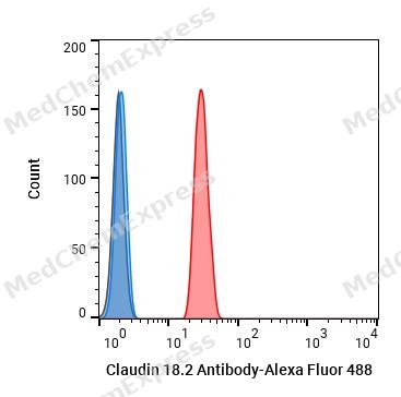 Flow cytometric analysis of 1X10^6 F9 cells labeling Claudin 18.2 Antibody (HY-P81773, red). Cells were fixed with 4% paraformaldehyde. Then stained with the primary antibody at 1/50 dilution for an hour at 4℃. Alexa Fluor® 488-conjugated AffiniPure Goat Anti-Rabbit IgG H&L (HY-P8002) was used as the secondary antibody at 1/1,000 dilution for 30 minutes at 4℃. Rabbit IgG Isotype Control (HY-P80879, blue) was used as the isotype control, cells without incubation with primary antibody were used as the unlabeled control (black).


Background
-
Function
Claudin 18.2 is involved in alveolar fluid homeostasis via regulation of alveolar epithelial tight junction composition and therefore ion transport and solute permeability, potentially via downstream regulation of the actin cytoskeleton organization and beta-2-adrenergic signaling. Required for lung alveolarization and maintenance of the paracellular alveolar epithelial barrier. Acts to maintain epithelial progenitor cell proliferation and organ size, via regulation of YAP1 localization away from the nucleus and thereby restriction of YAP1 target gene transcription. Acts as a negative regulator of RANKL-induced osteoclast differentiation, potentially via relocation of TJP2/ZO-2 away from the nucleus, subsequently involved in bone resorption in response to calcium deficiency. Mediates the osteoprotective effects of estrogen, potentially via acting downstream of estrogen signaling independently of RANKL signaling pathways; Involved in the maintenance of homeostasis of the alveolar microenvironment via regulation of pH and subsequent T-cell activation in the alveolar space, is therefore indirectly involved in limiting C.neoformans infection; Required for the formation of the gastric paracellular barrier via its role in tight junction formation, thereby involved in the response to gastric acidification
-
Subcellular Localization
Cell junction, tight junction; Cell membrane; Multi-pass membrane protein; Cell junction, tight junction; Cell junction, tight junction; Lateral cell membrane
-
Expression
Tissue_specificity:The expression of this gene is limited to the lungs; the expression of this gene is limited to the gastric mucosa, mainly found in the epithelial cells of the gastric pits and the epithelial cells of the base of the gastric glands, including exocrine and endocrine cells (at the protein level) . -
Isoforms & Post-Translational Modification
P56856 has 2 isomers: P56856-1: 27856 Da (predicted); P56856-2: 27720 Da (predicted).
-
Subunit
Interacts with TJP2/ZO-2 (By similarity). Interacts with TJP1/ZO-1 (By similarity).
-
SwissProt ID
-
Synonyms
CLDN 18.2,Claudin 18.2
-
Research Field
Cell Biology


Documentation
-
Data Sheet (235 KB)
-
SDS (251 KB)
- English - EN (251 KB)
- Français - FR (251 KB)
- Deutsch - DE (251 KB)
- Norwegian - NO (251 KB)
- Español - ES (251 KB)
- Swedish - SV (251 KB)
- Italian - IT (251 KB)
- Korean - KR (251 KB)
- Portuguese - PT (251 KB)
-
User Guide for Antibodies (1077 KB)

 Powered by Bioz
