



HSPA1A (Hsp70) Antibody (YA8425)
(Synonyms: HEL-S-103; HSP70-1; HSP70-1A; HSP70.1; HSP70I; HSP72; HSPA1)
Based on 1 Customer Validation
HSPA1A (Hsp70) Antibody (YA8425) is a Mouse-derived and non-conjugated IgG1 monoclonal antibody, targeting to HSPA1A (Hsp70).
-
Host:
Mouse
-
Isotype:
IgG1
-
Application:
WB, IHC-P, ICC/IF, FC
-
Reactivity :
Human, Dog, Monkey, Mouse, Rat
-
Formulation:
Spplied in PBS (pH 7.3) containing 1% BSA, 50% glycerol and 0.02% sodium azide.
-
Conjugation:
Non-conjugated


To place orders, for customer services and technical support, please contact: MedChemExpress USA
Tel: 609-228-6898 E-mail: [email protected] [email protected]





Applications
| Application |
WB

WB: Western Blot
|
IHC-P
IHC-P: Immunohistochemistry-Paraffin
|
ICC/IF
ICC/IF: Immunocytochemistry/
Immunofluorescence |
FC
FC: Flow Cytometry
|
|---|---|---|---|---|
| Dilution Ratio | 1:500-2000 | 1:50-500 | 1:100-250 | 1:100 |


Product Details
HSPA1A (Hsp70) Antibody (YA8425) is a Mouse-derived and non-conjugated IgG1 monoclonal antibody, targeting to HSPA1A (Hsp70).
-
Host Mouse

-
Clonality Monoclonal

-
Species ReactivityHuman, Dog, Monkey, Mouse, Rat
-
Calculated Molecular Weight Predicted band size: 70.0 kDa

Full length human recombinant protein of human HSPA1A produced in HEK293T cell.
Endogenous
Affinity purified
Non-conjugated
Unmodified
IgG1


Product Properties
-
Appearance
Liquid
-
Formulation
Spplied in PBS (pH 7.3) containing 1% BSA, 50% glycerol and 0.02% sodium azide.
-
Concentration
Batch-dependent, Please check the COA for the concentration of each lot. Check Lot Concentration
-
Storage & Stability
Stored at -20°C for 1 year. Avoid repeated freeze / thaw cycles.
-
Shipping
Shipping with blue ice.


Verification Images
-
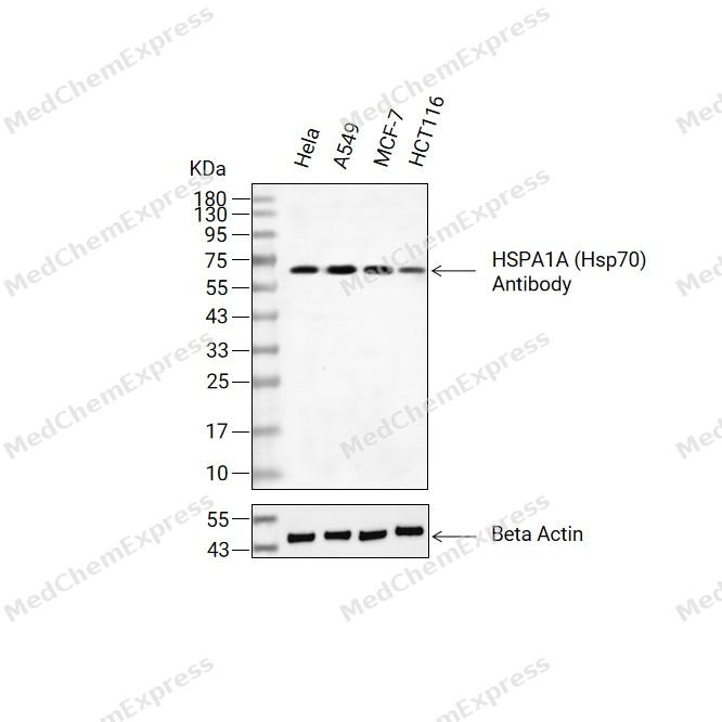 Western blot analysis of extracts from Hela(lane 2(20ug) , A549(lane 3(20ug) and MCF-7(lane 4(20ug),HCT116(lane 5(20ug) using HSPA1A Antibody (HY-P88741) Rabbit mAb. Proteins were transferred to a PVDF membrane and blocked with 5% non-fat milk in TBST for 2 hour at room temperature. The primary antibody (1/1000) and Loading control antibody (Beta Actin, HY-P83730, 1/10000) was used in 5% non-fat milk in TBST at 4°C overnight. Goat Anti-Mouse/Rabbit IgG-HRP Secondary Antibody (1/10000) was used for 1 hour at room temperature.
 -
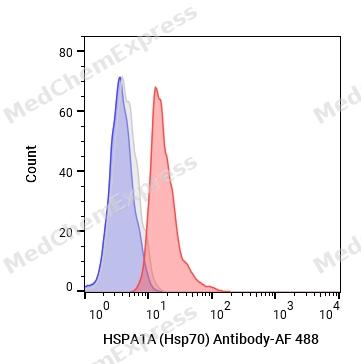 Flow cytometric analysis of 1X10^6 Jurkat cells labeling HSPA1A (Hsp70) Antibody (HY-P88741, red). Cells were fixed with 4% paraformaldehyde. Then stained with the primary antibody at 1/50 dilution for an hour at 4℃. AF 488-conjugated AffiniPure Goat Anti- Mouse IgG H&L (HY-P8005) was used as the secondary antibody at 1/1,000 dilution for 30 minutes at 4℃. Mouse IgG Isotype Control (HY-P80757, blue) was used as the isotype control, cells without incubation with primary antibody were used as the unlabeled control (black).
 -
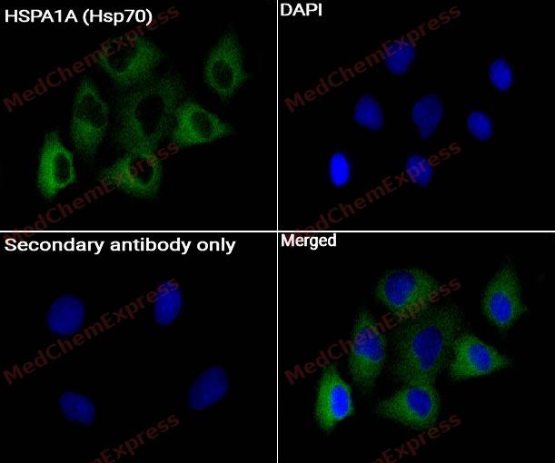 Immunocytochemistry analysis of HeLa cells labeling HSPA1A (Hsp70) Antibody (HY-P88741) at 1/200 dilution. Cells were fixed in 4% paraformaldehyde for 15 minutes at room temperature, permeabilized with 0.1% Triton X-100 for 10 minutes at room temperature, then blocked with BSA for Immunol Staining for 10 min at room temperature. Cells were then incubated with HSPA1A (Hsp70) Antibody (HY-P88741) at 1/50 dilution in BSA for Immunol Staining at 4 ℃ overnight. AF488-conjugated AffiniPure Goat Anti-Rabbit /Mouse IgG H&L(HY-P8002/HY-P8005, Green) was used as the secondary antibody at 1/1,000 dilution. PBS instead of the primary antibody was used as the secondary antibody only control. The Nuclear counterstain was DAPI (Blue).


Background
-
Function
HSPA1A is a Molecular chaperone implicated in a wide variety of cellular processes, including protection of the proteome from stress, folding and transport of newly synthesized polypeptides, activation of proteolysis of misfolded proteins and the formation and dissociation of protein complexes. Plays a pivotal role in the protein quality control system, ensuring the correct folding of proteins, the re-folding of misfolded proteins and controlling the targeting of proteins for subsequent degradation. This is achieved through cycles of ATP binding, ATP hydrolysis and ADP release, mediated by co-chaperones. The co-chaperones have been shown to not only regulate different steps of the ATPase cycle, but they also have an individual specificity such that one co-chaperone may promote folding of a substrate while another may promote degradation. The affinity for polypeptides is regulated by its nucleotide bound state. In the ATP-bound form, it has a low affinity for substrate proteins. However, upon hydrolysis of the ATP to ADP, it undergoes a conformational change that increases its affinity for substrate proteins. It goes through repeated cycles of ATP hydrolysis and nucleotide exchange, which permits cycles of substrate binding and release. The co-chaperones are of three types: J-domain co-chaperones such as HSP40s (stimulate ATPase hydrolysis by HSP70), the nucleotide exchange factors (NEF) such as BAG1/2/3 (facilitate conversion of HSP70 from the ADP-bound to the ATP-bound state thereby promoting substrate release), and the TPR domain chaperones such as HOPX and STUB1. Maintains protein homeostasis during cellular stress through two opposing mechanisms: protein refolding and degradation. Its acetylation/deacetylation state determines whether it functions in protein refolding or protein degradation by controlling the competitive binding of co-chaperones HOPX and STUB1. During the early stress response, the acetylated form binds to HOPX which assists in chaperone-mediated protein refolding, thereafter, it is deacetylated and binds to ubiquitin ligase STUB1 that promotes ubiquitin-mediated protein degradation. Regulates centrosome integrity during mitosis, and is required for the maintenance of a functional mitotic centrosome that supports the assembly of a bipolar mitotic spindle. Enhances STUB1-mediated SMAD3 ubiquitination and degradation and facilitates STUB1-mediated inhibition of TGF-beta signaling. Essential for STUB1-mediated ubiquitination and degradation of FOXP3 in regulatory T-cells (Treg) during inflammation. Required as a co-chaperone for optimal STUB1/CHIP ubiquitination of NFATC3. Negatively regulates heat shock-induced HSF1 transcriptional activity during the attenuation and recovery phase period of the heat shock response. Involved in the clearance of misfolded PRDM1/Blimp-1 proteins. Sequesters them in the cytoplasm and promotes their association with SYNV1/HRD1, leading to proteasomal degradation |(Microbial infection) In case of rotavirus A infection, serves as a post-attachment receptor for the virus to facilitate entry into the cell[1][2][3][4][5][6][7][8][9].
-
Subcellular Localization
Cytoplasm,Nucleus,Cytoplasm, cytoskeleton, microtubule organizing center, centrosome,Secreted
-
Expression
Induction: By heat shock
-
Isoforms & Post-Translational Modification
P0DMV8 has two isomers: P0DMV8-1: 70052 Da (predicted); P0DMV8-2: 63937 Da (predicted).
In response to cellular stress, acetylated at Lys-77 by NA110 and then gradually deacetylated by HDAC4 at later stages -
Subunit
Component of the CatSper complex
-
SwissProt ID
-
Synonyms
HEL-S-103; HSP70-1; HSP70-1A; HSP70.1; HSP70I; HSP72; HSPA1


Documentation
-
Data Sheet (231 KB)
-
SDS (251 KB)
- English - EN (251 KB)
- Français - FR (251 KB)
- Deutsch - DE (251 KB)
- Norwegian - NO (251 KB)
- Español - ES (251 KB)
- Swedish - SV (251 KB)
- Italian - IT (251 KB)
- Korean - KR (251 KB)
- Portuguese - PT (251 KB)
-
User Guide for Antibodies (1077 KB)

 Powered by Bioz
