



JAK2 Antibody (YA721)
(Synonyms: Tyrosine-protein kinase JAK2, Janus kinase 2, JAK-2, JAK2)
Based on 1 Customer Validation
JAK2 Antibody (YA721) is a Mouse-derived and non-conjugated IgG2b monoclonal antibody, targeting to JAK2.
-
Host:
Mouse
-
Isotype:
IgG
-
Application:
WB, ICC/IF, IHC-P, FC
-
Reactivity :
Human, Mouse, Rat
-
Formulation:
Supplied in 1*PBS (pH7.4), 0.2% BSA and 40% Glycerol. Preservative: 0.05% Sodium Azide.
-
Conjugation:
Non-conjugated


To place orders, for customer services and technical support, please contact: MedChemExpress USA
Tel: 609-228-6898 E-mail: [email protected] [email protected]



-

HY-P80467
Phospho-JAK2 (Tyr1007+Tyr1008) Antibody (YA181) -
HY-P80829A
Phospho-JAK2 (Tyr1007/1008) Antibody (YA6449) -
HY-P80344
STAT3 Antibody (YA056) -
HY-P8004
HRP-conjugated Goat Anti-Mouse IgG H&L -
HY-P80867
PI3 Kinase p85 alpha Antibody (YA689) -
HY-P80857
Phospho-STAT3 (Tyr705) Antibody -
HY-P80282
Phospho-STAT3 (Tyr705) Antibody (YA146) -
HY-P80281
Phospho-STAT3 (Ser727) Antibody (YA147) -
HY-P80347
STAT6 Antibody (YA051)


Applications
| Application |
WB

WB: Western Blot
|
ICC/IF
ICC/IF: Immunocytochemistry/
Immunofluorescence |
FC
FC: Flow Cytometry
|
IHC-P
IHC-P: Immunohistochemistry-Paraffin
|
|---|---|---|---|---|
| Dilution Ratio | 1:2000 | 1:50 | 1:200 | 1:200 |


Product Details
JAK2 Antibody (YA721) is a Mouse-derived and non-conjugated IgG2b monoclonal antibody, targeting to JAK2.
-
Host Mouse

-
Clonality Monoclonal

-
Species ReactivityHuman, Mouse, Rat
-
Observed Molecular WeightObserved band size: 130 kDa
Note: Due to possible protein modifications or aggregation, the molecular weight should be confirmed by actual measurement, and the predicted value is for reference only.

-
Calculated Molecular Weight Predicted band size: 130kDa

Entrez Gene: 3717 Human ; 16452 Mouse ; 24514 Rat
SwissProt: O60674 Human ; Q62120 Mouse ; Q62689 Rat
OMIM: 600880 Human
Synthetic peptide corresponding to Human JAK2.AA range:883-1132.
Endogenous
Protein A affinity purified.
Non-conjugated
Unmodified
IgG


Product Properties
-
Appearance
Solution
-
Formulation
Supplied in 1*PBS (pH7.4), 0.2% BSA and 40% Glycerol. Preservative: 0.05% Sodium Azide.
-
Concentration
Batch-dependent, Please check the COA for the concentration of each lot. Check Lot Concentration
-
Storage & Stability
Stored at -20°C for 1 year. Avoid repeated freeze / thaw cycles.
-
Shipping
Shipping with blue ice.


Verification Images
-
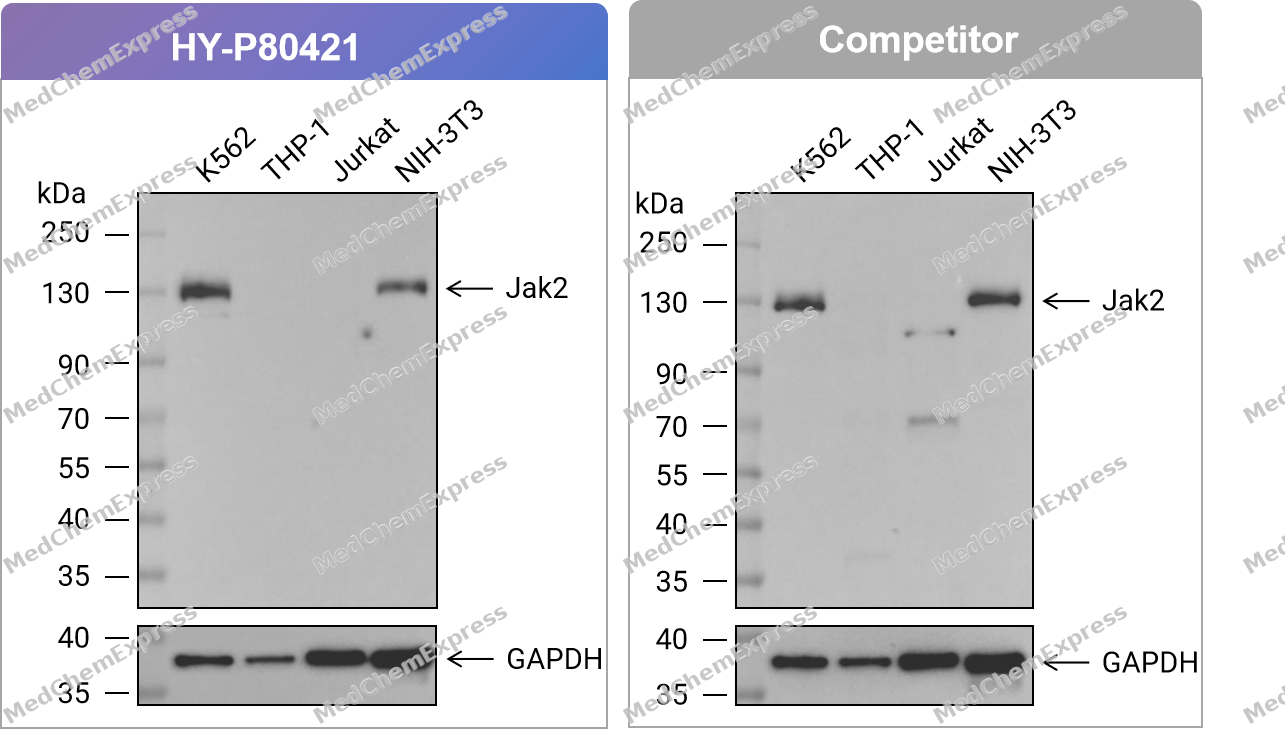 Western blot analysis of extracts from K562 (lane 1(20μg)) 、THP-1 (lane 2(20μg)) 、Jurkat (lane 3(20μg)) and NIH-3T3 (lane 4(20μg)) using Jak2 Antibody . Proteins were transferred to a PVDF membrane and blocked with 5% nonfat dry milk in TBST for 1.5 hour at room temperature. The primary antibody (HY-P80421, 1/1000) , competitor's antibody (1/1000) and Loading control antibody (GAPDH, 1/10000) was used in 5% nonfat dry milk in TBST at 4℃ overnight. Goat Anti-Mouse IgG-HRP Secondary Antibody (1/10,000) was used for 1 hour at room temperature.
 -
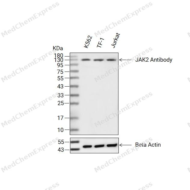 Western blot analysis of extracts from K562 (lane 2(20μg), RAW264.7 (lane 3(20μg) and C6(lane 4(40μg) using JAK2 (HY-P80421) Mouse mAb. Proteins were transferred to a PVDF membrane and blocked with 5% non-fat milk in TBST for 2 hour at room temperature. The primary antibody (1/1000) and Loading control antibody (Beta Actin, HY-P80438, 1/10000) was used in 5% non-fat milk in TBST at 4°C overnight. Goat Anti-Mouse IgG-HRP Secondary Antibody (1/10000) was used for 1 hour at room temperature.
 -
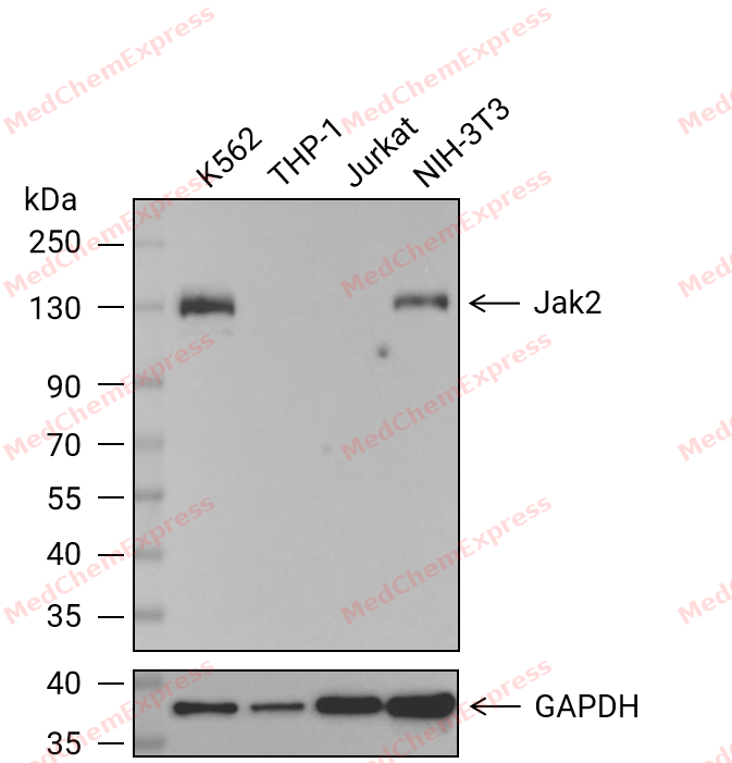 Western blot analysis of extracts from K562 (lane 1(20μg)) 、THP-1 (lane 2(20μg)) 、Jurkat (lane 3(20μg)) and NIH-3T3 (lane 4(20μg)) using Jak2 Antibody (HY-P80421) . Proteins were transferred to a PVDF membrane and blocked with 5% nonfat dry milk in TBST for 1.5 hour at room temperature. The primary antibody (1/1000) and Loading control antibody (GAPDH, 1/10000) was used in 5% nonfat dry milk in TBST at 4℃ overnight. Goat Anti-Mouse IgG-HRP Secondary Antibody (1/10,000) was used for 1 hour at room temperature.
 -
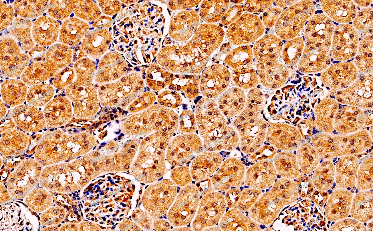 Immunohistochemical analysis of paraffin-embedded Rat kidney tissue using JAK2 Antibody (YA721). The section was pre-treated using heat mediated antigen retrieval with sodium citrate buffer (pH 6.0) for 8 minutes. The tissues were blocked in QuickBlock for 20 minutes at room temperature, washed with ddH2O and PBS, and then probed with the primary antibody (HY-P80421, 1/200) in 4℃ overnight. The detection was performed using an HRP conjugated compact polymer system. DAB was used as the chromogen. Tissues were counterstained with hematoxylin and mounted with DPX.
 -
Immunohistochemical analysis of paraffin-embedded Rat kidney tissue using JAK2 Antibody (YA721). The section was pre-treated using heat mediated antigen retrieval with sodium citrate buffer (pH 6.0) for 8 minutes. The tissues were blocked in QuickBlock for 20 minutes at room temperature, washed with ddH2O and PBS, and then probed with the primary antibody (HY-P80421, 1/200) in 4℃ overnight. The detection was performed using an HRP conjugated compact polymer system. DAB was used as the chromogen. Tissues were counterstained with hematoxylin and mounted with DPX.
-
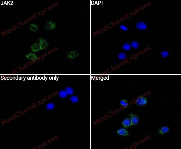 Immunocytochemistry analysis of RAW264.7 cells labeling JAK2 with JAK2 Antibody (HY-P80421)at 1/50 dilution. Cells were fixed in 4% paraformaldehyde for 15 minutes at room temperature, permeabilized with 0.1% Triton X-100 for 10 minutes at room temperature, then blocked with QuickBlock™ Blocking Buffer for Immunol Staining for 10 min at room temperature. Cells were then incubated with JAK2 Antibody (HY-P80421) at 1/50 dilution in QuickBlock™ Blocking Buffer for Immunol Staining at 4 ℃. Alexa Fluor® 488-conjugated AffiniPure Goat Anti-Mouse IgG H&L(HY-P8005, Green) was used as the secondary antibody at 1/1,000 dilution. PBS instead of the primary antibody was used as the secondary antibody only control. The Nuclear counterstain was DAPI (Blue).
 -
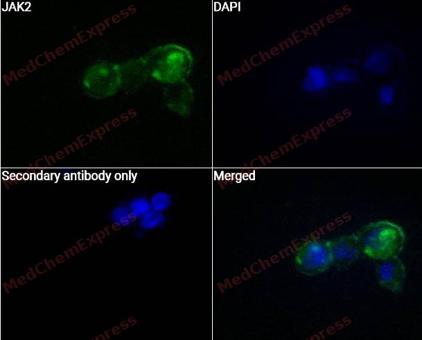 Immunocytochemistry analysis of MCF-7 cells labeling JAK2 with JAK2 Antibody (HY-P80421) at 1/50 dilution. Cells were fixed in 4% paraformaldehyde for 15 minutes at room temperature, permeabilized with 0.1% Triton X-100 for 10 minutes at room temperature, then blocked with QuickBlock™ Blocking Buffer for Immunol Staining for 10 min at room temperature. Cells were then incubated with JAK2 Antibody (HY-P80421) at 1/50 dilution in QuickBlock™ Blocking Buffer for Immunol Staining at 4 ℃. Alexa Fluor® 488-conjugated AffiniPure Goat Anti-Mouse IgG H&L(HY-P8005, Green) was used as the secondary antibody at 1/1,000 dilution. PBS instead of the primary antibody was used as the secondary antibody only control. The Nuclear counterstain was DAPI (Blue).


Background
-
Function
JAK2 is a non-receptor tyrosine kinase in the JAK1-JAK2-JAK3-TYK2 family and acts as a cytoplasmic signaling component of cytokine receptors[1]. Mechanistically, cytokine-mediated activation of JAK2 promotes receptor phosphorylation, recruits STAT proteins, and drives STAT phosphorylation-dependent transcriptional signaling[1][2]. This pathway supports cell growth, survival, development, differentiation, hematopoiesis, immune regulation, and homeostasis[2][3]. In disease models, gain-of-function JAK2 V617F in the pseudokinase domain causes constitutive activity and is strongly linked to myeloproliferative neoplasms and leukemias[1][4]. Compared with related isoforms, JAK3 is essential for common γ-chain cytokine receptor signaling in T- and B-cell development, whereas complete mouse deletion of JAK1 or JAK2 is not compatible with life[5][6]. For experimental applications, ruxolitinib is a selective oral JAK1/JAK2 inhibitor, and JAK2-focused compounds support studies of mutant signaling, cytokine responses, splenomegaly, angiogenesis, and inhibitor resistance[7][8][9].
-
Subcellular Localization
Endomembrane system; Peripheral membrane protein; Cytoplasm; Nucleus
-
Expression
Tissue_specificity:Commonly expressed in most organizations -
Subunit
Interacts with EPOR, LYN, SIRPA, SH2B1 and TEC (By similarity). Interacts with IL23R (PubMed:12023369). Interacts with SKB1 (PubMed:10531356). Interacts with STAM2 (PubMed:10899310). Interacts with IFNGR2 (via intracellular domain) (PubMed:7615558, PubMed:7673114). Interacts with LEPR (Isoform B) (By similarity). Interacts with HSP90AB1; promotes functional activation in a heat shock-dependent manner (PubMed:20353823). Interacts with STRA6 (PubMed:21368206). Interacts with RHEX; this interaction occurs in a erythropoietin (EPO)-dependent manner (PubMed:25092874). Interacts with ASB2; the interaction targets JAK2 for Notch-induced proteasomal degradation (PubMed:21119685). Interacts with MPL/TPOR (PubMed:15899890)
-
SwissProt ID
-
Synonyms
Tyrosine-protein kinase JAK2, Janus kinase 2, JAK-2, JAK2
-
Research Field
Cell Biology


Documentation
-
Data Sheet (263 KB)
-
SDS (251 KB)
- English - EN (251 KB)
- Français - FR (251 KB)
- Deutsch - DE (251 KB)
- Norwegian - NO (251 KB)
- Español - ES (251 KB)
- Swedish - SV (251 KB)
- Italian - IT (251 KB)
- Korean - KR (251 KB)
- Portuguese - PT (251 KB)
-
User Guide for Antibodies (1077 KB)
References
[1]. Hubbard SR. Mechanistic Insights into Regulation of JAK2 Tyrosine Kinase. Front Endocrinol (Lausanne). 2018 Jan 5;8:361. doi: 10.3389/fendo.2017.00361. PMID: 29379470; PMCID: PMC5770812. et al. Mechanistic Insights into Regulation of JAK2 Tyrosine Kinase. Front Endocrinol (Lausanne). 2018 Jan 5;8:361. [Content Brief]
[2]. Ihle JN, et al. Jaks and Stats in signaling by the cytokine receptor superfamily. Trends Genet. 1995 Feb;11(2):69-74. [Content Brief]
[3]. Rawlings JS, et al. The JAK/STAT signaling pathway. J Cell Sci. 2004 Mar 15;117(Pt 8):1281-3. [Content Brief]
[4]. Gäbler K, et al. JAK2 mutants (e.g., JAK2V617F) and their importance as drug targets in myeloproliferative neoplasms. JAKSTAT. 2013 Jul 1;2(3):e25025. [Content Brief]
[5]. Suzuki K, et al. Janus kinase 3 (Jak3) is essential for common cytokine receptor gamma chain (gamma(c))-dependent signaling: comparative analysis of gamma(c), Jak3, and gamma(c) and Jak3 double-deficient mice. Int Immunol. 2000 Feb;12(2):123-32. [Content Brief]
[6]. Ghoreschi K, et al. Janus kinases in immune cell signaling. Immunol Rev. 2009 Mar;228(1):273-87. [Content Brief]
[7]. Naqvi K, et al. A potential role of ruxolitinib in leukemia. Expert Opin Investig Drugs. 2011 Aug;20(8):1159-66. [Content Brief]
[8]. Hart S, et al. SB1518, a novel macrocyclic pyrimidine-based JAK2 inhibitor for the treatment of myeloid and lymphoid malignancies. Leukemia. 2011 Nov;25(11):1751-9. [Content Brief]
[9]. Cheng Z, et al. Angiogenesis in JAK2 V617F positive myeloproliferative neoplasms and ruxolitinib decrease VEGF, HIF-1 enesis in JAK2 V617F positive cells. Leuk Lymphoma. 2018 Jan;59(1):196-203. [Content Brief]

 Powered by Bioz
