



NADPH Oxidase 4 Antibody (YA4640)
(Synonyms: NADPH oxidase 4; Kidney oxidase-1; KOX-1; KOX1; Kidney superoxide-producing NADPH oxidase; Renal NAD(P)H-oxidase; NOX4; RENOX)Based on 1 publication(s) in Google Scholar
NADPH Oxidase 4 Antibody (YA4640) is a Rabbit-derived and non-conjugated IgG monoclonal antibody, targeting to NADPH Oxidase 4.
-
Host:
Rabbit
-
Isotype:
IgG
-
Application:
WB, IHC-P, ICC/IF, IP
-
Reactivity :
Human, Mouse, Rat
-
Formulation:
Supplied in Rabbit IgG in 10mM phosphate buffered saline , pH 7.4, 150mM sodium chloride, 0.05% BSA, 0.02% sodium azide and 50% glycerol.
-
Conjugation:
Non-conjugated


To place orders, for customer services and technical support, please contact: MedChemExpress USA
Tel: 609-228-6898 E-mail: [email protected] [email protected]



Publications Citing Use of MedChemExpress (MCE) NADPH Oxidase 4 Antibody (YA4640)
More

Applications
| Application |
WB

WB: Western Blot
|
IHC-P
IHC-P: Immunohistochemistry-Paraffin
|
ICC/IF
ICC/IF: Immunocytochemistry/
Immunofluorescence |
IP
IP: Immunoprecipitation
|
|---|---|---|---|---|
| Dilution Ratio | 1:500-1:1000 | 1:50-1:100 | 1:50-1:200 | 1:20 |


Product Details
NADPH Oxidase 4 Antibody (YA4640) is a Rabbit-derived and non-conjugated IgG monoclonal antibody, targeting to NADPH Oxidase 4.
-
Host Rabbit

-
Clonality Monoclonal,Recombinant

-
Species ReactivityHuman, Mouse, Rat
-
Calculated Molecular Weight Predicted band size: 67 kDa

Entrez Gene: 50507 Human
SwissProt: Q9NPH5 Human
Synthetic peptide corresponding to Human NOX4 aa510-578.
affinity purified
Non-conjugated
Unmodified
IgG


Product Properties
-
Appearance
Solution
-
Formulation
Supplied in Rabbit IgG in 10mM phosphate buffered saline , pH 7.4, 150mM sodium chloride, 0.05% BSA, 0.02% sodium azide and 50% glycerol.
-
Concentration
Batch-dependent, Please check the COA for the concentration of each lot. Check Lot Concentration
-
Storage & Stability
Stored at -20°C for 1 year. Avoid repeated freeze / thaw cycles.
-
Shipping
Shipping with blue ice.


Publications (1)
-
Journal Impact Factor
-
Most Recent


Verification Images
-
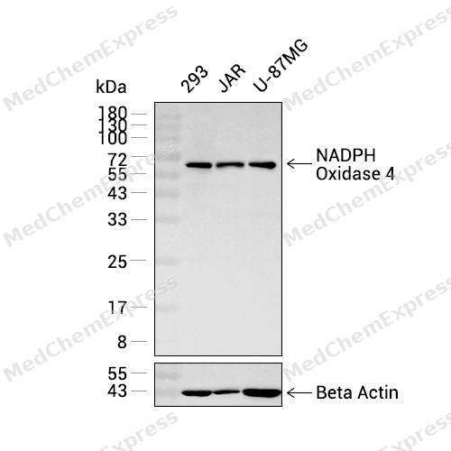 Western blot analysis was performed on extracts from 293 (lane 1, 15 μg), JAR (lane 2, 15 μg), and U-87MG (lane 3, 15 μg) using NADPH Oxidase 4 Rabbit mAb.Proteins were transferred to a PVDF membrane and blocked with 5% non-fat milk in TBST at 4°C overnight.The primary antibody (1:1000 dilution) and the loading control antibody (beta-Actin, HY-P83730, 1:20000 dilution) were incubated in 5% non-fat milk in TBST for 1 hour at 37°C.Goat Anti-Rabbit IgG-HRP Secondary Antibody (1:20000 dilution) was then applied for 40 minutes at 37°C.
 -
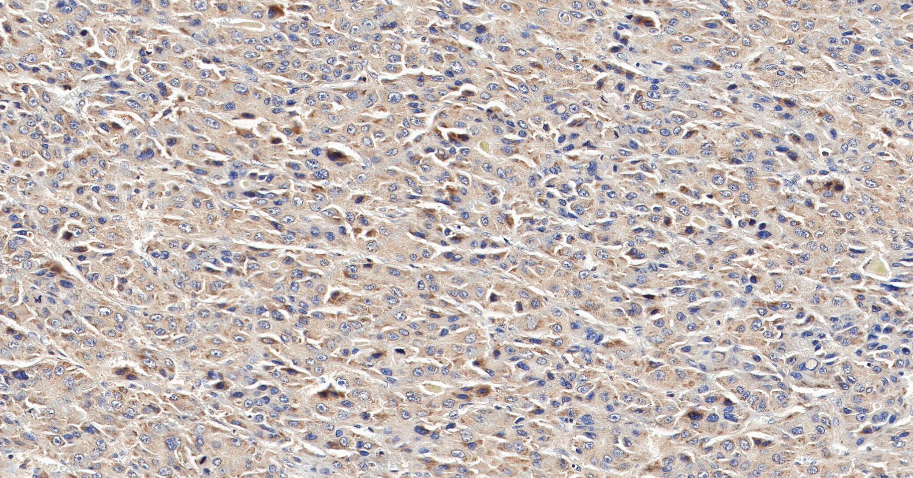 Immunohistochemical analysis of paraffin-embedded human hepatocellular carcinoma tissue using NADPH Oxidase 4 Antibody (YA4640). The section was pre-treated using heat mediated antigen retrieval with Tris/EDTA buffer (pH 9.0) for 20 minutes. The tissues were probed with the primary antibody (HY-P80762A, 1/100) overnight at 4℃. The detection was performed using an HRP conjugated compact polymer system. DAB was used as the chromogen. Tissues were counterstained with hematoxylin and mounted with neutral balsam.
 -
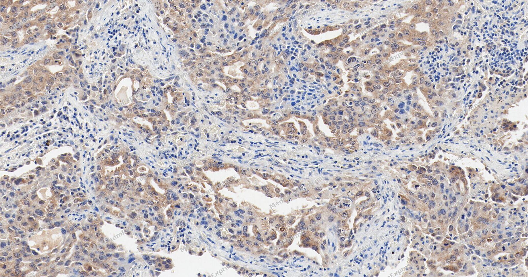 Immunohistochemical analysis of paraffin-embedded human lung adenocarcinoma tissue using NADPH Oxidase 4 Antibody (YA4640). The section was pre-treated using heat mediated antigen retrieval with Tris/EDTA buffer (pH 9.0) for 20 minutes. The tissues were probed with the primary antibody (HY-P80762A, 1/100) overnight at 4℃. The detection was performed using an HRP conjugated compact polymer system. DAB was used as the chromogen. Tissues were counterstained with hematoxylin and mounted with neutral balsam.
 -
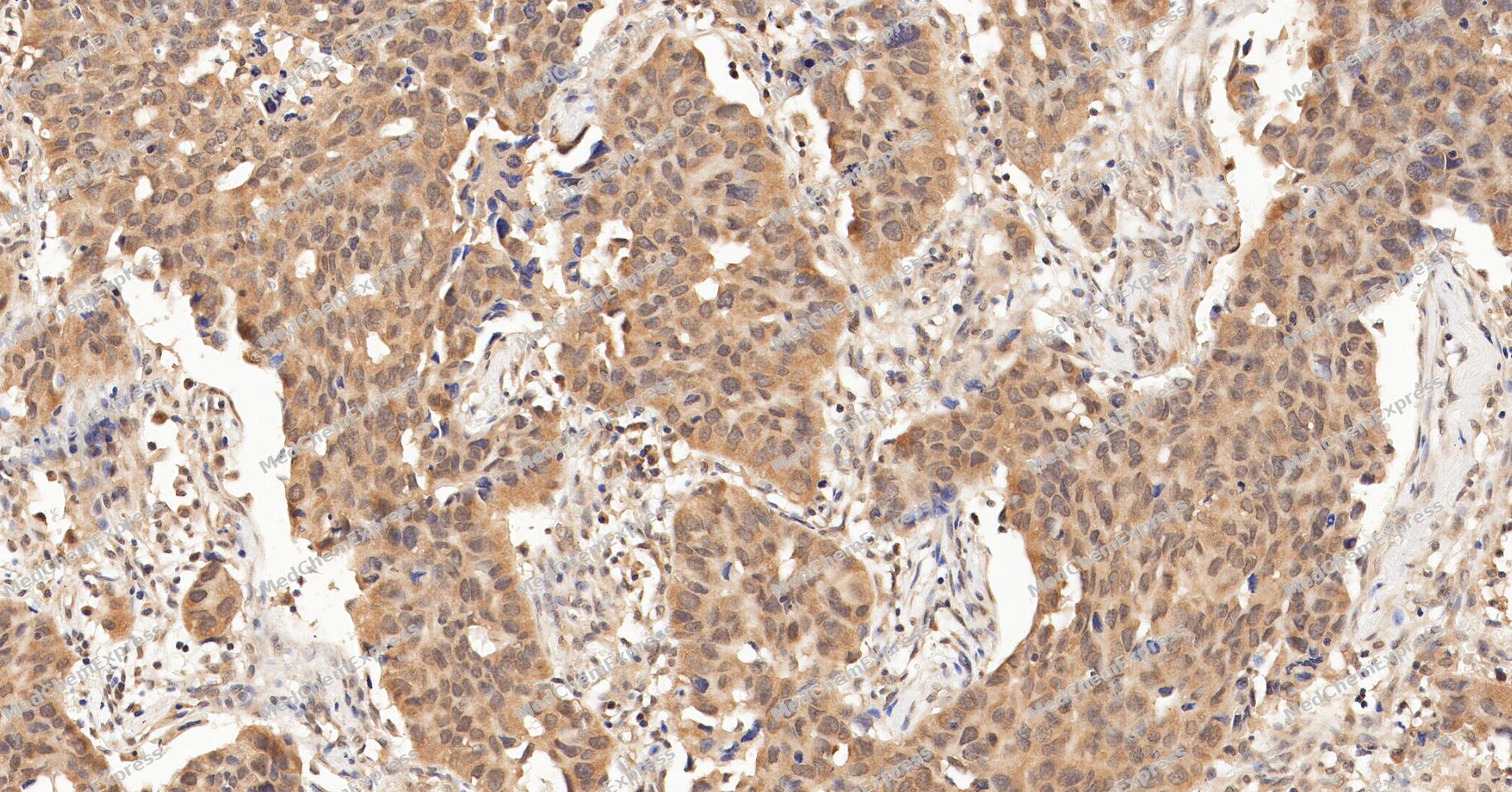 Immunohistochemical analysis of paraffin-embedded human lung cancer tissue using NADPH Oxidase 4 Antibody (YA4640). The section was pre-treated using heat mediated antigen retrieval with Tris/EDTA buffer (pH 9.0) for 20 minutes. The tissues were probed with the primary antibody (HY-P80762A, 1/100) overnight at 4℃. The detection was performed using an HRP conjugated compact polymer system. DAB was used as the chromogen. Tissues were counterstained with hematoxylin and mounted with neutral balsam.
 -
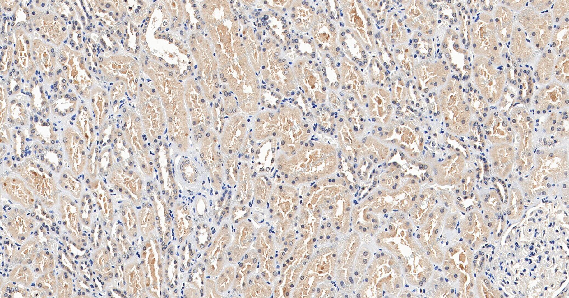 Immunohistochemical analysis of paraffin-embedded human kidney tissue using NADPH Oxidase 4 Antibody (YA4640). The section was pre-treated using heat mediated antigen retrieval with Tris/EDTA buffer (pH 9.0) for 20 minutes. The tissues were probed with the primary antibody (HY-P80762A, 1/100) overnight at 4℃. The detection was performed using an HRP conjugated compact polymer system. DAB was used as the chromogen. Tissues were counterstained with hematoxylin and mounted with neutral balsam.
 -
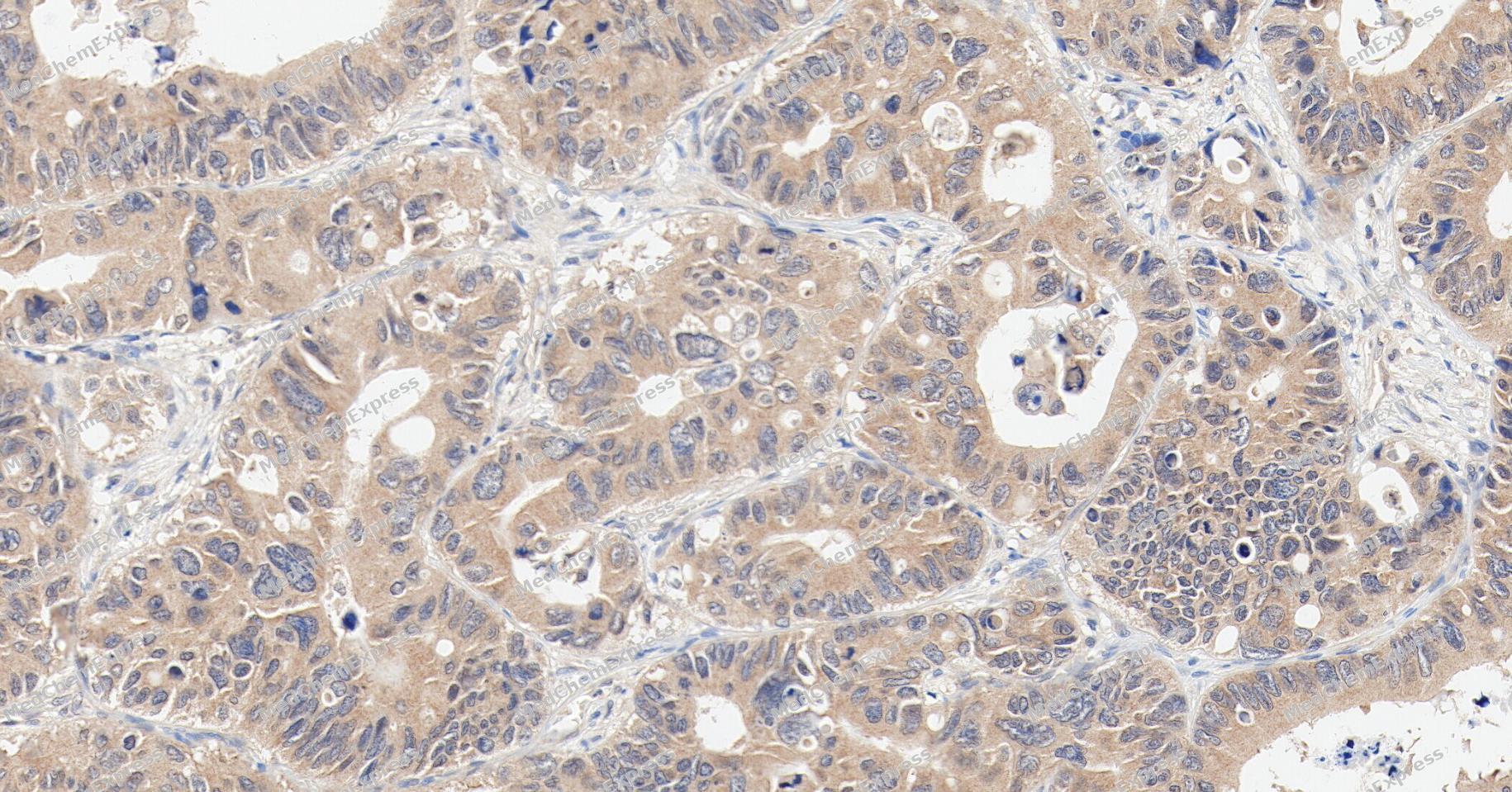 Immunohistochemical analysis of paraffin-embedded human liver cancer tissue using NADPH Oxidase 4 Antibody (YA4640). The section was pre-treated using heat mediated antigen retrieval with Tris/EDTA buffer (pH 9.0) for 20 minutes. The tissues were probed with the primary antibody (HY-P80762A, 1/100) overnight at 4℃. The detection was performed using an HRP conjugated compact polymer system. DAB was used as the chromogen. Tissues were counterstained with hematoxylin and mounted with neutral balsam.
 -
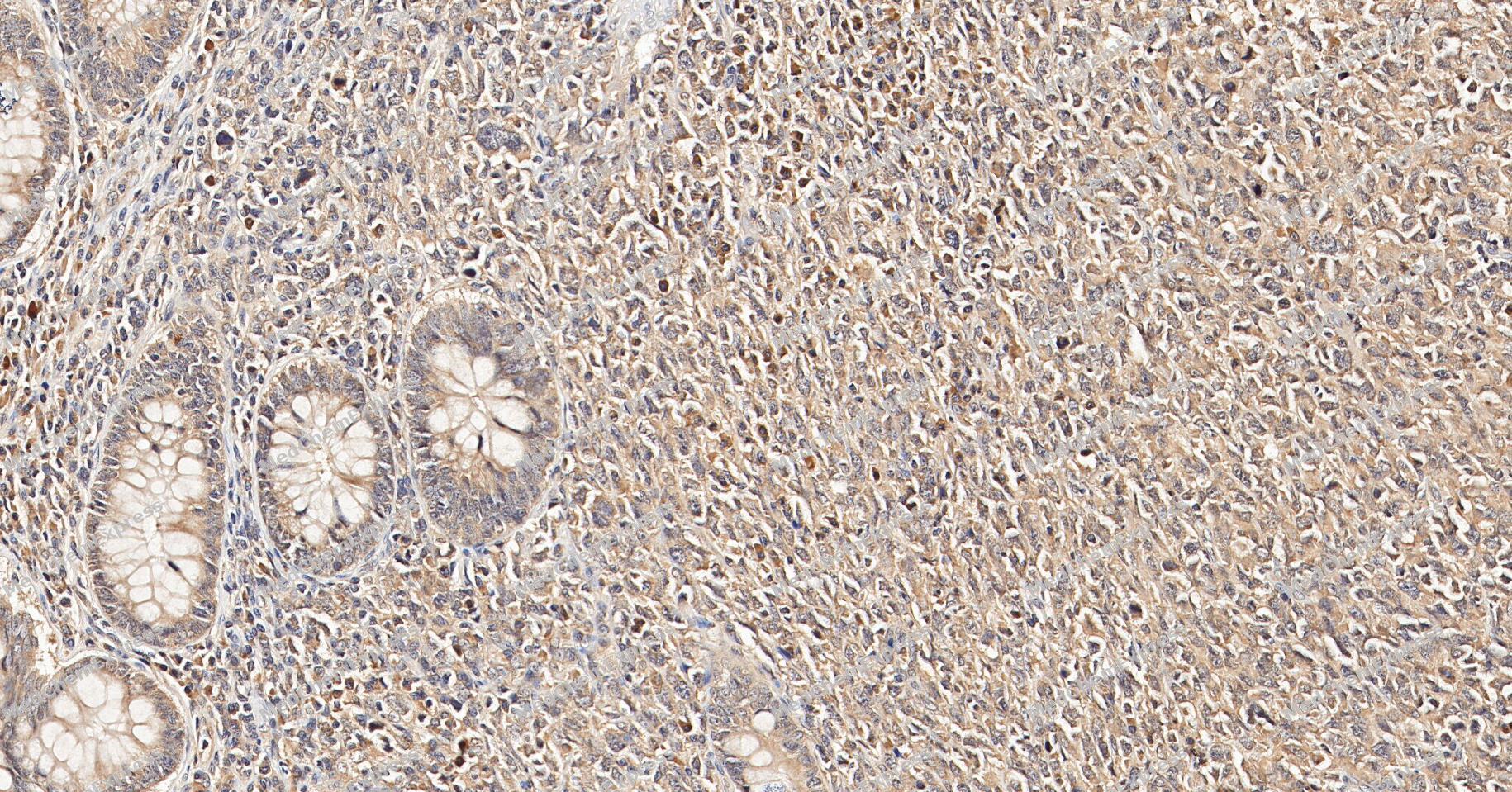 Immunohistochemical analysis of paraffin-embedded human colon cancer tissue using NADPH Oxidase 4 Antibody (YA4640). The section was pre-treated using heat mediated antigen retrieval with Tris/EDTA buffer (pH 9.0) for 20 minutes. The tissues were probed with the primary antibody (HY-P80762A, 1/100) overnight at 4℃. The detection was performed using an HRP conjugated compact polymer system. DAB was used as the chromogen. Tissues were counterstained with hematoxylin and mounted with neutral balsam.
 -
 Immunocytochemistry analysis of Hela cells labeling NADPH Oxidase 4 with NADPH Oxidase 4 Antibody (HY-P80762A) at 1/100 dilution. Cells were fixed in 4% paraformaldehyde for 15 minutes at room temperature, permeabilized with 0.1% Triton X-100 in PBS for 15 minutes at room temperature, then blocked with quick block buffer for 10 minutes at room temperature. Cells were then incubated with NADPH Oxidase 4 Antibody (HY-P80762A) at 1/100 dilution in quick block buffer overnight at 4 ℃. AF488-conjugated Goat Anti-Rabbit IgG H&L(HY-P8002, Green) was used as the secondary antibody at 1/1,000 dilution. PBS instead of the primary antibody was used as the secondary antibody only control. The Nuclear counterstain was DAPI (Blue).
 -
 Immunocytochemistry analysis of HepG2 cells labeling NADPH Oxidase 4 with NADPH Oxidase 4 Antibody (HY-P80762A) at 1/100 dilution. Cells were fixed in 4% paraformaldehyde for 15 minutes at room temperature, permeabilized with 0.1% Triton X-100 in PBS for 15 minutes at room temperature, then blocked with quick block buffer for 10 minutes at room temperature. Cells were then incubated with NADPH Oxidase 4 Antibody (HY-P80762A) at 1/100 dilution in quick block buffer overnight at 4 ℃. AF488-conjugated Goat Anti-Rabbit IgG H&L(HY-P8002, Green) was used as the secondary antibody at 1/1,000 dilution. PBS instead of the primary antibody was used as the secondary antibody only control. The Nuclear counterstain was DAPI (Blue).


Background
-
Function
NADPH Oxidase 4 is a NADPH oxidase that catalyzes predominantly the reduction of oxygen to H2O2. Can also catalyze to a smaller extent, the reduction of oxygen to superoxide. May function as an oxygen sensor regulating the KCNK3/TASK-1 potassium channel and HIF1A activity. May regulate insulin signaling cascade. May play a role in apoptosis, bone resorption and lipolysaccharide-mediated activation of NFKB. May produce superoxide in the nucleus and play a role in regulating gene expression upon cell stimulation. Promotes ferroptosis, reactive oxygen species production and reduced glutathione (GSH) levels by activating NLRP3 inflammasome activation and cytokine release; NADPH oxidase that catalyzes the generation of superoxide from molecular oxygen utilizing NADPH as an electron donor. Involved in redox signaling in vascular cells. Modulates the nuclear activation of ERK1/2 and the ELK1 transcription factor, and is capable of inducing nuclear DNA damage; Lacks superoxide-generating NADPH oxidase activity[1][2][3][4][5][6][7][8][9][10][11][12][13][14][15][16].
-
Subcellular Localization
Cytoplasm; Endoplasmic reticulum membrane; Multi-pass membrane protein; Cell membrane; Multi-pass membrane protein; Cell junction, focal adhesion; Nucleus; Nucleus; Nucleus, nucleolus; Cytoplasm; Cytoplasm, perinuclear region; Cytoplasm; Cytoplasm, perinuclear region; Cytoplasm; Cytoplasm, perinuclear region; Cytoplasm; Cytoplasm, perinuclear region
-
Expression
Tissue_specificity:This gene is expressed (protein level) in distal renal tubular cells and endothelial cells of the renal cortex. Expression is widespread. It is highly expressed in the kidneys, and expressed at lower levels in the heart, adipocytes, liver cancer cells, endothelial cells, skeletal muscle, brain, various brain tumor cell lines, and airway epithelial cells.
Induction:By 7-ketocholesterol (at protein level) -
Isoforms & Post-Translational Modification
Q9NPH5 has 9 isomers: Q9NPH5-1: 66932 Da (predicted); Q9NPH5-2: 58410 Da (predicted); Q9NPH5-3: 27625 Da (predicted); Q9NPH5-4: 31812 Da (predicted); Q9NPH5-5: 25761 Da (predicted); Q9NPH5-6: 62745 Da (predicted); Q9NPH5-7: 6516 Da (predicted); Q9NPH5-8: 64102 Da (predicted); Q9NPH5-9: 59915 Da (predicted).
Deubiquitinated by USP19;N-glycosylated and glycosylation is required for its proper function;N-glycosylated -
Subunit
Interacts with protein disulfide isomerase (By similarity). Interacts with, relocalizes and stabilizes CYBA/p22phox. Interacts with TLR4. Interacts with PPP1R15A (PubMed:26742780). Interacts with LRRC8A; this interaction prevents the ubiquitin-mediated degradation of LRRC8A (PubMed:39909992)
-
SwissProt ID
-
Synonyms
NADPH oxidase 4; Kidney oxidase-1; KOX-1; KOX1; Kidney superoxide-producing NADPH oxidase; Renal NAD(P)H-oxidase; NOX4; RENOX
-
Research Field
Cardiovascular
Documentation
-
Data Sheet (234 KB)
-
SDS (252 KB)
- English - EN (252 KB)
- Français - FR (252 KB)
- Deutsch - DE (252 KB)
- Norwegian - NO (252 KB)
- Español - ES (252 KB)
- Swedish - SV (252 KB)
- Italian - IT (252 KB)
- Korean - KR (252 KB)
- Portuguese - PT (252 KB)
-
User Guide for Antibodies (1077 KB)

 Powered by Bioz
