



PARK7/DJ1 Antibody (YA4445)
(Synonyms: DJ1; DJ-1; GATD2; HEL-S-67p)Based on 1 Customer Validation
PARK7/DJ1 Antibody (YA4445) is a Mouse-derived and non-conjugated IgG1 monoclonal antibody, targeting to PARK7/DJ1.
-
Host:
Mouse
-
Isotype:
IgG1
-
Application:
WB, IHC-P, FC, ELISA
-
Reactivity :
Human
-
Formulation:
Supplied in PBS with 0.05% sodium azide
-
Conjugation:
Non-conjugated


To place orders, for customer services and technical support, please contact: MedChemExpress USA
Tel: 609-228-6898 E-mail: [email protected] [email protected]





Applications
| Application |
WB

WB: Western Blot
|
IHC-P
IHC-P: Immunohistochemistry-Paraffin
|
FC
FC: Flow Cytometry
|
ELISA
ELISA: Enzyme Linked Immunosorbent Assay
|
|---|---|---|---|---|
| Dilution Ratio | 1:500-1:2000 | 1:200-1:1000 | 1:200-1:400 | 1:10000 |


Product Details
PARK7/DJ1 Antibody (YA4445) is a Mouse-derived and non-conjugated IgG1 monoclonal antibody, targeting to PARK7/DJ1.
-
Host Mouse

-
Clonality Monoclonal

-
Species ReactivityHuman
-
Observed Molecular WeightObserved band size: 20 kDa
Note: Due to possible protein modifications or aggregation, the molecular weight should be confirmed by actual measurement, and the predicted value is for reference only.

-
Calculated Molecular Weight Predicted band size: 20 kDa

Purified recombinant fragment of human PARK7 (AA: 1-189) expressed in E. Coli.
affinity purified.
Non-conjugated
Unmodified
IgG1


Product Properties
-
Appearance
Liquid
-
Formulation
Supplied in PBS with 0.05% sodium azide
-
Concentration
Batch-dependent, Please check the COA for the concentration of each lot. Check Lot Concentration
-
Storage & Stability
Stored at -20°C for 1 year. Avoid repeated freeze / thaw cycles.
-
Shipping
Shipping with blue ice.


Verification Images
-
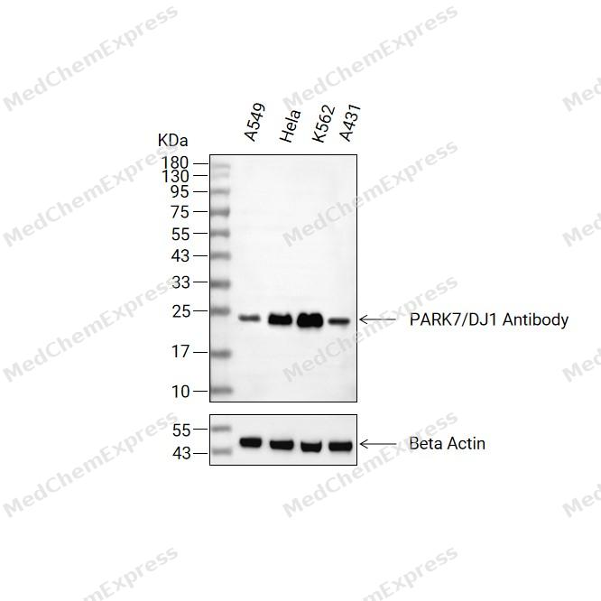 Western blot analysis of extracts from A549(lane 2(20ug) , Hela(lane 3(20ug) and K562(lane 4(20ug),A431(lane 5(20ug) using PARK7 Antibody(HY-P84748) Mouse mAb. Proteins were transferred to a PVDF membrane and blocked with 5% non-fat milk in TBST for 2 hour at room temperature. The primary antibody (1/1000) and Loading control antibody (Beta Actin, HY-P83730, 1/10000) was used in 5% non-fat milk in TBST at 4°C overnight. Goat Anti-Mouse/Rabbit IgG-HRP Secondary Antibody (1/10000) was used for 1 hour at room temperature.
 -
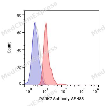 Flow cytometric analysis of 1X10^6 Jurkat cells labeling PARK7 Antibody (HY-P84748, red). Cells were fixed with 4% paraformaldehyde. Then stained with the primary antibody at 1/50 dilution for an hour at 4℃. AF 488-conjugated AffiniPure Goat Anti- Mouse IgG H&L (HY-P8005) was used as the secondary antibody at 1/1,000 dilution for 30 minutes at 4℃. Mouse IgG Isotype Control (HY-P80757, blue) was used as the isotype control, cells without incubation with primary antibody were used as the unlabeled control (black).


Background
-
Function
PARK7/DJ1 multifunctional protein with controversial molecular function which plays an important role in cell protection against oxidative stress and cell death acting as oxidative stress sensor and redox-sensitive chaperone and protease. It is involved in neuroprotective mechanisms like the stabilization of NFE2L2 and PINK1 proteins, male fertility as a positive regulator of androgen signaling pathway as well as cell growth and transformation through, for instance, the modulation of NF-kappa-B signaling pathway. Has been described as a protein and nucleotide deglycase that catalyzes the deglycation of the Maillard adducts formed between amino groups of proteins or nucleotides and reactive carbonyl groups of glyoxals. But this function is rebuted by other works. As a protein deglycase, repairs methylglyoxal- and glyoxal-glycated proteins, and releases repaired proteins and lactate or glycolate, respectively. Deglycates cysteine, arginine and lysine residues in proteins, and thus reactivates these proteins by reversing glycation by glyoxals. Acts on early glycation intermediates (hemithioacetals and aminocarbinols), preventing the formation of advanced glycation endproducts (AGE) that cause irreversible damage. Also functions as a nucleotide deglycase able to repair glycated guanine in the free nucleotide pool (GTP, GDP, GMP, dGTP) and in DNA and RNA. Is thus involved in a major nucleotide repair system named guanine glycation repair (GG repair), dedicated to reversing methylglyoxal and glyoxal damage via nucleotide sanitization and direct nucleic acid repair. Protects histones from adduction by methylglyoxal, controls the levels of methylglyoxal-derived argininine modifications on chromatin. Able to remove the glycations and restore histone 3, histone glycation disrupts both local and global chromatin architecture by altering histone-DNA interactions as well as histone acetylation and ubiquitination levels. Displays a very low glyoxalase activity that may reflect its deglycase activity. Eliminates hydrogen peroxide and protects cells against hydrogen peroxide-induced cell death. Required for correct mitochondrial morphology and function as well as for autophagy of dysfunctional mitochondria. Plays a role in regulating expression or stability of the mitochondrial uncoupling proteins SLC25A14 and SLC25A27 in dopaminergic neurons of the substantia nigra pars compacta and attenuates the oxidative stress induced by calcium entry into the neurons via L-type channels during pacemaking. Regulates astrocyte inflammatory responses, may modulate lipid rafts-dependent endocytosis in astrocytes and neuronal cells. In pancreatic islets, involved in the maintenance of mitochondrial reactive oxygen species (ROS) levels and glucose homeostasis in an age- and diet dependent manner. Protects pancreatic beta cells from cell death induced by inflammatory and cytotoxic setting. Binds to a number of mRNAs containing multiple copies of GG or CC motifs and partially inhibits their translation but dissociates following oxidative stress. Metal-binding protein able to bind copper as well as toxic mercury ions, enhances the cell protection mechanism against induced metal toxicity. In macrophages, interacts with the NADPH oxidase subunit NCF1 to direct NADPH oxidase-dependent ROS production, and protects against sepsis[1][2][3][4][5][6][7][8][9][10][11][12][13][14][15][16][17][18][19][20][21][22][23][24].
-
Subcellular Localization
Cell membrane; Lipid-anchor; Cytoplasm; Nucleus; Membrane raft; Mitochondrion; Endoplasmic reticulum
-
Expression
Tissue_specificity:It is highly expressed in the pancreas, kidneys, skeletal muscle, liver, testes, and heart. Slightly lower expression levels were detected in the placenta and brain (protein level) . It was also detected in astrocytes, Sertoli cells, spermatogonia, spermatids, and sperm. Expression levels in the islets of Langerhans were higher than in surrounding exocrine tissues (PubMed: 22611253) .
Induction:By hydrogen peroxide and UV irradiation (PubMed:14749723, PubMed:15976810) . In pancreatic islets, expression increases under hyperglycemic conditions (PubMed:22611253) . Expression is also induced by sulforaphane, an isothiocyanate obtained from cruciferous vegetables (PubMed:26995087) -
Subunit
Homodimer (PubMed:12796482, PubMed:12851414, PubMed:12855764, PubMed:31653696). Binds EFCAB6/DJBP and PIAS2 (PubMed:11477070, PubMed:12612053, PubMed:12851414). Part of a ternary complex containing PARK7, EFCAB6/DJBP and AR (PubMed:12612053). Interacts (via N-terminus) with OTUD7B (PubMed:21097510). Interacts with BBS1, HIPK1, CLCF1 and MTERF (PubMed:16390825, PubMed:21097510). Forms a complex with PINK1 and PRKN (PubMed:19229105). Interacts (via C-terminus) with NCF1; the interaction is enhanced by LPS and modulates NCF1 phosphorylation and membrane translocation (By similarity). Interacts with NENF (PubMed:31536960)
-
SwissProt ID
-
Synonyms
DJ1; DJ-1; GATD2; HEL-S-67p


Documentation
-
Data Sheet (235 KB)
-
SDS (251 KB)
- English - EN (251 KB)
- Français - FR (251 KB)
- Deutsch - DE (251 KB)
- Norwegian - NO (251 KB)
- Español - ES (251 KB)
- Swedish - SV (251 KB)
- Italian - IT (251 KB)
- Korean - KR (251 KB)
- Portuguese - PT (251 KB)
-
User Guide for Antibodies (1077 KB)

 Powered by Bioz
