



PIAS1 Antibody (YA2217)
(Synonyms: PIAS1; DDXBP1; E3 SUMO-protein ligase PIAS1; DEAD/H box-binding protein 1; Gu-binding protein; GBP; Protein inhibitor of activated STAT protein 1; RNA helicase II-binding protein)
Based on 1 Customer Validation
PIAS1 Antibody (YA2217) is a Rabbit-derived and non-conjugated IgG monoclonal antibody, targeting to PIAS1.
-
Host:
Rabbit
-
Isotype:
IgG
-
Application:
WB, IHC-P, ICC/IF, FC
-
Reactivity :
Human, Mouse, Rat
-
Formulation:
Supplied in Rabbit IgG in 10mM phosphate buffered saline , pH 7.4, 150mM sodium chloride, 0.05% BSA, 0.02% sodium azide and 50% glycerol.
-
Conjugation:
Non-conjugated


To place orders, for customer services and technical support, please contact: MedChemExpress USA
Tel: 609-228-6898 E-mail: [email protected] [email protected]





Applications
| Application |
WB

WB: Western Blot
|
IHC-P
IHC-P: Immunohistochemistry-Paraffin
|
ICC/IF
ICC/IF: Immunocytochemistry/
Immunofluorescence |
FC
FC: Flow Cytometry
|
|---|---|---|---|---|
| Dilution Ratio | 1:500-1:1000 | 1:50-1:100 | 1:50-1:200 | 1:50-1:100 |


Product Details
PIAS1 Antibody (YA2217) is a Rabbit-derived and non-conjugated IgG monoclonal antibody, targeting to PIAS1.
-
Host Rabbit

-
Clonality Recombinant,Monoclonal

-
Species ReactivityHuman, Mouse, Rat
-
Observed Molecular WeightObserved band size: 76 kDa
Note: Due to possible protein modifications or aggregation, the molecular weight should be confirmed by actual measurement, and the predicted value is for reference only.

-
Calculated Molecular Weight Predicted band size: 72 kDa

Entrez Gene: 8554 Human ; 56469 Mouse ; 300772 Rat
SwissProt: O75925 Human ; O88907 Mouse ;
OMIM: 603566 Human
A synthesized peptide derived from human PIAS1 aa63-117.
Endogenous
Affinity Chromatography
Non-conjugated
Unmodified
IgG


Product Properties
-
Appearance
Liquid
-
Formulation
Supplied in Rabbit IgG in 10mM phosphate buffered saline , pH 7.4, 150mM sodium chloride, 0.05% BSA, 0.02% sodium azide and 50% glycerol.
-
Concentration
Batch-dependent, Please check the COA for the concentration of each lot. Check Lot Concentration
-
Storage & Stability
Stored at -20°C for 1 year. Avoid repeated freeze / thaw cycles.
-
Shipping
Shipping with blue ice.


Verification Images
-
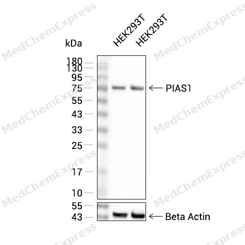 Western blot analysis of extracts from HEK293T (lane 2(20μg), HEK293T (lane 3(40μg), using PIAS1 Antibody. Proteins were transferred to a PVDF membrane and blocked with 5% BSA in TBST for 2 hour at room temperature. The primary antibody and Loading control antibody (Beta Actin, HY-P80438, 1/3000) was used in 5% BSA in TBST at 4°C overnight. Goat Anti-Mouse/Rabbit IgG-HRP Secondary Antibody (HY-P8004/HY-P8001, 1/10,000) was used for 1 hour at room temperature.
 -
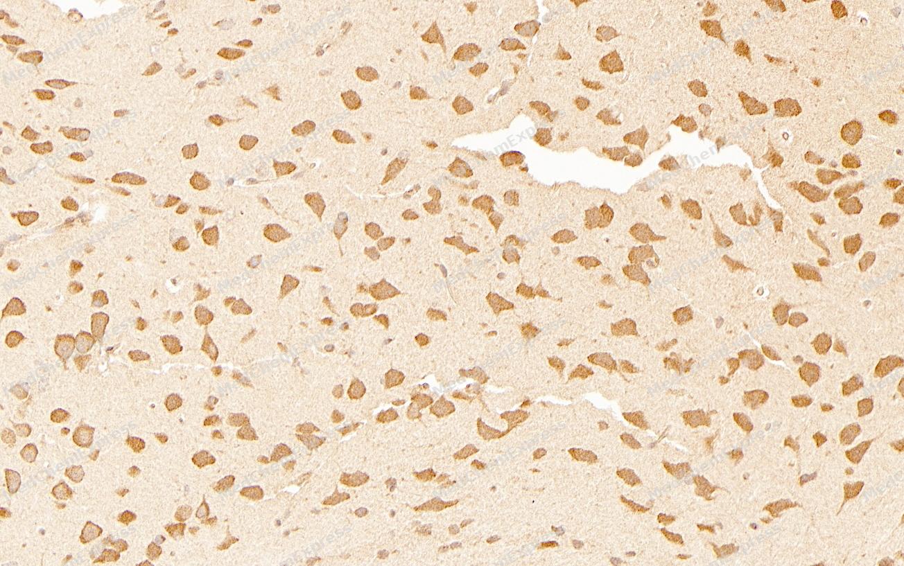 Immunohistochemical analysis of paraffin-embedded Mouse brain tissue using PIAS1 Antibody (YA2217). The section was pre-treated using heat mediated antigen retrieval with sodium citrate buffer (pH 6.0) for 2 minutes. The tissues were blocked in QuickBlock for 20 minutes at room temperature, washed with ddH2O and PBS, and then probed with the primary antibody (HY-P82472, 1/100) in 4℃ overnight. The detection was performed using an HRP conjugated compact polymer system. DAB was used as the chromogen. Tissues were counterstained with hematoxylin and mounted with DPX.
 -
Immunohistochemical analysis of paraffin-embedded Mouse brain tissue using PIAS1 Antibody (YA2217). The section was pre-treated using heat mediated antigen retrieval with sodium citrate buffer (pH 6.0) for 2 minutes. The tissues were blocked in QuickBlock for 20 minutes at room temperature, washed with ddH2O and PBS, and then probed with the primary antibody (HY-P82472, 1/100) in 4℃ overnight. The detection was performed using an HRP conjugated compact polymer system. DAB was used as the chromogen. Tissues were counterstained with hematoxylin and mounted with DPX.
-
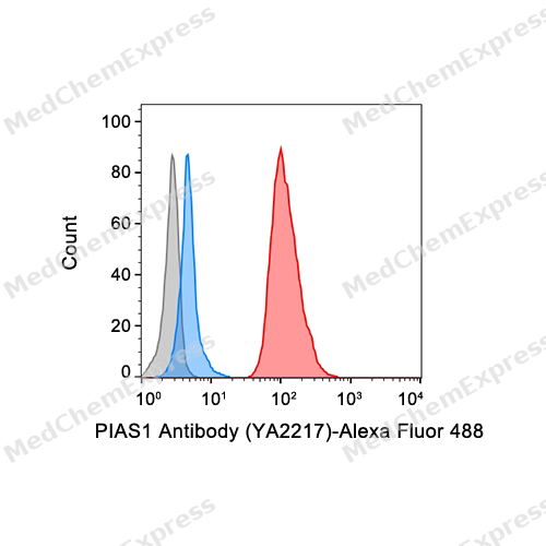 Flow cytometric analysis of 1X10^6 Hela cells labeling PIAS1 Antibody (YA2217) (HY-P82472, red). Cells were fixed with 4% paraformaldehyde and permeabilised with intranuclear rupture fluid. Then stained with the primary antibody at 1/50 dilution for an hour at 4℃. Alexa Fluor® 488-conjugated AffiniPure Goat Anti-Rabbit IgG H&L (HY-P8002) was used as the secondary antibody at 1/1,000 dilution for 30 minutes at 4℃. Rabbit IgG Isotype Control (HY-P80879, blue) was used as the isotype control, cells without incubation with primary antibody were used as the unlabeled control (black).
 -
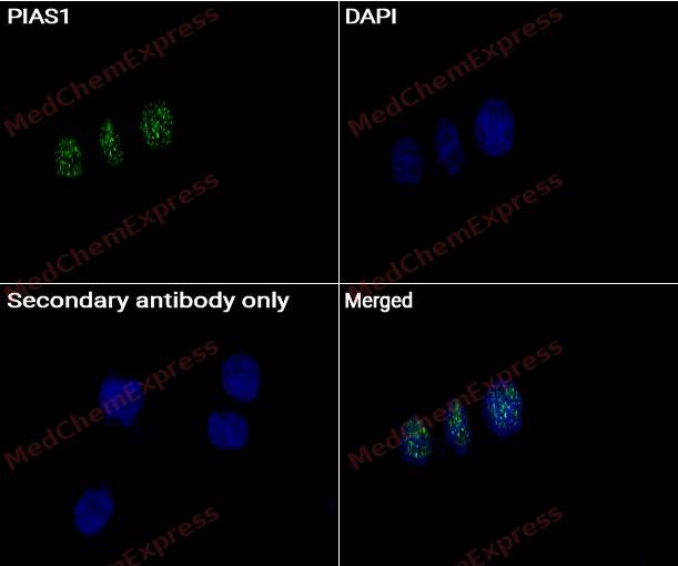 Immunocytochemistry analysis of HeLa cells labeling PIAS1 with PIAS1 antibody (HY-P82472) at 1/50 dilution. Cells were fixed in 4% paraformaldehyde for 15 minutes at room temperature, permeabilized with 0.1% Triton X-100 for 10 minutes at room temperature, then blocked with BSA for Immunol Staining for 10 min at room temperature. Cells were then incubated with PIAS1 antibody (HY-P82472) at 1/50 dilution in BSA for Immunol Staining at 4 ℃, Stay overnight. AF488-conjugated AffiniPure Goat Anti-Rabbit IgG H&L(HY-P8002, Green) was used as the secondary antibody at 1/1,000 dilution. PBS instead of the primary antibody was used as the secondary antibody only control. The Nuclear counterstain was DAPI (Blue).
 -
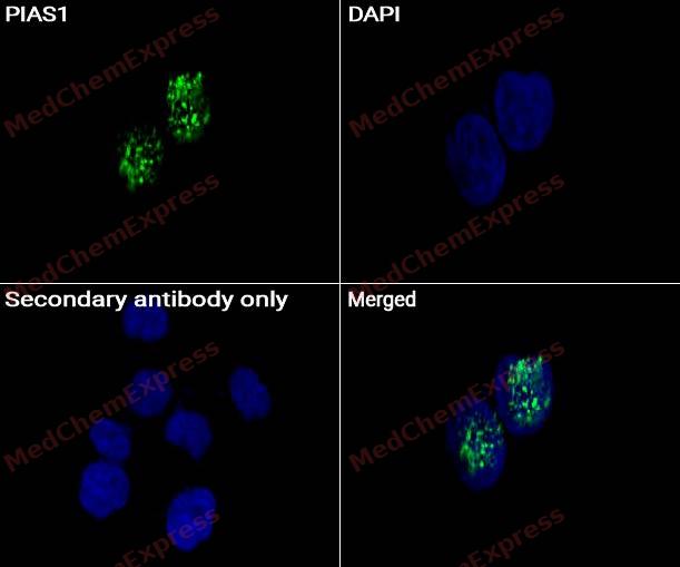 Immunocytochemistry analysis of HeLa cells labeling PIAS1 with PIAS1 antibody (HY-P82472) at 1/100 dilution. Cells were fixed in 4% paraformaldehyde for 15 minutes at room temperature, permeabilized with 0.1% Triton X-100 for 10 minutes at room temperature, then blocked with BSA for Immunol Staining for 10 min at room temperature. Cells were then incubated with PIAS1 antibody (HY-P82472) at 1/100 dilution in BSA for Immunol Staining at 4℃, Stay overnight. AF488-conjugated AffiniPure Goat Anti-Rabbit IgG H&L(HY-P8002, Green) was used as the secondary antibody at 1/1,000 dilution. PBS instead of the primary antibody was used as the secondary antibody only control. The Nuclear counterstain was DAPI (Blue).


Background
-
Function
PIAS1 functions as an E3-type small ubiquitin-like modifier (SUMO) ligase, stabilizing the interaction between UBE2I and the substrate, and as a SUMO-tethering factor. Catalyzes sumoylation of various proteins, such as CEBPB, MRE11, MTA1, PTK2 and PML. Plays a crucial role as a transcriptional coregulation in various cellular pathways, including the STAT pathway, the p53 pathway and the steroid hormone signaling pathway. In vitro, binds A/T-rich DNA. The effects of this transcriptional coregulation, transactivation or silencing, may vary depending upon the biological context. Mediates sumoylation of MRE11, stabilizing MRE11 on chromatin during end resection. Sumoylates PML (at 'Lys-65' and 'Lys-160') and PML-RAR and promotes their ubiquitin-mediated degradation. PIAS1-mediated sumoylation of PML promotes its interaction with CSNK2A1/CK2 which in turn promotes PML phosphorylation and degradation. Enhances the sumoylation of MTA1 and may participate in its paralog-selective sumoylation. Plays a dynamic role in adipogenesis by promoting the SUMOylation and degradation of CEBPB. Mediates the nuclear mobility and localization of MSX1 to the nuclear periphery, whereby MSX1 is brought into the proximity of target myoblast differentiation factor genes. Also required for the binding of MSX1 to the core enhancer region in target gene promoter regions, independent of its sumoylation activity. Capable of binding to the core enhancer region TAAT box in the MYOD1 gene promoter; (Microbial infection) Restricts Epstein-Barr virus (EBV) lytic replication by acting as an inhibitor for transcription factors involved in lytic gene expression. The virus can use apoptotic caspases to antagonize PIAS1-mediated restriction and express its lytic genes[1][2][3][4][5][6][7].
-
Subcellular Localization
Nucleus; Nucleus speckle; Nucleus, PML body; Cytoplasm, cytoskeleton
-
Expression
Tissue_specificity:It is expressed in multiple tissues, with the highest expression level in the testes. -
Isoforms & Post-Translational Modification
O75925 has 3 isomers: O75925-1: 71836 Da (predicted); O75925-2: 72203 Da (predicted); O75925-3: 36204 Da (predicted).
Sumoylated -
Subunit
Interacts with NCOA2 and AR. Interacts with NR2C1; the interaction promotes its sumoylation (By similarity). Interacts with DDX21, CSRP2, AXIN1, JUN, UBE2I, SUMO1, SATB2, PLAG1, TP53 and STAT1 (dimer), following IFNA1-stimulation. Interacts with SP3 (preferentially when SUMO-modified). Interacts with KLF8; the interaction results in SUMO ligation and repression of KLF8 transcriptional activity and of its cell cycle progression into G(1) phase. Interacts with CHUK/IKKA; this interaction induces PIAS1 phosphorylation. Interacts with PTK2/FAK1; the interaction promotes its sumoylation. Interacts with DDX5. Interacts with PML (By similarity). Interacts with MTA1. Interacts with SUMO1P1/SUMO5 (PubMed:27211601). Interacts with PRDM1/Blimp-1 (PubMed:28842558). Interacts (via N-terminus) with MSX1 (via C-terminus); the interaction is required for the localization of both proteins to the nuclear periphery and specific binding of MSX1 to the core enhancer region in target gene promoters (PubMed:16600910)
-
SwissProt ID
-
Synonyms
PIAS1; DDXBP1; E3 SUMO-protein ligase PIAS1; DEAD/H box-binding protein 1; Gu-binding protein; GBP; Protein inhibitor of activated STAT protein 1; RNA helicase II-binding protein
-
Research Field
Epigenetics and Nuclear Signaling


Documentation
-
Data Sheet (234 KB)
-
SDS (252 KB)
- English - EN (252 KB)
- Français - FR (252 KB)
- Deutsch - DE (252 KB)
- Norwegian - NO (252 KB)
- Español - ES (252 KB)
- Swedish - SV (252 KB)
- Italian - IT (252 KB)
- Korean - KR (252 KB)
- Portuguese - PT (252 KB)
-
User Guide for Antibodies (1077 KB)

 Powered by Bioz
