



TXNIP Antibody (YA024)
(Synonyms: VDUP1, TXNIP, Thioredoxin-interacting protein, Thioredoxin-binding protein 2, Vitamin D3 up-regulated protein 1)Based on 2 publication(s) in Google Scholar
TXNIP Antibody (YA024) is a Rabbit-derived and non-conjugated IgG monoclonal antibody, targeting to TXNIP.
-
Host:
Rabbit
-
Isotype:
IgG
-
Application:
WB, ICC/IF, IHC-P, FC
-
Reactivity :
Human, Mouse, Rat
-
Formulation:
Supplied in 1*TBS (pH7.4), 0.05% BSA and 40% Glycerol. Preservative: 0.05% Sodium Azide.
-
Conjugation:
Non-conjugated


To place orders, for customer services and technical support, please contact: MedChemExpress USA
Tel: 609-228-6898 E-mail: [email protected] [email protected]



Publications Citing Use of MedChemExpress (MCE) TXNIP Antibody (YA024)
More

Applications
| Application |
WB

WB: Western Blot
|
ICC/IF
ICC/IF: Immunocytochemistry/
Immunofluorescence |
IHC-P
IHC-P: Immunohistochemistry-Paraffin
|
FC
FC: Flow Cytometry
|
|---|---|---|---|---|
| Dilution Ratio | 1:500-1:5000 | 1:50-1:200 | 1:50-1:400 | 1:50-1:100 |


Product Details
TXNIP Antibody (YA024) is a Rabbit-derived and non-conjugated IgG monoclonal antibody, targeting to TXNIP.
-
Host Rabbit

-
Clonality Recombinant,Monoclonal

-
Species ReactivityHuman, Mouse, Rat
-
Observed Molecular WeightObserved band size: 50 kDa
Note: Due to possible protein modifications or aggregation, the molecular weight should be confirmed by actual measurement, and the predicted value is for reference only.

-
Calculated Molecular Weight Predicted band size: 44 kDa

Entrez Gene: 10628 Human ; 56338 Mouse ; 117514 Rat
SwissProt: Q9H3M7 Human ; Q8BG60 Mouse ; Q5M7W1 Rat
OMIM: 606599 Human
Synthetic peptide corresponding to Human TXNIP.AA range:50-99.
Endogenous
Protein A affinity purified.
Non-conjugated
Unmodified
IgG


Product Properties
-
Appearance
Solution
-
Formulation
Supplied in 1*TBS (pH7.4), 0.05% BSA and 40% Glycerol. Preservative: 0.05% Sodium Azide.
-
Concentration
Batch-dependent, Please check the COA for the concentration of each lot. Check Lot Concentration
-
Storage & Stability
Stored at -20°C for 1 year. Avoid repeated freeze / thaw cycles.
-
Shipping
Shipping with blue ice.


Publications (2)
-
Journal Impact Factor
-
Most Recent
-
iScience
CRISPR-Cas9 screening identifies INTS3 as an anti-apoptotic RNA-binding protein and therapeutic target for colorectal cancer. [Abstract]2024 Apr 6;27(5):109676. PMID: 38665208 -
Biochem Biophys Res Commun
TXNIP participates in the pathogenesis of PCOS by regulating glycolysis in granulosa cells. [Abstract]2025 Aug 15:775:152149. PMID: 40499496


Verification Images
-
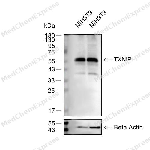 Western blot analysis of extracts from Hela (lane 2(20μg) or lane 3(40μg)) and NIH3T3 (lane 4(20μg) or lane 5(40μg)) using TXNIP (HY-P80361) Rabbit mAb. Proteins were transferred to a PVDF membrane and blocked with 5% BSA in TBST for 2 hour at room temperature. The primary antibody (1/1000) and Loading control antibody (Beta Actin, HY-P80438, 1/10000) was used in 5% BSA in TBST at 4°C overnight. Goat Anti-Rabbit IgG-HRP Secondary Antibody (1/10,000) was used for 1 hour at room temperature.
 -
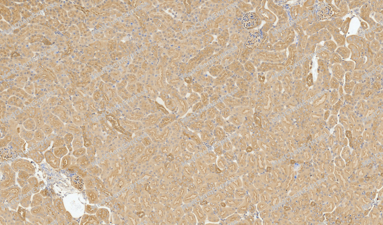 Immunohistochemical analysis of paraffin-embedded Mouse kidney tissue using TXNIP Antibody. The section was pre-treated using heat mediated antigen retrieval with sodium citrate buffer (pH 6.0) for 8 minutes. The tissues were blocked in QuickBlock for 20 minutes at room temperature, washed with ddH2O and PBS, and then probed with the primary antibody (HY-P80361, 1/1000) in 4℃ overnight. The detection was performed using an HRP conjugated compact polymer system. DAB was used as the chromogen. Tissues were counterstained with hematoxylin and mounted with DPX.
 -
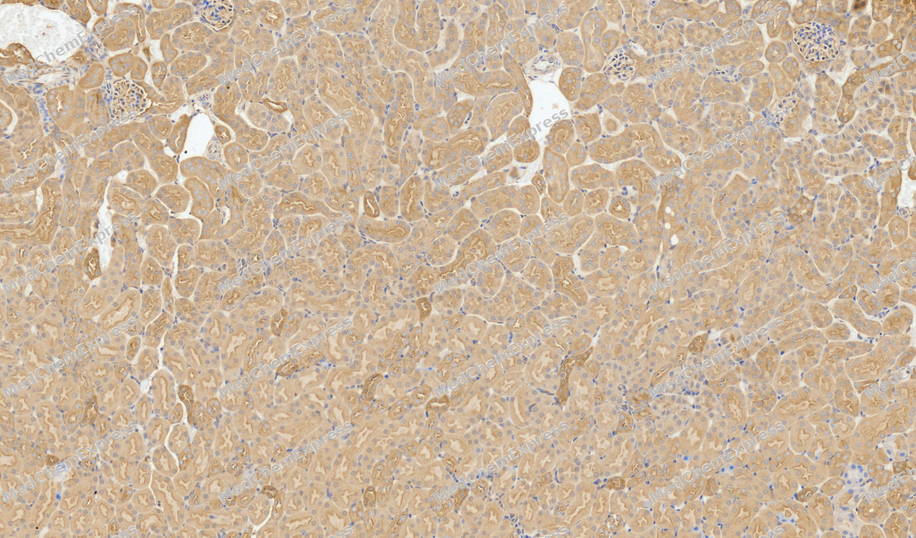 Immunohistochemical analysis of paraffin-embedded Mouse kidney tissue using TXNIP Antibody. The section was pre-treated using heat mediated antigen retrieval with sodium citrate buffer (pH 6.0) for 8 minutes. The tissues were blocked in QuickBlock for 20 minutes at room temperature, washed with ddH2O and PBS, and then probed with the primary antibody (HY-P80361, 1/1000) in 4℃ overnight. The detection was performed using an HRP conjugated compact polymer system. DAB was used as the chromogen. Tissues were counterstained with hematoxylin and mounted with DPX.
 -
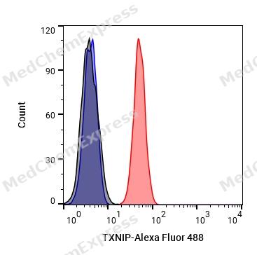 Flow cytometric analysis of 1X10^6 Hela cells labeling TXNIP Antibody (HY-P80361, red). Cells were fixed with 4% paraformaldehyde and permeabilised with 90% methanol. Then stained with the primary antibody at 1/50 dilution for an hour at 4℃. Alexa Fluor® 488-conjugated AffiniPure Goat Anti-Rabbit IgG H&L (HY-P8002) was used as the secondary antibody at 1/1,000 dilution for 30 minutes at 4℃. Rabbit IgG Isotype Control (HY-P80879, blue) was used as the isotype control, cells without incubation with primary antibody were used as the unlabeled control (black).
 -
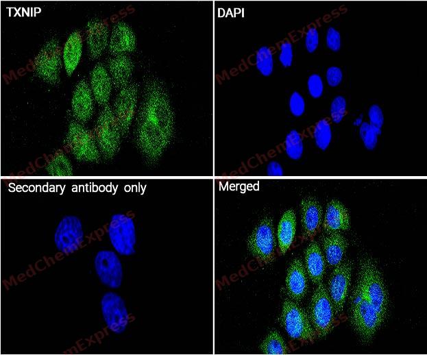 Immunocytochemistry analysis of Hela cells labeling TXNIP with TXNIP Antibody (HY-P80361) at 1/100 dilution. Cells were fixed in 4% paraformaldehyde for 15 minutes at room temperature, permeabilized with 0.1% Triton X-100 in PBS for 15 minutes at room temperature, then blocked with quick block buffer for 10 minutes at room temperature. Cells were then incubated with TXNIP Antibody (HY-P80361) at 1/100 dilution in quick block buffer overnight at 4 ℃. AF488-conjugated Goat Anti-Rabbit IgG H&L(HY-P8002, Green) was used as the secondary antibody at 1/1,000 dilution. PBS instead of the primary antibody was used as the secondary antibody only control. The Nuclear counterstain was DAPI (Blue).
 -
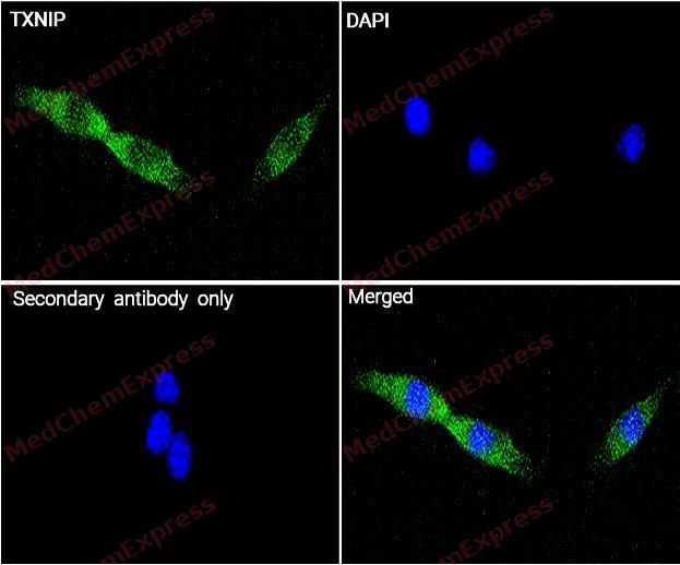 Immunocytochemistry analysis of NIH-3T3 cells labeling TXNIP with TXNIP Antibody (HY-P80361) at 1/100 dilution. Cells were fixed in 4% paraformaldehyde for 15 minutes at room temperature, permeabilized with 0.1% Triton X-100 in PBS for 15 minutes at room temperature, then blocked with quick block buffer for 10 minutes at room temperature. Cells were then incubated with TXNIP Antibody (HY-P80361) at 1/100 dilution in quick block buffer overnight at 4 ℃. AF488-conjugated Goat Anti-Rabbit IgG H&L(HY-P8002, Green) was used as the secondary antibody at 1/1,000 dilution. PBS instead of the primary antibody was used as the secondary antibody only control. The Nuclear counterstain was DAPI (Blue).


Background
-
Function
TXNIP may act as an oxidative stress mediator by inhibiting thioredoxin activity or by limiting its bioavailability. Interacts with COPS5 and restores COPS5-induced suppression of CDKN1B stability, blocking the COPS5-mediated translocation of CDKN1B from the nucleus to the cytoplasm. Functions as a transcriptional repressor, possibly by acting as a bridge molecule between transcription factors and corepressor complexes, and over-expression will induce G0/G1 cell cycle arrest. Required for the maturation of natural killer cells. Acts as a suppressor of tumor cell growth. Inhibits the proteasomal degradation of DDIT4, and thereby contributes to the inhibition of the mammalian target of rapamycin complex 1 (mTORC1)[1][2][3][4].
-
Subcellular Localization
Cytoplasm; Nucleus
-
Expression
Induction:By 1,25-dihydroxyvitamin D-3 and TGFB1. Down-regulated in response to oxidative stress -
Subunit
Homodimer; disulfide-linked (PubMed:23519408). Interacts with TXN/thioredoxin through its redox-active site (PubMed:17603038). Interacts with transcriptional repressors ZBTB16, ZBTB32 and HDAC1 (PubMed:12821938). Interacts with DDIT4 (PubMed:21460850)
-
SwissProt ID
-
Synonyms
VDUP1, TXNIP, Thioredoxin-interacting protein, Thioredoxin-binding protein 2, Vitamin D3 up-regulated protein 1
-
Research Field
Signal Transduction
Documentation
-
Data Sheet (262 KB)
-
SDS (251 KB)
- English - EN (251 KB)
- Français - FR (251 KB)
- Deutsch - DE (251 KB)
- Norwegian - NO (251 KB)
- Español - ES (251 KB)
- Swedish - SV (251 KB)
- Italian - IT (251 KB)
- Korean - KR (251 KB)
- Portuguese - PT (251 KB)
-
User Guide for Antibodies (1077 KB)
References
[1]. Liyanage NP, et al. Regulation of the bioavailability of thioredoxin in the lens by a specific thioredoxin-binding protein (TBP-2). Exp Eye Res. 2007 Aug;85(2):270-9. [Content Brief]
[2]. Han SH, et al. VDUP1 upregulated by TGF-beta1 and 1,25-dihydorxyvitamin D3 inhibits tumor cell growth by blocking cell-cycle progression. Oncogene. 2003 Jun 26;22(26):4035-46. [Content Brief]
[3]. Shin KH, et al. hnRNP G elicits tumor-suppressive activity in part by upregulating the expression of Txnip. Biochem Biophys Res Commun. 2008 Aug 8;372(4):880-5. [Content Brief]
[4]. Jin HO, et al. TXNIP potentiates Redd1-induced mTOR suppression through stabilization of Redd1. Oncogene. 2011 Sep 1;30(35):3792-801. [Content Brief]

 Powered by Bioz
