



HJC0416
Based on 1 publication(s) in Google Scholar
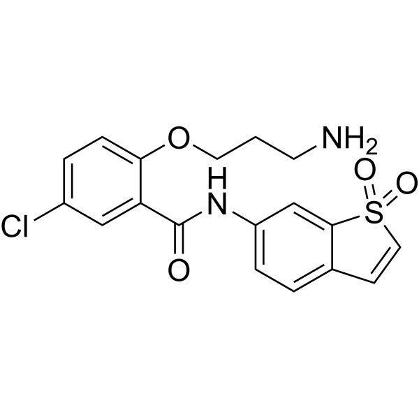
HJC0416 is a potent and orally active STAT3 inhibitor. HJC0416 shows antiprolifeative activity and induces Apoptosis. HJC0416 decreases the expression of p-STAT3 (Tyr-705), Cyclin D1 and increases the expression of cleaved caspase-3 protein. HJC0416 shows anti-tumor activity.
Nur für Forschungszwecke. Wir verkaufen nicht an Patienten.
- CAS. Nr.: 1617518-22-5
- Formel: C18H17ClN2O4S
- Molecular Weight:392.86
-
Speicherung:
Please store the product under the recommended conditions in the Certificate of Analysis.

To place orders, for customer services and technical support, please contact: MedChemExpress USA
Tel: 609-228-6898 E-mail: [email protected] [email protected]


-
 Biologische Aktivität
-
Chemical Information
- Protokoll
- Reinheit & Dokumentation
- Verweise
-
 Help & FAQs


Publications Citing Use of MedChemExpress (MCE) HJC0416
More

Biologische Aktivität
Beschreibung
IC50 & Target
[1]|
STAT3 |
Cellular Effect
|
Cell Line

|
Type | Value | Description | References |
|---|---|---|---|---|
| ASPC1 | IC50 |
0.04 μM
Compound: 12, HJC0416
|
Antiproliferative activity against human AsPC1 cells assessed as inhibition of cell proliferation after 72 hrs by MTS assay
Antiproliferative activity against human AsPC1 cells assessed as inhibition of cell proliferation after 72 hrs by MTS assay
|
[PMID: 24904966] |
| MCF7 | IC50 |
1.76 μM
Compound: 12, HJC0416
|
Antiproliferative activity against ER-positive human MCF7 cells assessed as inhibition of cell proliferation after 72 hrs by MTS assay
Antiproliferative activity against ER-positive human MCF7 cells assessed as inhibition of cell proliferation after 72 hrs by MTS assay
|
[PMID: 24904966] |
| MDA-MB-231 | IC50 |
1.97 μM
Compound: 12, HJC0416
|
Antiproliferative activity against ER-negative and triple-negative human MDA-MB-231 cells assessed as inhibition of cell proliferation after 72 hrs by MTS assay
Antiproliferative activity against ER-negative and triple-negative human MDA-MB-231 cells assessed as inhibition of cell proliferation after 72 hrs by MTS assay
|
[PMID: 24904966] |
| PANC-1 | IC50 |
1.88 μM
Compound: 12, HJC0416
|
Antiproliferative activity against human PANC1 cells assessed as inhibition of cell proliferation after 72 hrs by MTS assay
Antiproliferative activity against human PANC1 cells assessed as inhibition of cell proliferation after 72 hrs by MTS assay
|
[PMID: 24904966] |
In Vitro
HJC0416 (0-10 µM; 48 h) shows antiprolifeative activity and induces apoptosis in MDA-MB-231 cells[1].
HJC0416 (0-10 µM; 12 h) decreases the expression of p-STAT3 (Tyr-705), Cyclin D1 and increases the expression of cleaved caspase-3 protein[1].
MedChemExpress (MCE) has not independently confirmed the accuracy of these methods. They are for reference only.
-
Cell Line:MCF-7, MDA-MB-231, AsPC1, Panc-1 cells
-
Concentration:0-100 µM
-
Incubation Time:72 h
-
Result:Showed antiproliferative activity with IC50s 1.76, 1.97, 0.04, 1.88 µM for MCF-7, MDA-MB-231, AsPC1, Panc-1 cells, respectively.
-
Cell Line:MDA-MB-231 cells
-
Concentration:1, 5, 10 µM
-
Incubation Time:48 h
-
Result:Induced cell apoptosis.
-
Cell Line:MDA-MB-231 cells
-
Concentration:0, 1, 5, 10 µM
-
Incubation Time:12 h
-
Result:Suppressed the expression of phosphorylated STAT3 at Tyr-705, Cyclin D1, increased the expression of cleaved caspase-3.
In Vivo
MedChemExpress (MCE) has not independently confirmed the accuracy of these methods. They are for reference only.
-
Animal Model:6 weeks, female nude mice (MDA-MB-231 cells)[1]
-
Dosage:10 mg/kg for i.p.; 100 mg/kg for p.o.
-
Administration:I.p. or p.o.; daily for 7 days
-
Result:Decreased the tumor volume for 67% as compared to the control mice for i.p.; the growth of xenograft tumors in mice was also significantly reduced at a dose of 100 mg/kg by 46%.


Chemical Information
-
CAS. Nr. 1617518-22-5
-
Molecular Weight 392.86
-
Formel C18H17ClN2O4S
-
SMILES
O=C(NC1=CC=C(C=CS2(=O)=O)C2=C1)C3=CC(Cl)=CC=C3OCCCN
-
Versand
Room temperature in continental US; may vary elsewhere.
-
Speicherung
Please store the product under the recommended conditions in the Certificate of Analysis.


Publications (1)
-
Journal Impact Factor
-
Most Recent
-
Cell Mol Gastroenterol Hepatol
Siglec-H-/- Plasmacytoid Dendritic Cells Protects Against Acute Liver Injury by Suppressing IFN-γ/Th1 Response and Promoting IL-21+ CD4 T Cells. [Abstract]2024;18(3):101367. PMID: 38849082


Reinheit & Dokumentation
Verweise

Calculators
Konzentration (Stammlösung) × Volumen (Stammlösung) = Konzentration (Ziellösung) × Volumen (Ziellösung)


HJC0416 Related Classifications
HY-12352 Related Classifications
 Powered by Bioz