



Capadenoson
Based on 11 publication(s) in Google Scholar
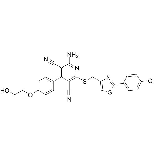
Capadenoson is a selective agonist of adenosine-A1 receptor.
For research use only. We do not sell to patients.
- Purity : 98.43%
- CAS No.: 544417-40-5
- Formula: C25H18ClN5O2S2
- Molecular Weight:520.03
-
Storage:Powder -20°C, 3 years , 4°C, 2 years ; In solvent -80°C, 2 years , -20°C, 1 year

To place orders, for customer services and technical support, please contact: MedChemExpress USA
Tel: 609-228-6898 E-mail: [email protected] [email protected]


-
 Biological Activity
-
Chemical Information
-
Solvent & Solubility
- Purity & Documentation
- References
-
 Help & FAQs


-
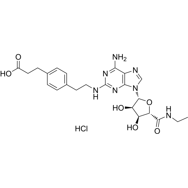
HY-13201A
CGS 21680 HydrochlorideAdenosine A2A Receptor Agonist -
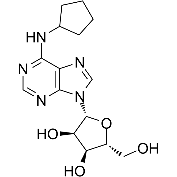
HY-103181
N6-CyclopentyladenosineAdenosine A1 Receptor Agonist -
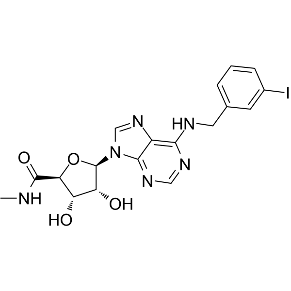
HY-13591
PiclidenosonA3AR Agonist -
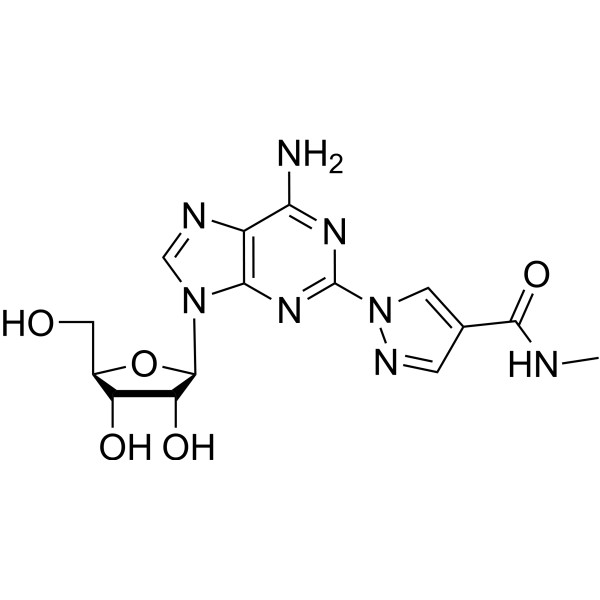
HY-A0168
RegadenosonVasodilator/A2A Adenosine Receptor Agonist -
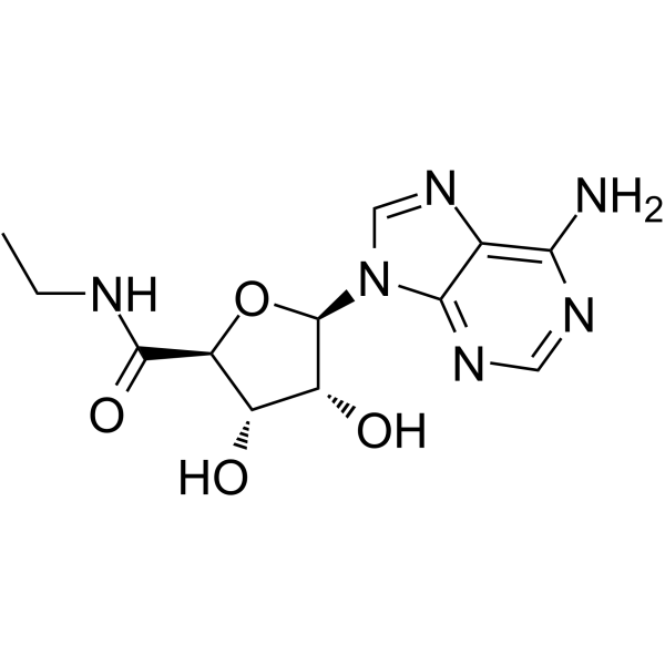
HY-103173
5'-N-EthylcarboxamidoadenosineAdenosine Receptor Agonist -
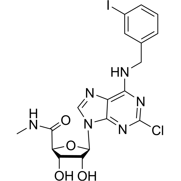
HY-12365
NamodenosonA3AR Agonist -
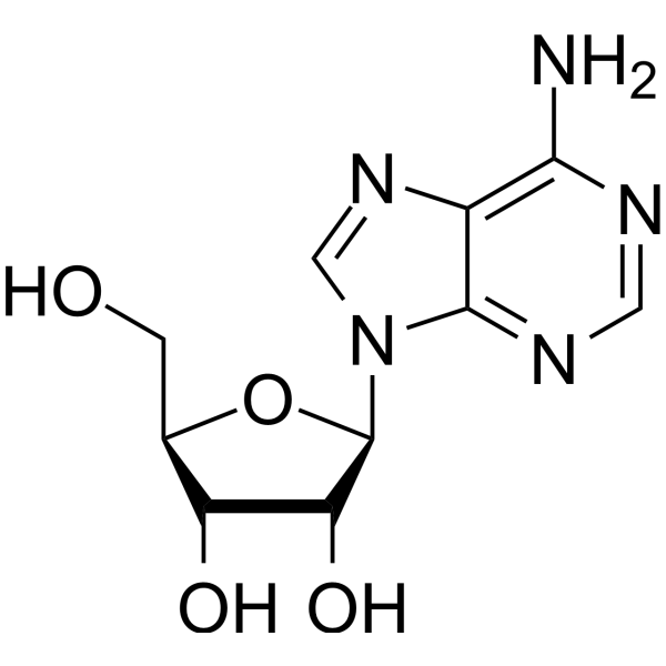
HY-B0228
AdenosineEndogenous Autacoid -
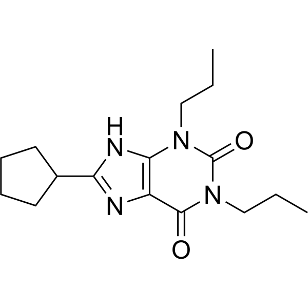
HY-100937
DPCPXA1 Receptor Antagonist -
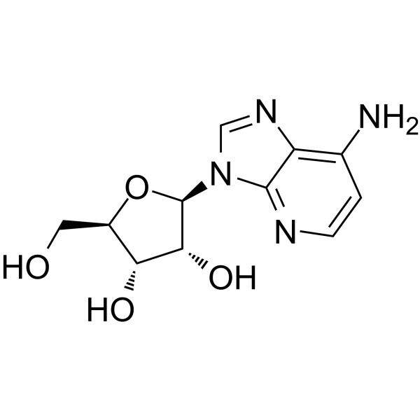
HY-103159
1-DeazaadenosineADA Inhibitor -
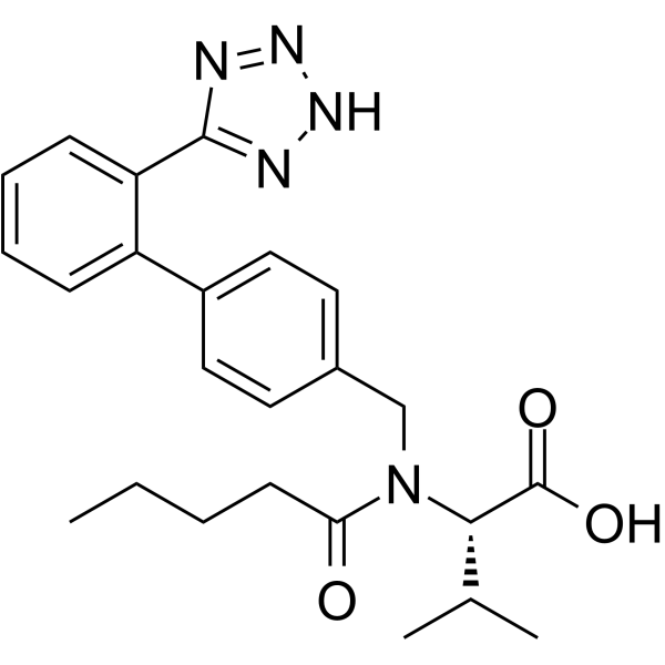
HY-18204
ValsartanAngiotensin II Receptor Antagonist -
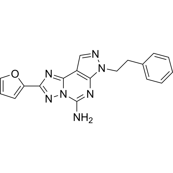
HY-19533
SCH 58261A2A Receptor Antagonist -

HY-B0228R
Adenosine (Standard)Endogenous Autacoid
Publications Citing Use of MedChemExpress (MCE) Capadenoson
More- Proc Natl Acad Sci U S A. 2025 Dec 2;122(48):e2517552122. [Abstract]
- Cancer Immunol Res. 2026 Aug 26.
- Br J Pharmacol. 2020 Jan;177(2):346-359. [Abstract]
- Br J Pharmacol. 2019 Apr;176(7):864-878. [Abstract]
- Biochem Pharmacol. 2017 Jul 1:135:79-89. [Abstract]
- Biochem Pharmacol. 2016 Jan 1:99:101-12. [Abstract]
- J Pharmacol Exp Ther. 2017 Sep;362(3):424-430. [Abstract]
- SLAS Discov. 2020 Feb;25(2):186-194. [Abstract]
- Purinergic Signal. 2021 Sep;17(3):503-514. [Abstract]
- bioRxiv. 2025 Jun 26:2025.06.23.660251. [Abstract]
- University of Alaska Fairbanks. 2017 Oct.
All Adenosine Receptor Isoforms
More

Biological Activity
Description
IC50 & Target
Adenosine A1 receptor[1]
Cellular Effect
|
Cell Line

|
Type | Value | Description | References |
|---|---|---|---|---|
| CHO | EC50 |
0.1 nM
Compound: Capadenoson
|
Agonist activity at adenosine A1 receptor (unknown origin) expressed in CHO cells assessed as increase in intracellular cAMP level by RIA
Agonist activity at adenosine A1 receptor (unknown origin) expressed in CHO cells assessed as increase in intracellular cAMP level by RIA
|
[PMID: 24669958] |
| CHO | EC50 |
1.1 nM
Compound: 4, page S2,26
|
Agonist activity at human adenosine A1 receptor expressed in CHO cell membranes by [35S]GTPgammaS binding assay
Agonist activity at human adenosine A1 receptor expressed in CHO cell membranes by [35S]GTPgammaS binding assay
|
[PMID: 26210506] |
In Vitro
To further elucidate the pharmacological properties of Capadenson, GTP shift assays are performed with the standard full A1-agonist CCPA and the A1-antagonist 8-cyclopentyl-1,3-dipropylxanthine (DPCPX). CCPA shows a Ki value of 4.2 nM in the binding assay on rat cortical brain membranes. In the presence of 1 mM GTP this Ki value shifts to a value of 64 nM. Therefore the GTP shift for CCPA is 15. DPCPX shows a GTP shift of 1 with virtually identical Ki values in the absence and presence of GTP. Capadenson shows a Ki value of 24 nM in the binding assay. In the presence of 1 mM GTP this Ki value shifts to a value of 116 nM resulting in a GTP shift of 5 for Capadenoson[1].
MedChemExpress (MCE) has not independently confirmed the accuracy of these methods. They are for reference only.
In Vivo
MedChemExpress (MCE) has not independently confirmed the accuracy of these methods. They are for reference only.
Clinical Trial
| NCT Number | Sponsor | Condition | Start Date |
Phase
|
|---|---|---|---|---|
| NCT01329991 | Plexxikon| | 2011-05 | PHASE1 |


Chemical Information
-
CAS No. 544417-40-5
-
Appearance Solid
-
Molecular Weight 520.03
-
Formula C25H18ClN5O2S2
-
Color Light yellow to yellow
-
SMILES
N#CC1=C(C2=CC=C(OCCO)C=C2)C(C#N)=C(SCC3=CSC(C4=CC=C(Cl)C=C4)=N3)N=C1N
-
Synonyms
BAY 68-4986
-
Shipping
Room temperature in continental US; may vary elsewhere.
-
Storage
Powder -20°C 3 years 4°C 2 years In solvent -80°C 2 years -20°C 1 year


Publications (11)
-
Journal Impact Factor
-
Most Recent
-
Proc Natl Acad Sci U S A
Drug repurposing screen identifies an HRI activating compound that promotes adaptive mitochondrial remodeling in MFN2-deficient cells. [Abstract]2025 Dec 2;122(48):e2517552122. PMID: 41289394 -
-
Br J Pharmacol
The effect of two selective A1 -receptor agonists and the bitopic ligand VCP746 on heart rate and regional vascular conductance in conscious rats. [Abstract]2020 Jan;177(2):346-359. PMID: 31596949 -
Br J Pharmacol
Probe dependence of allosteric enhancers on the binding affinity of adenosine A1 -receptor agonists at rat and human A1 -receptors measured using NanoBRET. [Abstract]2019 Apr;176(7):864-878. PMID: 30644086 -
Biochem Pharmacol
Capadenoson, a clinically trialed partial adenosine A1 receptor agonist, can stimulate adenosine A2B receptor biased agonism. [Abstract]2017 Jul 1:135:79-89. PMID: 28344125 -
Biochem Pharmacol
Quantification of adenosine A(1) receptor biased agonism: Implications for drug discovery. [Abstract]2016 Jan 1:99:101-12. PMID: 26581123 -
J Pharmacol Exp Ther
2017 Sep;362(3):424-430. PMID: 28652388 -
SLAS Discov
NanoBiT Complementation to Monitor Agonist-Induced Adenosine A1 Receptor Internalization. [Abstract]2020 Feb;25(2):186-194. PMID: 31583945 -
Purinergic Signal
Lack of efficacy of a partial adenosine A1 receptor agonist in neuropathic pain models in mice. [Abstract]2021 Sep;17(3):503-514. PMID: 34313915 -
bioRxiv
Drug Repurposing Screen Identifies an HRI Activating Compound that Promotes Adaptive Mitochondrial Remodeling in MFN2-deficient Cells. [Abstract]2025 Jun 26:2025.06.23.660251. PMID: 40666974 -


Solvent & Solubility
In Vitro:
DMSO : ≥ 50 mg/mL (96.15 mM; Hygroscopic DMSO has a significant impact on the solubility of product, please use newly opened DMSO)
* "≥" means soluble, but saturation unknown.
Please refer to the solubility information to select the appropriate solvent. Once prepared, please aliquot and store the solution to prevent product inactivation from repeated freeze-thaw cycles.
Storage method and period of stock solution: -80°C, 2 years; -20°C, 1 year. When stored at -80°C, please use it within 2 years. When stored at -20°C, please use it within 1 year.
Please refer to the solubility information to select the appropriate solvent. Once prepared, please aliquot and store the solution to prevent product inactivation from repeated freeze-thaw cycles.
Storage method and period of stock solution: -80°C, 2 years; -20°C, 1 year. When stored at -80°C, please use it within 2 years. When stored at -20°C, please use it within 1 year.
Concentration (start) × Volume (start) = Concentration (final) × Volume (final)
In Vivo:
Select the appropriate dissolution method based on your experimental animal and administration route.
- For the following dissolution methods, please ensure to first prepare a clear stock solution using an In Vitro approach and then sequentially add co-solvents:
- To ensure reliable experimental results, the clarified stock solution can be appropriately stored based on storage conditions. As for the working solution for In Vivo experiments, it is recommended to prepare freshly and use it on the same day.
- The percentages shown for the solvents indicate their volumetric ratio in the final prepared solution. If precipitation or phase separation occurs during preparation, heat and/or sonication can be used to aid dissolution.
Add each solvent one by one: 10% DMSO 40% PEG300 5% Tween-80 45% Saline
Solubility: 2.5 mg/mL (4.81 mM); Suspended solution; Need ultrasonic and warming
This protocol yields a suspended solution of 2.5 mg/mL. Suspended solution can be used for oral and intraperitoneal injection.
Taking 1 mL working solution as an example, add 100 μL DMSO stock solution (25.0 mg/mL) to 400 μL PEG300, and mix evenly; then add 50 μL Tween-80 and mix evenly; then add 450 μL Saline to adjust the volume to 1 mL.
Preparation of Saline: Dissolve 0.9 g sodium chloride in ddH₂O and dilute to 100 mL to obtain a clear Saline solution.
In Vivo Dissolution Calculator
Please enter the basic information of animal experiments:
-
-
-
-
Recommended: Prepare an additional quantity of animals to account for potential losses during experiments.
Please enter your animal formula composition:
-
%DMSO +
Recommended: Keep the proportion of DMSO in working solution below 2% if your animal is weak.
-
%+
-
+%Tween-80 + +
-
%Saline +
The co-solvents required include: DMSO, . All of co-solvents are available by MedChemExpress (MCE). , Tween 80. All of co-solvents are available by MedChemExpress (MCE).
Working solution concentration: 0.22 mg/mL
Method for preparing stock solution: mg drug dissolved in μL DMSO. Stock solution concentration: mg/mL.
1. Take μL DMSO stock solution;
2. Add μL .
μL , mix evenly;
3. Then add μL Tween 80, mix evenly;
4. Then add μL
Please ensure that the stock solution in the first step is dissolved to a clear state, and add co-solvents in sequence. You can use ultrasonic heating (ultrasonic cleaner, recommended frequency 20-40 kHz), vortexing, etc. to assist dissolution.


Purity & Documentation
-
Data Sheet (278 KB)
-
SDS (393 KB)
- English - EN (393 KB)
- Français - FR (393 KB)
- Deutsch - DE (393 KB)
- Norwegian - NO (393 KB)
- Español - ES (393 KB)
- Swedish - SV (393 KB)
- Italian - IT (393 KB)
- Korean - KR (393 KB)
- Portuguese - PT (393 KB)
-
Handling Instructions (2659 KB)
References
[1]. Bott-Flügel L, et al. Selective attenuation of norepinephrine release and stress-induced heart rate increase by partial adenosine A1 agonism. PLoS One. 2011 Mar 28;6(3):e18048. [Content Brief]
[2]. Bailey IR, et al. Optimization of Thermolytic Response to A1 Adenosine Receptor Agonists in Rats. J Pharmacol Exp Ther. 2017 Sep;362(3):424-430. [Content Brief]


Complete Stock Solution Preparation Table
Please refer to the solubility information to select the appropriate solvent. Once prepared, please aliquot and store the solution to prevent product inactivation from repeated freeze-thaw cycles.
Storage method and period of stock solution: -80°C, 2 years; -20°C, 1 year. When stored at -80°C, please use it within 2 years. When stored at -20°C, please use it within 1 year.
| Optional Solvent | Concentration Solvent Mass | 1 mg | 5 mg | 10 mg | 25 mg |
|---|---|---|---|---|---|
| DMSO | 1 mM | 1.9230 mL | 9.6148 mL | 19.2297 mL | 48.0741 mL |
| 5 mM | 0.3846 mL | 1.9230 mL | 3.8459 mL | 9.6148 mL | |
| 10 mM | 0.1923 mL | 0.9615 mL | 1.9230 mL | 4.8074 mL | |
| 15 mM | 0.1282 mL | 0.6410 mL | 1.2820 mL | 3.2049 mL | |
| 20 mM | 0.0961 mL | 0.4807 mL | 0.9615 mL | 2.4037 mL | |
| 25 mM | 0.0769 mL | 0.3846 mL | 0.7692 mL | 1.9230 mL | |
| 30 mM | 0.0641 mL | 0.3205 mL | 0.6410 mL | 1.6025 mL | |
| 40 mM | 0.0481 mL | 0.2404 mL | 0.4807 mL | 1.2019 mL | |
| 50 mM | 0.0385 mL | 0.1923 mL | 0.3846 mL | 0.9615 mL | |
| 60 mM | 0.0320 mL | 0.1602 mL | 0.3205 mL | 0.8012 mL | |
| 80 mM | 0.0240 mL | 0.1202 mL | 0.2404 mL | 0.6009 mL |


Capadenoson Related Classifications
HY-14917 Related Classifications
 Powered by Bioz