



ER-Tracker Green
Based on 8 publication(s) in Google Scholar
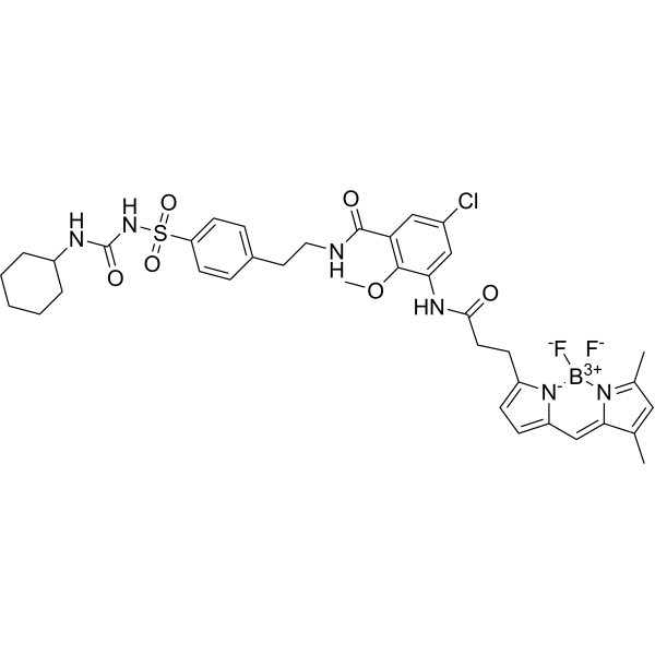
ER-Tracker dye is a derivative of BODIPY series dyes coupled with Glibenclamide (HY-15206), highly selective binding to the endoplasmic reticulum, non-toxic to cells at low concentrations, this type of dye is an environmentally sensitive probe, and formaldehyde treatment can still retain part of the fluorescence, with high fluorescence life, good extinction coefficient and other characteristics. Glibenclamide is an atp-dependent K+ channel blocker (Kir6, KATP) and CFTR Cl-channel blocker that binds in the endoplasmic reticulum. ER-Tracker is not suitable for staining cells after fixation.
For research use only. We do not sell to patients.
- Purity: 98.03%
- CAS No.: 730931-46-1
- Formula: C37H42BClF2N6O6S
- Molecular Weight:783.09
-
Storage:
4°C, sealed storage, away from moisture and light
* In solvent : -80°C, 6 months; -20°C, 1 month (sealed storage, away from moisture and light)

To place orders, for customer services and technical support, please contact: MedChemExpress USA
Tel: 609-228-6898 E-mail: [email protected] [email protected]


-
 Biological Activity
-
Chemical Information
-
Solvent & Solubility
- Purity & Documentation
- References
-
 Help & FAQs


-
HY-D1783
MitoTracker Deep Red FM -
HY-135056
MitoTracker Green FM -
HY-D1300
LysoTracker Red -
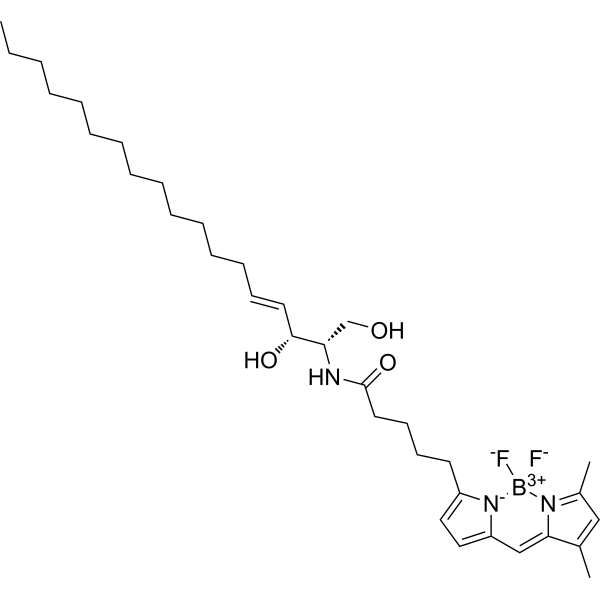
HY-D1612
BODIPY FL C5-Ceramide -
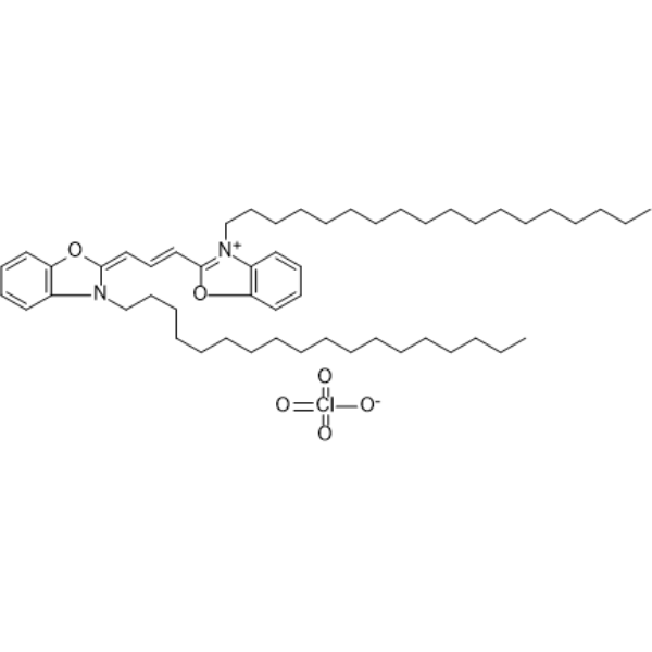
HY-D0969
DiO -
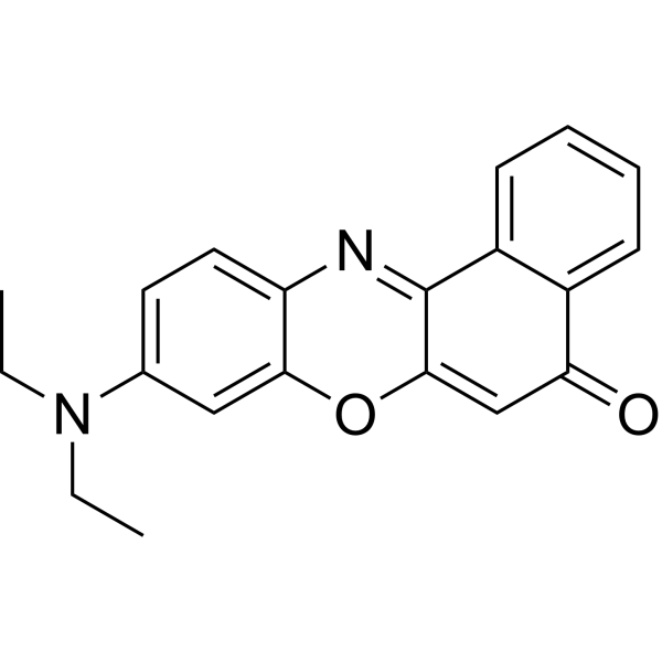
HY-D0718
Nile Red -
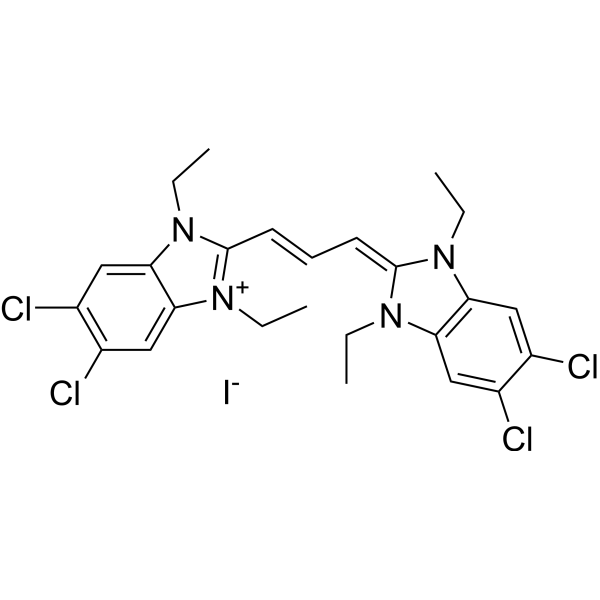
HY-15534
JC-1 -
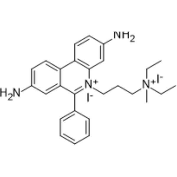
HY-D0815
Propidium Iodide -
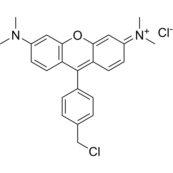
HY-D1696
MitoTracker Orange CMTMRos -

HY-DY1040
LysoTracker Red (solution) -
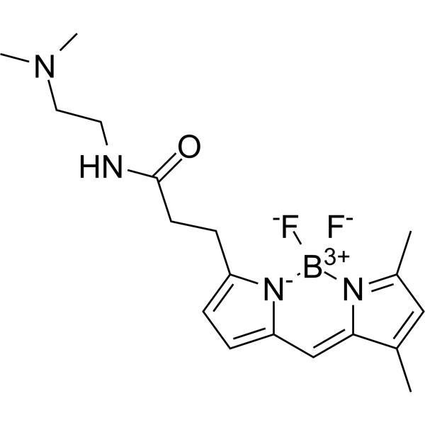
HY-D1296
Green DND-26 -

HY-DY1032
MitoTracker Green FM (solution)
Publications Citing Use of MedChemExpress (MCE) ER-Tracker Green
More- Cell. 2026 Jul 9;189(14):4396-4414.e32. [Abstract]
- Innovation (Camb). 2025 Jan 12;6(2):100757. [Abstract]
- Cell Host Microbe. 2026 Jan 14;34(1):35-51.e9. [Abstract]
- Adv Healthc Mater. 2025 Sep 4:e02881. [Abstract]
- Food Chem. 2026 Jun 15:514:149116. [Abstract]
- ACS Appl Mater Interfaces. 2026 Feb 26. [Abstract]
- Bioorg Chem. 2025 Sep 1:165:108951. [Abstract]
- Res Sq. 2025 Sep 12.
 Customer Validation & Images

-
Cell Imaging/Staining
-
Cell Imaging/Staining
-
Cell Imaging/Staining
-
Cell Imaging/Staining
-
Cell Imaging/Staining


Biological Activity
Description
In Vitro
Guide (The following is our recommended protocol. This protocol only provides guidance and should be modified according to your specific needs).
1. Preparation of ER-Tracker working solution
1.1 Preparation of stock solution
Dissolve 100 μg ER-Tracker in 128 μL DMSO to obtain a 1 mM stock solution.
Note: It is recommended to store the stock solution at -20°C or -80°C, protected from light, and avoid repeated freeze-thaw cycles.
1.2 Preparation of ER-Tracker working solution
Dilute the stock solution with serum-free cell culture medium or PBS to obtain a 100 nM-1 μM working solution.
Note: The concentration of the ER-Tracker working solution should be adjusted according to actual conditions.
2. Cell staining
2.1 Suspension cells (6-well plate)
a. Centrifuge at 1000 g at 4°C for 3-5 minutes, discard the supernatant. Wash twice with PBS, 5 minutes each time. Adjust the cell density to 1×10⁶/mL.
b. Add 1 mL working solution and incubate at room temperature for 5-30 minutes.
c. Centrifuge at 400 g at 4°C for 3-4 minutes and discard the supernatant.
d. Wash twice with PBS, 5 minutes each time.
e. Resuspend the cells in serum-free cell culture medium or PBS. Observe using a fluorescence microscope or flow cytometry.
2.2 Adherent cells
a. Culture adherent cells on sterile coverslips.
b. Remove the coverslips from the culture medium and aspirate excess medium.
c. Add 100 μL working solution, gently shake to fully cover the cells, and incubate at room temperature for 5-30 minutes.
d. Wash twice with culture medium, 5 minutes each time. Observe using a fluorescence microscope or flow cytometer.
Note: If flow cytometry is used, cells need to be resuspended before staining.
MedChemExpress (MCE) has not independently confirmed the accuracy of these methods. They are for reference only.
Emission (Em)
520
Excitation (Ex)
489


Chemical Information
-
CAS No. 730931-46-1
-
Appearance Solid
-
Molecular Weight 783.09
-
Formula C37H42BClF2N6O6S
-
Color Brown to reddish brown
-
SMILES
O=C(NS(=O)(C1=CC=C(CCNC(C2=C(OC)C(NC(CCC3=CC=C4C=C5C(C)=CC(C)=[N]5[B+3]([F-])([N-]43)[F-])=O)=CC(Cl)=C2)=O)C=C1)=O)NC6CCCCC6
-
Shipping
Room temperature in continental US; may vary elsewhere.
-
Storage
4°C, sealed storage, away from moisture and light
* In solvent : -80°C, 6 months; -20°C, 1 month (sealed storage, away from moisture and light)


Publications (8)
-
Journal Impact Factor
-
Most Recent
-
Cell
2026 Jul 9;189(14):4396-4414.e32. PMID: 42143020 -
Innovation (Camb)
Fast reconstruction and optical-sectioning three-dimensional structured illumination microscopy. [Abstract]2025 Jan 12;6(2):100757. PMID: 39991474
ER-Tracker Green purchased from MedChemExpress. Usage Cited in: Innovation (Camb). 2025 Jan 12;6(2):100757. [Abstract]
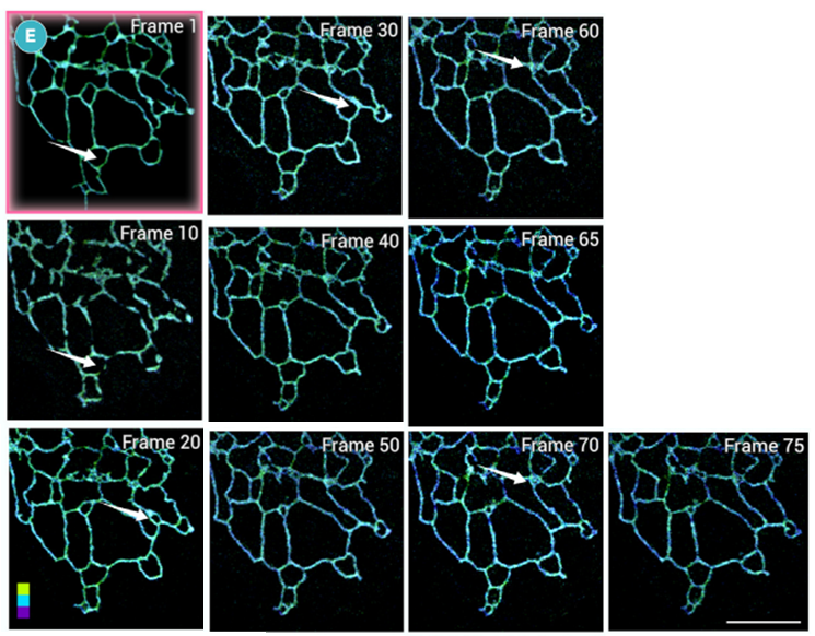
Magnified zone of 3-layer 3DSIM imaging reveals the motion of the ER. Live-cell endoplasmic reticulum (ER) imaging experiments were performed using ER-Tracker Green (1 μM; 30 min). Scale bar, 4 μm.
-
Cell Host Microbe
Rothia mucilaginosa membrane vesicles stabilize labile iron to alleviate UVB-induced ferroptosis. [Abstract]2026 Jan 14;34(1):35-51.e9. PMID: 41500222
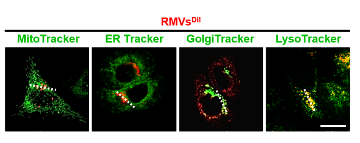
ER-Tracker Green purchased from MedChemExpress. Usage Cited in: Cell Host Microbe. 2026 Jan 14;34(1):35-51.e9. [Abstract]
Representative fluorescence images of RMVs (red) and mitochondria (MitoTracker, green), endoplasmic reticulum (ER-Tracker Green (1 μM), green), Golgi apparatus (GolgiTracker, green) and lysosomes (LysoTracker, green) in HaCaT cells. Fluorescence-probe staining showed that RMVs predominantly co-localized with lysosomes, rather than with mitochondria, endoplasmic reticulum, or Golgi apparatus. Scale bar, 10 μm.
-
Adv Healthc Mater
Targeted Reprogramming of Tumor Cells by Digoxin-Loaded Immunogenic Nanoparticles Enhances Immunity Against Disseminated Tumor Cells. [Abstract]2025 Sep 4:e02881. PMID: 40904220
ER-Tracker Green purchased from MedChemExpress. Usage Cited in: Adv Healthc Mater. 2025 Sep 4:e02881. [Abstract]
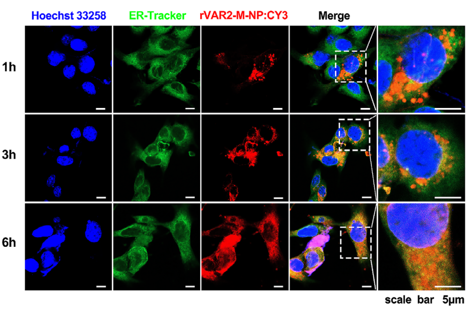
ER targeting of rVAR2-M-NPs in 4T1 cells. CLSM images of 4T1 cell by cultured with rVAR2-M-NPs CY3 for 12 h. Hoechst 33258 (blue) used for staining nucleus, ER-Tracker Green (green) used for staining ER, and CY3 (red) used for representing the rVAR2-M-NP core, respectively.
-
Food Chem
Two-step single B cell technology for high-affinity rabbit monoclonal antibodies: development of a highly sensitive immunoassay for monensin in raw milk. [Abstract]2026 Jun 15:514:149116. PMID: 41956048 -
ACS Appl Mater Interfaces
A Near-Infrared Emitting Aggregation-Induced Emission Photosensitizer with Endoplasmic Reticulum Targeting Ability for Breast Cancer Photodynamic Therapy. [Abstract]2026 Feb 26. PMID: 41747230
ER-Tracker Green purchased from MedChemExpress. Usage Cited in: ACS Appl Mater Interfaces. 2026 Feb 26. [Abstract]
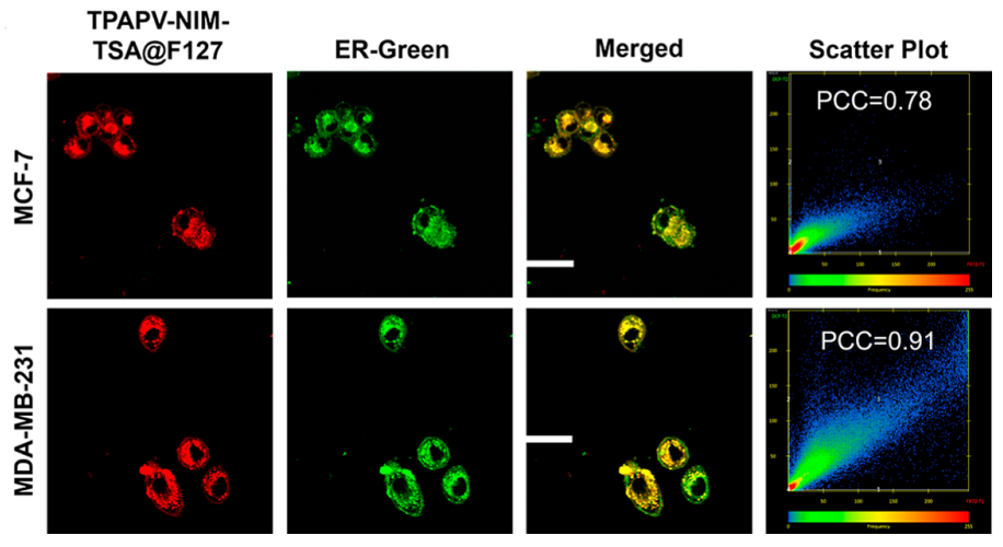
Confocal fluorescence images of MCF-7 and MDA-MB-231 cancer cells costained with TPAPV-NIM-TSA@F127 (10 μM) and ER-Tracker Green (ER-Green) (3 μM; 30 min). TPAPV-NIM-TSA@F127 was excited with a 488 nm laser, with emission collected between 610 and 700 nm. Hoechst 33342 was excited with a 405 nm laser, and emission was collected between 400 and 515 nm. ER-Green was excited with 488 nm, with emission collected between 490 and 560 nm. Scale bar: 10 μm.
-
Bioorg Chem
Two in one: Acceptor engineering strategy to construct efficient type I aggregation induced emission photosensitizer for photodynamic antitumor and antibacterial therapy. [Abstract]2025 Sep 1:165:108951. PMID: 40902511
ER-Tracker Green purchased from MedChemExpress. Usage Cited in: Bioorg Chem. 2025 Sep 1:165:108951. [Abstract]
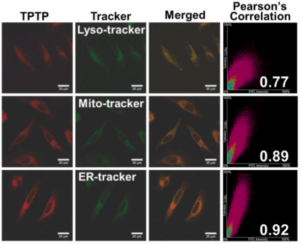
Co-localization of TPTP (10 μM) with Lyso-Tracker Green (75 nM), ER-Tracker Green (100 nM; 30 min; HeLa cells) and Mito-Tracker Green (100 nM), respectively. Green channel (λex = 488 nm, λem = 500-550 nm), and red channel (λex = 488 nm, λem = 600-700 nm), scale bar: 20 μm.
-


Solvent & Solubility
In Vitro:
DMSO : 50 mg/mL (63.85 mM; Need ultrasonic; Hygroscopic DMSO has a significant impact on the solubility of product, please use newly opened DMSO)
Please refer to the solubility information to select the appropriate solvent. Once prepared, please aliquot and store the solution to prevent product inactivation from repeated freeze-thaw cycles.
Storage method and period of stock solution: -80°C, 6 months; -20°C, 1 month (sealed storage, away from moisture and light). When stored at -80°C, please use it within 6 months. When stored at -20°C, please use it within 1 month.
Please refer to the solubility information to select the appropriate solvent. Once prepared, please aliquot and store the solution to prevent product inactivation from repeated freeze-thaw cycles.
Storage method and period of stock solution: -80°C, 6 months; -20°C, 1 month (sealed storage, away from moisture and light). When stored at -80°C, please use it within 6 months. When stored at -20°C, please use it within 1 month.
Concentration (start) × Volume (start) = Concentration (final) × Volume (final)


Purity & Documentation
-
Data Sheet (275 KB)
-
SDS (252 KB)
- English - EN (252 KB)
- Français - FR (252 KB)
- Deutsch - DE (252 KB)
- Norwegian - NO (252 KB)
- Español - ES (252 KB)
- Swedish - SV (252 KB)
- Italian - IT (252 KB)
- Korean - KR (252 KB)
- Portuguese - PT (252 KB)
-
Handling Instructions (2659 KB)
References
[1]. Tanuja T Merianda, et al. A functional equivalent of endoplasmic reticulum and Golgi in axons for secretion of locally synthesized proteins. Mol Cell Neurosci. 2009 Feb;40(2):128-42. [Content Brief]
[3]. J Jacob Strouse, et al. Fluorescent substrates for flow cytometric evaluation of efflux inhibition in ABCB1, ABCC1, and ABCG2 transporters. Anal Biochem. 2013 Jun 1;437(1):77-87. [Content Brief]


Complete Stock Solution Preparation Table
Please refer to the solubility information to select the appropriate solvent. Once prepared, please aliquot and store the solution to prevent product inactivation from repeated freeze-thaw cycles.
Storage method and period of stock solution: -80°C, 6 months; -20°C, 1 month (sealed storage, away from moisture and light). When stored at -80°C, please use it within 6 months. When stored at -20°C, please use it within 1 month.
| Optional Solvent | Concentration Solvent Mass | 1 mg | 5 mg | 10 mg | 25 mg |
|---|---|---|---|---|---|
| DMSO | 1 mM | 1.2770 mL | 6.3850 mL | 12.7699 mL | 31.9248 mL |
| 5 mM | 0.2554 mL | 1.2770 mL | 2.5540 mL | 6.3850 mL | |
| 10 mM | 0.1277 mL | 0.6385 mL | 1.2770 mL | 3.1925 mL | |
| 15 mM | 0.0851 mL | 0.4257 mL | 0.8513 mL | 2.1283 mL | |
| 20 mM | 0.0638 mL | 0.3192 mL | 0.6385 mL | 1.5962 mL | |
| 25 mM | 0.0511 mL | 0.2554 mL | 0.5108 mL | 1.2770 mL | |
| 30 mM | 0.0426 mL | 0.2128 mL | 0.4257 mL | 1.0642 mL | |
| 40 mM | 0.0319 mL | 0.1596 mL | 0.3192 mL | 0.7981 mL | |
| 50 mM | 0.0255 mL | 0.1277 mL | 0.2554 mL | 0.6385 mL | |
| 60 mM | 0.0213 mL | 0.1064 mL | 0.2128 mL | 0.5321 mL |


ER-Tracker Green Related Classifications
HY-D1297 Related Classifications
 Powered by Bioz