



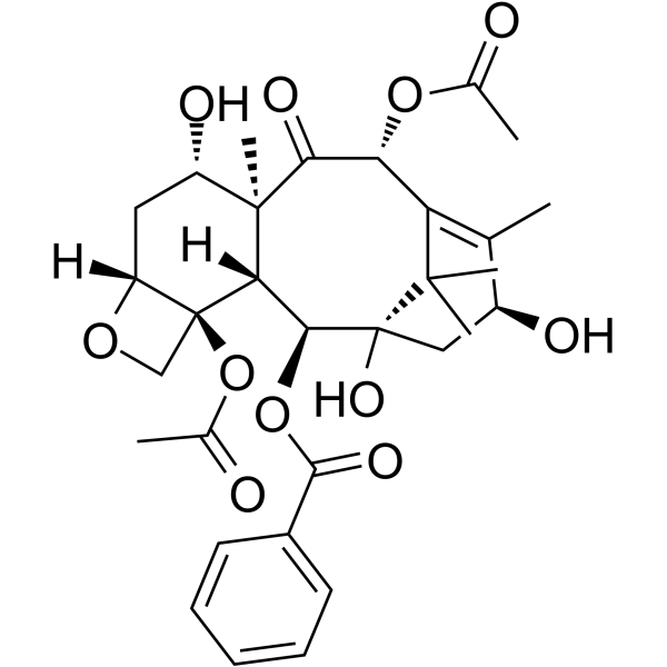
Baccatin III
Based on 2 publication(s) in Google Scholar
Baccatin III is an orally available, selective inhibitor of the TGF-β1 signaling pathway and myeloid-derived suppressor cell (MDSC) activation. Baccatin III targets the AKT/STAT6 and Smad2/3 pathways, blocking TGF-β1-induced fibroblast differentiation and MDSC-mediated immunosuppression. Baccatin III exerts anti-inflammatory and anti-fibrotic effects by inhibiting macrophage activation and extracellular matrix deposition, and shows potential in the treatment of pulmonary fibrosis and cancer in terms of regulating the tumor immune microenvironment.
For research use only. We do not sell to patients.
- Purity : 99.82%
- CAS No.: 27548-93-2
- Formula: C31H38O11
- Molecular Weight:586.63
-
Storage:Powder -20°C, 3 years , 4°C, 2 years ; In solvent -80°C, 6 months , -20°C, 1 month

To place orders, for customer services and technical support, please contact: MedChemExpress USA
Tel: 609-228-6898 E-mail: [email protected] [email protected]


-
 Biological Activity
-
Chemical Information
-
Solvent & Solubility
- Protocols
- Purity & Documentation
- References
-
 Help & FAQs


-

HY-B0015
PaclitaxelAntitumor Agent -
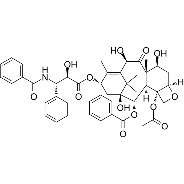
HY-N1391
10-DeacetyltaxolTaxane Derivative -
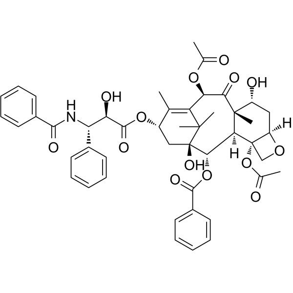
HY-N0227
7-epi-TaxolActive Metabolite of Taxol -
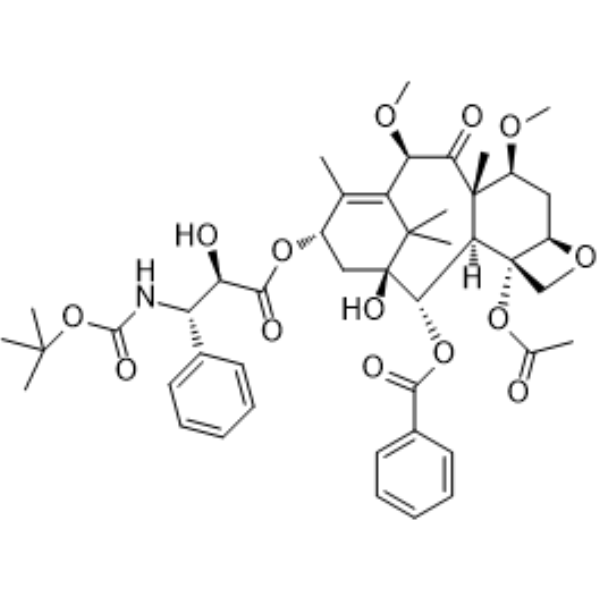
HY-15459
Cabazitaxel -
HY-16565
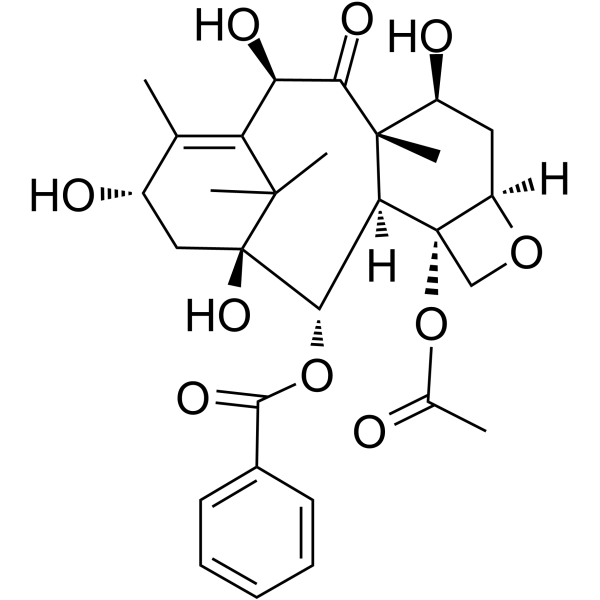
10-Deacetylbaccatin IIIIntermediate -

HY-16565R
10-Deacetylbaccatin III (Standard)Intermediate -
HY-20584
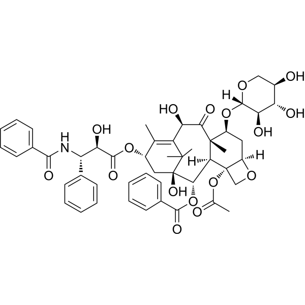
10-Deacetyl-7-xylosyl paclitaxelMicrotubule/Tubulin Inhibitor -
HY-77572
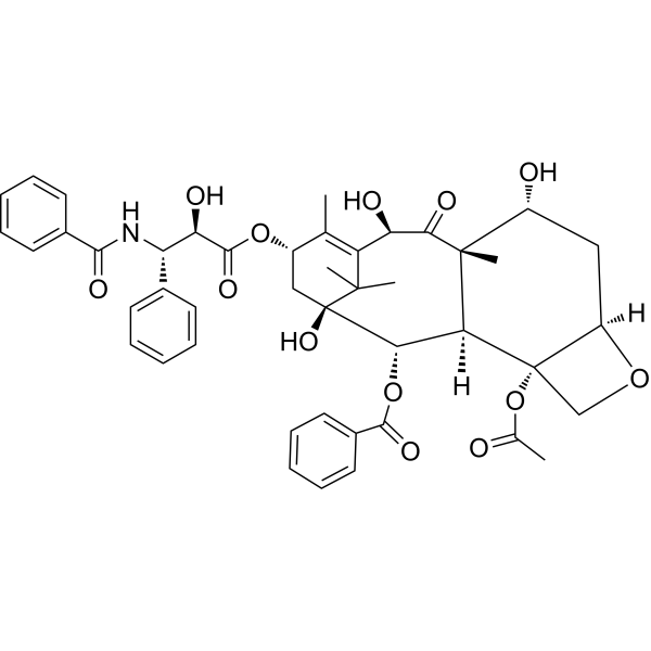
7-Epi 10-desacetyl paclitaxelNatural Product -

HY-N6985R
Baccatin III (Standard)Anticancer Agent
Publications Citing Use of MedChemExpress (MCE) Baccatin III
More

Biological Activity
Description
Cellular Effect
|
Cell Line

|
Type | Value | Description | References |
|---|---|---|---|---|
| A2780 | IC50 |
>25 μM
Compound: 15
|
Antiproliferative activity against human A2780 cells assessed as growth inhibition after 48 hrs by sulforhodamine B assay
Antiproliferative activity against human A2780 cells assessed as growth inhibition after 48 hrs by sulforhodamine B assay
|
[PMID: 25682561] |
| A549 | IC50 |
>25 μM
Compound: 15
|
Antiproliferative activity against human A549 cells assessed as growth inhibition after 48 hrs by sulforhodamine B assay
Antiproliferative activity against human A549 cells assessed as growth inhibition after 48 hrs by sulforhodamine B assay
|
[PMID: 25682561] |
| A549 | IC50 |
24 μM
Compound: 15
|
Antiproliferative activity against human A549/CDDP cells assessed as growth inhibition after 48 hrs by sulforhodamine B assay
Antiproliferative activity against human A549/CDDP cells assessed as growth inhibition after 48 hrs by sulforhodamine B assay
|
[PMID: 25682561] |
| B16 | ED50 |
>1000 nM
Compound: 4
|
Cytotoxicity against B16 melanoma cell line.40% inhibition at 1 uM
Cytotoxicity against B16 melanoma cell line.40% inhibition at 1 uM
|
[PMID: 1359143] |
| HCT-8 | IC50 |
12 μM
Compound: 15
|
Antiproliferative activity against human HCT8 cells assessed as growth inhibition after 48 hrs by sulforhodamine B assay
Antiproliferative activity against human HCT8 cells assessed as growth inhibition after 48 hrs by sulforhodamine B assay
|
[PMID: 25682561] |
| HepG2 | IC50 |
9.91 μg/mL
Compound: 4
|
Cytotoxicity againt human HepG2 cells
Cytotoxicity againt human HepG2 cells
|
[PMID: 17239597] |
| KB | IC50 |
10 μg/mL
Compound: 10
|
Cytotoxic activity was evaluated against Epidermoid Carcinoma KB cells
Cytotoxic activity was evaluated against Epidermoid Carcinoma KB cells
|
[PMID: 9873389] |
| L1210 | IC50 |
>10 μg/mL
Compound: 10
|
Cytotoxic activity was evaluated against murine Leukemia L1210 cells
Cytotoxic activity was evaluated against murine Leukemia L1210 cells
|
[PMID: 9873389] |
| MCF7 | IC50 |
7.4 μM
Compound: 15
|
Antiproliferative activity against human MCF7 cells assessed as growth inhibition after 48 hrs by sulforhodamine B assay
Antiproliferative activity against human MCF7 cells assessed as growth inhibition after 48 hrs by sulforhodamine B assay
|
[PMID: 25682561] |
| NIH3T3 | IC50 |
>25 μM
Compound: 15
|
Antiproliferative activity against mouse NIH/3T3 cells assessed as growth inhibition after 48 hrs by sulforhodamine B assay
Antiproliferative activity against mouse NIH/3T3 cells assessed as growth inhibition after 48 hrs by sulforhodamine B assay
|
[PMID: 25682561] |
| WI-38 | IC50 |
16.1 μg/mL
Compound: 4
|
Cytotoxicity againt human WI38 cells
Cytotoxicity againt human WI38 cells
|
[PMID: 17239597] |
| WI-38 VA13 | IC50 |
0.84 μg/mL
Compound: 4
|
Cytotoxicity againt human VA13 cells
Cytotoxicity againt human VA13 cells
|
[PMID: 17239597] |
In Vitro
Baccatin III (10-100 μM; 16 h) significantly reduces the production of reactive oxygen species (ROS) and nitric oxide (NO) in myeloid-derived suppressor cells (MDSCs) stimulated by LPS/IFN-γ[1].
Baccatin III (3-30 μM; 24 h) dose-dependently inhibits IL-13-induced TGF-β1 mRNA expression in primary mouse lung macrophages[2].
Baccatin III (3-30 μM; 24 h) inhibits TGF-β1-induced mRNA expression of α-SMA, fibronectin and collagen I/III in primary mouse lung fibroblasts[2].
MedChemExpress (MCE) has not independently confirmed the accuracy of these methods. They are for reference only. Further protocols information, click here.
In Vivo
Baccatin III (5-10 mg/kg; intraperitoneally; single; 21 days) alleviates Bleomycin (HY-108345)-induced lung fibrosis in C57BL/6 mice by inhibiting TGF-β1[2].
MedChemExpress (MCE) has not independently confirmed the accuracy of these methods. They are for reference only.


Chemical Information
-
CAS No. 27548-93-2
-
Appearance Solid
-
Molecular Weight 586.63
-
Formula C31H38O11
-
Color White to off-white
-
SMILES
O=C1[C@H](OC(C)=O)C(C2(C)C)=C(C)[C@@H](O)C[C@@]2(O)[C@@H](OC(C3=CC=CC=C3)=O)[C@@]4([H])[C@@]1(C)[C@@H](O)C[C@@]5([H])OC[C@]54OC(C)=O
-
Structure Classification
-
Initial Source
-
Shipping
Room temperature in continental US; may vary elsewhere.
-
Storage
Powder -20°C 3 years 4°C 2 years In solvent -80°C 6 months -20°C 1 month


Publications (2)
-
Journal Impact Factor
-
Most Recent
-
Nature
2025 Jul;643(8071):582-592. PMID: 40500440 -
bioRxiv
Multiplexed perturbation of yew reveals cryptic proteins that enable a total biosynthesis of baccatin III and Taxol precursors. [Abstract]2024 Nov 16:2024.11.06.622305. PMID: 39574719


Solvent & Solubility
In Vitro:
DMSO : 33.33 mg/mL (56.82 mM; Need ultrasonic; Hygroscopic DMSO has a significant impact on the solubility of product, please use newly opened DMSO)
Please refer to the solubility information to select the appropriate solvent. Once prepared, please aliquot and store the solution to prevent product inactivation from repeated freeze-thaw cycles.
Storage method and period of stock solution: -80°C, 6 months; -20°C, 1 month. When stored at -80°C, please use it within 6 months. When stored at -20°C, please use it within 1 month.
Please refer to the solubility information to select the appropriate solvent. Once prepared, please aliquot and store the solution to prevent product inactivation from repeated freeze-thaw cycles.
Storage method and period of stock solution: -80°C, 6 months; -20°C, 1 month. When stored at -80°C, please use it within 6 months. When stored at -20°C, please use it within 1 month.
Concentration (start) × Volume (start) = Concentration (final) × Volume (final)
In Vivo:
Select the appropriate dissolution method based on your experimental animal and administration route.
- For the following dissolution methods, please ensure to first prepare a clear stock solution using an In Vitro approach and then sequentially add co-solvents:
- To ensure reliable experimental results, the clarified stock solution can be appropriately stored based on storage conditions. As for the working solution for In Vivo experiments, it is recommended to prepare freshly and use it on the same day.
- The percentages shown for the solvents indicate their volumetric ratio in the final prepared solution. If precipitation or phase separation occurs during preparation, heat and/or sonication can be used to aid dissolution.
Add each solvent one by one: 10% DMSO 40% PEG300 5% Tween-80 45% Saline
Solubility: ≥ 2.08 mg/mL (3.55 mM); Clear solution
This protocol yields a clear solution of ≥ 2.08 mg/mL (saturation unknown).
Taking 1 mL working solution as an example, add 100 μL DMSO stock solution (20.8 mg/mL) to 400 μL PEG300, and mix evenly; then add 50 μL Tween-80 and mix evenly; then add 450 μL Saline to adjust the volume to 1 mL.
Preparation of Saline: Dissolve 0.9 g sodium chloride in ddH₂O and dilute to 100 mL to obtain a clear Saline solution.
Add each solvent one by one: 10% DMSO 90% (20% SBE-β-CD in Saline)
Solubility: ≥ 2.08 mg/mL (3.55 mM); Clear solution
This protocol yields a clear solution of ≥ 2.08 mg/mL (saturation unknown).
Taking 1 mL working solution as an example, add 100 μL DMSO stock solution (20.8 mg/mL) to 900 μL 20% SBE-β-CD in Saline, and mix evenly.
Preparation of 20% SBE-β-CD in Saline (4°C, storage for one week): 2 g SBE-β-CD powder is dissolved in 10 mL Saline, completely dissolve until clear.
In Vivo Dissolution Calculator
Please enter the basic information of animal experiments:
-
-
-
-
Recommended: Prepare an additional quantity of animals to account for potential losses during experiments.
Please enter your animal formula composition:
-
%DMSO +
Recommended: Keep the proportion of DMSO in working solution below 2% if your animal is weak.
-
%+
-
+%Tween-80 + +
-
%Saline +
The co-solvents required include: DMSO, . All of co-solvents are available by MedChemExpress (MCE). , Tween 80. All of co-solvents are available by MedChemExpress (MCE).
Working solution concentration: 0.22 mg/mL
Method for preparing stock solution: mg drug dissolved in μL DMSO. Stock solution concentration: mg/mL.
1. Take μL DMSO stock solution;
2. Add μL .
μL , mix evenly;
3. Then add μL Tween 80, mix evenly;
4. Then add μL
Please ensure that the stock solution in the first step is dissolved to a clear state, and add co-solvents in sequence. You can use ultrasonic heating (ultrasonic cleaner, recommended frequency 20-40 kHz), vortexing, etc. to assist dissolution.

Protocols
-
Fibrosis/Collagen Morphometry
Fibrosis and collagen morphometry is based on the quantitative visualization of fibrillar collagen deposition in tissue sections using histochemical stains such as Sirius Red (Picrosirius Red) or Masson's trichrome, followed by image-based or polarization-enhanced analysis to estimate collagen proportional area as a surrogate of extracellular matrix accumulation during fibrotic remodeling. Sirius Red combined with polarized light microscopy enhances detection of collagen fibers due to birefringence properties, enabling more specific visualization of collagen type I and III fibrils compared to conventional bright-field histology, while whole-section or region-restricted digital morphometry reduces field-selection bias in fibrosis assessment. Alternative quantitative approaches include second harmonic generation (SHG) and two-photon excited fluorescence microscopy, which enable label-free detection of fibrillar collagen and have been validated against histological staining and biochemica
-
Collagen: Sirius Red Staining
Sirius Red or picrosirius red staining is a histochemical method for visualizing collagen-rich extracellular matrix in tissue sections, and collagen fibers are detected as red-stained structures under bright-field microscopy with enhanced birefringence under polarized light. Picrosirius red is useful for assessing total collagen organization, distribution, and fibrosis burden, but polarized color should not be interpreted as a definitive collagen type I versus type III readout because color is affected by fiber orientation, thickness, and packing.
-
ECM-Embedded Organoid (Matrigel/Dome) Culture
ECM-embedded organoid dome culture embeds epithelial stem cells, crypts, organoid fragments, or tumor-derived epithelial cells in a basement-membrane-like hydrogel such as Matrigel, allowing 3D growth, self-organization, lumen formation, budding or cystic morphogenesis, and lineage maintenance under defined niche-factor-containing medium. The primary readouts are organoid establishment efficiency, growth, morphology, passaging capacity, lineage-marker expression, and, when fluorescently labeled lines are used, microscopy- or flow-cytometry-based quantification of population behavior in 3D culture.
-
Connective Tissue: Masson's Trichrome/Collagen Trichrome Staining
Masson’s Trichrome (collagen/trichrome staining) is a histological technique that differentially stains tissue compartments using sequential acidic dyes to distinguish collagen from muscle and cytoplasmic components based on dye affinity and tissue permeability differences, enabling visualization of fibrosis and connective tissue architecture in histological sections. The classical formulation typically uses Weigert's iron hematoxylin for nuclear staining, Biebrich scarlet-acid fuchsin for cytoplasm and muscle, and aniline blue (or light green variants) for collagen, producing a characteristic blue/green collagen signal contrasted against red cytoplasm and dark nuclei. The staining principle relies on selective displacement of smaller dye molecules by larger anionic dyes in collagen-rich regions under controlled acidified conditions, which enhances collagen-specific dye retention. This property makes the method widely used for fibrosis assessment in organs such as heart, liver, lung, a
-
Matrigel/ECM Transwell Invasion Assay
The Matrigel/ECM Transwell invasion assay measures the ability of cells to move toward a chemoattractant while crossing an extracellular-matrix barrier placed on a porous membrane; therefore, the readout reflects both chemotactic motility and matrix invasion rather than migration alone. Matrigel is a basement-membrane-rich matrix derived from Engelbreth-Holm-Swarm mouse sarcoma and has been used as a reconstituted basement membrane barrier in chemoinvasion assays. The assay readout is generated by quantifying cells that reach the underside of the insert membrane or lower compartment after incubation, commonly by staining and counting invaded cells or by fluorescence-based quantification.
-
Research Protocol for Inflammation-related Diseases
The NLRP3 inflammasome is a cytosolic innate immune signaling platform that integrates priming signals and danger-signal activation to promote caspase-1 activation, maturation of IL-1β and IL-18, and gasdermin D-mediated pyroptotic cell death. The core experimental logic is to determine whether inflammatory disease phenotypes are driven by increased NLRP3 expression, ASC-containing inflammasome assembly, caspase-1 cleavage, GSDMD cleavage, and extracellular release of IL-1β/IL-18 rather than by nonspecific cell injury alone. The pathway is strongly linked to inflammation-related disease phenotypes because monosodium urate crystals activate NALP3/NLRP3 inflammasome signaling in gout-like crystal inflammation, cholesterol crystals activate NLRP3 inflammasomes in atherogenesis models, and DSS-induced intestinal inflammation has been reported to involve NLRP3 inflammasome activity. However, experimental colitis studies also show context-dependent protective effects of NLRP3 inflammasome co
-
Matrigel Transwell/Boyden Chamber Invasion Assay
Matrigel Transwell/Boyden chamber invasion assay measures the ability of cells to degrade or traverse an extracellular matrix-coated porous membrane and move from an upper chamber toward a chemoattractant in a lower chamber. Invasion is distinguished from migration by coating the membrane with Matrigel or basement membrane matrix; uncoated inserts measure migration, while coated inserts require cells to cross an ECM barrier before reaching the underside of the membrane.


Purity & Documentation
-
Data Sheet (285 KB)
-
SDS (644 KB)
- English - EN (644 KB)
- Français - FR (644 KB)
- Deutsch - DE (644 KB)
- Norwegian - NO (644 KB)
- Español - ES (644 KB)
- Swedish - SV (644 KB)
- Italian - IT (644 KB)
- Korean - KR (644 KB)
- Portuguese - PT (644 KB)
-
Handling Instructions (2659 KB)
References
[1]. Lee YH, et al. Baccatin III, a precursor for the semisynthesis of paclitaxel, inhibits the accumulation and suppressive activity of myeloid-derived suppressor cells in tumor-bearing mice. Int Immunopharmacol. 2014 Aug;21(2):487-93. [Content Brief]
[2]. Nie Y, et al. Baccatin III ameliorates bleomycin-induced pulmonary fibrosis via suppression of TGF-β1 production and TGF-β1-induced fibroblast differentiation. Int Immunopharmacol. 2019 Sep;74:105696. [Content Brief]


Complete Stock Solution Preparation Table
Please refer to the solubility information to select the appropriate solvent. Once prepared, please aliquot and store the solution to prevent product inactivation from repeated freeze-thaw cycles.
Storage method and period of stock solution: -80°C, 6 months; -20°C, 1 month. When stored at -80°C, please use it within 6 months. When stored at -20°C, please use it within 1 month.
| Optional Solvent | Concentration Solvent Mass | 1 mg | 5 mg | 10 mg | 25 mg |
|---|---|---|---|---|---|
| DMSO | 1 mM | 1.7047 mL | 8.5233 mL | 17.0465 mL | 42.6163 mL |
| 5 mM | 0.3409 mL | 1.7047 mL | 3.4093 mL | 8.5233 mL | |
| 10 mM | 0.1705 mL | 0.8523 mL | 1.7047 mL | 4.2616 mL | |
| 15 mM | 0.1136 mL | 0.5682 mL | 1.1364 mL | 2.8411 mL | |
| 20 mM | 0.0852 mL | 0.4262 mL | 0.8523 mL | 2.1308 mL | |
| 25 mM | 0.0682 mL | 0.3409 mL | 0.6819 mL | 1.7047 mL | |
| 30 mM | 0.0568 mL | 0.2841 mL | 0.5682 mL | 1.4205 mL | |
| 40 mM | 0.0426 mL | 0.2131 mL | 0.4262 mL | 1.0654 mL | |
| 50 mM | 0.0341 mL | 0.1705 mL | 0.3409 mL | 0.8523 mL |


Baccatin III Related Classifications
HY-N6985 Related Classifications
 Powered by Bioz