



Bavituximab
Based on 2 publication(s) in Google Scholar
Bavituximab (Anti-Human Phosphatidylserine Recombinant Antibody) is a phosphatidylserine (PS)-targeting monoclonal antibody, suppresses tumor growth by targeting tumor vasculature and reactivating antitumor immunity. Bavituximab plus Paclitaxel (HY-B0015) and Carboplatin (HY-17393), have enhanced inhibition on non-small-cell lung cancer.
For research use only. We do not sell to patients.
- Purity : 97.00%
- CAS No.: 648904-28-3
- Molecular Weight:145.336 kDa
-
Storage:
Please store the product under the recommended conditions in the Certificate of Analysis.


To place orders, for customer services and technical support, please contact: MedChemExpress USA
Tel: 609-228-6898 E-mail: [email protected] [email protected]


-
 Biological Activity
-
Chemical Information
- Protocols
- Purity & Documentation
- References
-
 Help & FAQs


-

HY-P99041
PanitumumabAnti-EGFR Monoclonal Antibody -

HY-P9908
AdalimumabTNF-α Inhibitor -

HY-141598
Datopotamab deruxtecan (solution)TROP2-Targeted ADC -

HY-P99001
Human IgG1 kappa, Isotype ControlIgG1κ Isotype Control -

HY-107541
O-1602GPR55 Agonist -

HY-P9918
TremelimumabAnti-CTLA-4 mAb -

HY-138298A
Trastuzumab deruxtecanHER2 ADC -

HY-P99355
BimagrumabAnti-ACVR2B Antibody -

HY-P99241
PonsegromabGDF15 Inhibitor
Publications Citing Use of MedChemExpress (MCE) Bavituximab
More Customer Validation & Images

-
Cell Imaging/Staining

-
Flow Cytometry


Biological Activity
Description
Isotype
Human IgG1 kappa
Recommend Isotype Controls
Species Reactivity
Human
IC50 & Target
Phosphatidylserine
In Vitro
Bavituximab binds to exposed phosphatidylserine (PS) molecules via the serum protein, β2-glycoprotein 1 (β2GP1)[1].
Bavituximab binds PS to induces antibody-dependent cellular cytotoxicity, resulting in tumor vessel destruction[1].
Bavituximab (10 μg/mL; 48 h) binds to exposed phosphatidylserine (PS) via 10 μM Sorafenib inducing exposure in HUVEC and bEnd.3 cells[2].
MedChemExpress (MCE) has not independently confirmed the accuracy of these methods. They are for reference only. Further protocols information, click here.
In Vivo
MedChemExpress (MCE) has not independently confirmed the accuracy of these methods. They are for reference only.
-
Animal Model:Mice bearing subcutaneous PLC/PRF/5, C3A, and Huh7 tumors[2]
-
Dosage:100 μg/mouse (Bavituximab/β2GP1)
-
Administration:Intravenous injection single dose; 48 h after sorafenib treatment (100 mg/kg; p.o.; single dose)
-
Result:Traced phosphatidylserine exposure in vivo in mice with tumors.
Clinical Trial
| NCT Number | Sponsor | Condition | Start Date |
Phase

|
|---|---|---|---|---|
| NCT01329991 | Plexxikon| | 2011-05 | PHASE1 |
Gene ID
Accession
Conjugated
Unconjugated
Reconsititution
The product can be reconstituted/diluted with sterile PBS or saline.
Format
-
Human IgG1 kappa
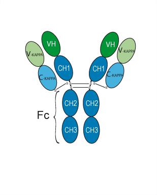
Application
ELISA, FACS, Functional assay


Chemical Information
-
CAS No. 648904-28-3
-
Appearance Liquid
-
Molecular Weight 145.336 kDa
-
Color Colorless to light yellow
-
SMILES
[Bavituximab]
-
Synonyms
Anti-Human Phosphatidylserine Recombinant Antibody; PGN-401; CH3G4
-
Shipping
Shipping with dry ice.
-
Formulation
Please refer to the lot-specific COA for specific buffer information.
-
Storage
Please store the product under the recommended conditions in the Certificate of Analysis.


Publications (2)
-
Journal Impact Factor
-
Most Recent
-
Nat Cancer
CD300ld on pathologically activated neutrophils promotes tumor immune suppression by binding phosphatidylserine on CD8+ T cells. [Abstract]2026 May 15. PMID: 42141174 -
Cell Rep Med
Macrophage efferocytosis mediated by the TP63-RAC2 pathway promotes immunosuppressive remodeling in esophageal cancer. [Abstract]2025 Dec 18:102529. PMID: 41418775
Bavituximab purchased from MedChemExpress. Usage Cited in: Cell Rep Med. 2025 Dec 18:102529. [Abstract]
Human primary macrophages were treated with Bavituximab and co-incubated with apoptotic EC9706 or KYSE180 cells; subsequently, the efferocytosis of apoptotic EC9706 and KYSE180 cells by the macrophages was assessed via confocal imaging.
Bavituximab purchased from MedChemExpress. Usage Cited in: Cell Rep Med. 2025 Dec 18:102529. [Abstract]
Human primary macrophages were treated with Bavituximab and incubated the apoptotic EC9706 or apoptotic KYSE180 cells. The efferocytosis of apoptotic EC9706 and KYSE180 cells by macrophages were detected by flow cytometry.

Protocols
-
Patient-Derived Xenograft (PDX)
Patient-derived xenograft (PDX) models are generated by engrafting primary human tumor tissue directly into immunodeficient mice, allowing in vivo propagation of patient tumor biology without initial in vitro adaptation. These models are used to preserve key histopathological and molecular characteristics of the original tumor and enable assessment of tumor growth dynamics and therapeutic response in a living organism. The biological readout is tumor engraftment and subsequent growth in the murine host, which reflects the ability of human tumor cells to survive, vascularize, and expand in an immunocompromised microenvironment.
-
Patient-Derived Orthotopic Xenograft (PDOX)
Patient-derived orthotopic xenograft (PDOX) modeling implants fresh patient tumor tissue or patient-derived tumor cells into the anatomically corresponding organ or tissue site of immunodeficient mice, usually by surgical orthotopic implantation, to preserve patient tumor histology, local microenvironmental context, invasion, metastatic behavior, and treatment-response features better than subcutaneous implantation. PDOX readouts include tumor engraftment, orthotopic tumor growth, local invasion, metastasis, recurrence after resection, histologic similarity to the donor tumor, biomarker retention, molecular concordance, survival, and response or resistance to therapy. PDOX models are used for preclinical drug testing and individualized therapy evaluation, but engraftment success varies by tumor type and specimen quality.
-
Recombinant antibody expression and Protein A/G purification
Recombinant antibody expression produces antibody chains from cloned antibody genes in mammalian cells, commonly HEK293-derived cells, so that secreted IgG or Fc-fusion antibodies can be collected from culture supernatant. Protein A/G purification is affinity chromatography in which immobilized Protein A, Protein G, or Protein A/G binds the antibody Fc region; nonbound culture components are washed away, and bound antibody is recovered by changing buffer conditions, commonly acidic elution followed by neutralization.
-
Subcutaneous Cell-Line-Derived Xenograft
Subcutaneous cell-line-derived xenograft (CDX) models are established by implanting cultured human cancer cell lines into immunodeficient mice, where the injected cells form localized tumors that can be monitored in vivo as a measure of tumorigenic potential, growth kinetics, and treatment response. These models are widely used in oncology research because they allow reproducible tumor formation and enable comparative assessment of tumor growth between different cell lines or genetic manipulations in a controlled in vivo microenvironment. Subcutaneous implantation of cancer cells in immunodeficient mice is a standard approach for evaluating tumor growth behavior and therapeutic response across multiple cancer types, including prostate, esophageal, pancreatic, and colon cancer models.
-
Orthotopic Cell-Line Xenograft
Orthotopic cell-line xenograft models involve implantation of human cancer cell lines into the anatomically corresponding organ of immunodeficient mice to reproduce tumor growth within a native microenvironment, enabling more clinically relevant tumor behavior compared with subcutaneous models. These models are widely used because orthotopic placement better recapitulates tumor progression, including invasion and metastatic spread, which are often underrepresented in heterotopic implantation systems. Compared with conventional xenografts, orthotopic implantation is described as more technically complex but provides improved simulation of tumor-microenvironment interactions and metastatic behavior, making it particularly valuable for translational oncology research. Surgical orthotopic implantation approaches have been emphasized as enabling faithful reproduction of clinical cancer features, including metastasis and disease progression patterns that align with the tumor’s organ of origi
-
Research Protocol for Cancer Immunology
Cancer immunology studies how the immune system recognizes, suppresses, edits, or fails to eliminate malignant cells through tumor antigen release, antigen presentation, T-cell priming, immune trafficking, tumor-cell killing, and feedback inhibition in the tumor microenvironment. The cancer-immunity cycle links tumor antigenicity, dendritic-cell priming, CD8+ T-cell infiltration, cytotoxic function, and immune-checkpoint regulation to tumor rejection or immune escape. Immune-checkpoint pathways such as PD-1/PD-L1 and CTLA-4 suppress antitumor T-cell activity and can be therapeutically blocked, but many tumors remain resistant because of poor antigen presentation, weak T-cell infiltration, suppressive myeloid cells, regulatory T cells, and tumor-intrinsic immune-exclusion programs. Unresolved questions include which immune-cell states predict response, how tumor-intrinsic pathways exclude immune cells, how myeloid suppression limits checkpoint blockade, and which combination strategies


Purity & Documentation
-
Data Sheet (261 KB)
-
SDS (252 KB)
- English - EN (252 KB)
- Français - FR (252 KB)
- Deutsch - DE (252 KB)
- Norwegian - NO (252 KB)
- Español - ES (252 KB)
- Swedish - SV (252 KB)
- Italian - IT (252 KB)
- Korean - KR (252 KB)
- Portuguese - PT (252 KB)
-
Inhibitory Antibodies User Guide (603 KB)
References
[1]. Digumarti R, et al. Bavituximab plus paclitaxel and carboplatin for the treatment of advanced non-small-cell lung cancer. Lung Cancer. 2014 Nov;86(2):231-6. [Content Brief]
[2]. Cheng X, et al. Antibody-Mediated Blockade of Phosphatidylserine Enhances the Antitumor Effect of Sorafenib in Hepatocellular Carcinomas Xenografts. Ann Surg Oncol. 2016 Dec;23(Suppl 5):583-591. [Content Brief]

Calculators
Concentration (start) × Volume (start) = Concentration (final) × Volume (final)


Bavituximab Related Classifications
HY-P99279 Related Classifications
 Powered by Bioz