



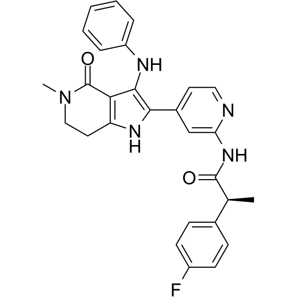
BAY-888

Based on 1 Customer Validation
BAY-888 is a selective CK1α/CSNK1A1 (casein kinase 1α) ATP-competitive inhibitor (IC50: 4 nM@10 μM ATP; 63 nM@1 mM ATP). BAY-888 blocks the negative regulation of p53 and other signaling pathways by CK1α, induces apoptosis and inhibits proliferation of tumor cells. BAY-888 has shown inhibitory efficacy against cancers such as acute myeloid leukemia (AML) in PRISM barcoded cell line screening and can mimic the effects of shRNA-mediated CK1α knockdown. BAY-888 is primarily used for the development of anticancer drugs for p53 wild-type tumors and for the study of the mechanisms of CK1α-related signaling pathways.
For research use only. We do not sell to patients.
- Purity : 98.27%
- CAS No.: 2468783-75-5
- Formula: C28H26FN5O2
- Molecular Weight:483.54
-
Storage:
4°C, sealed storage, away from moisture and light
* In solvent : -80°C, 6 months; -20°C, 1 month (sealed storage, away from moisture and light)

To place orders, for customer services and technical support, please contact: MedChemExpress USA
Tel: 609-228-6898 E-mail: [email protected] [email protected]


-
 Biological Activity
-
Chemical Information
-
Solvent & Solubility
- Protocols
- Purity & Documentation
- References
-
 Help & FAQs


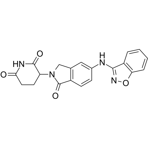

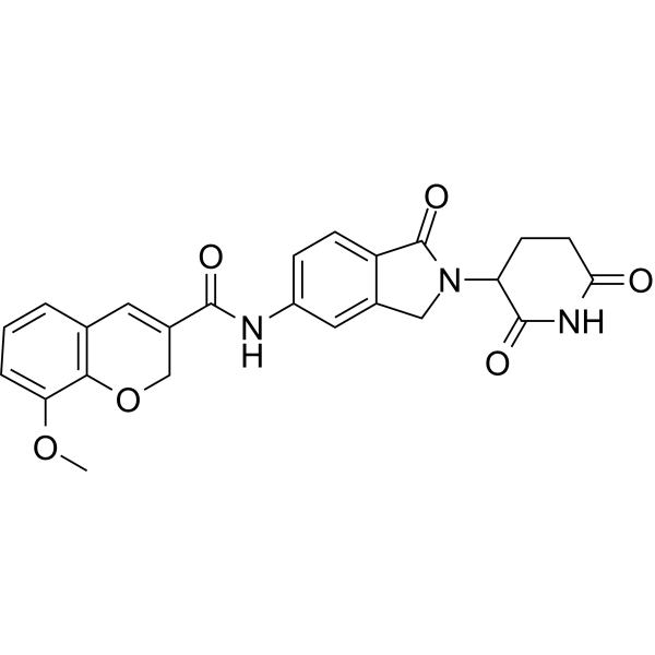
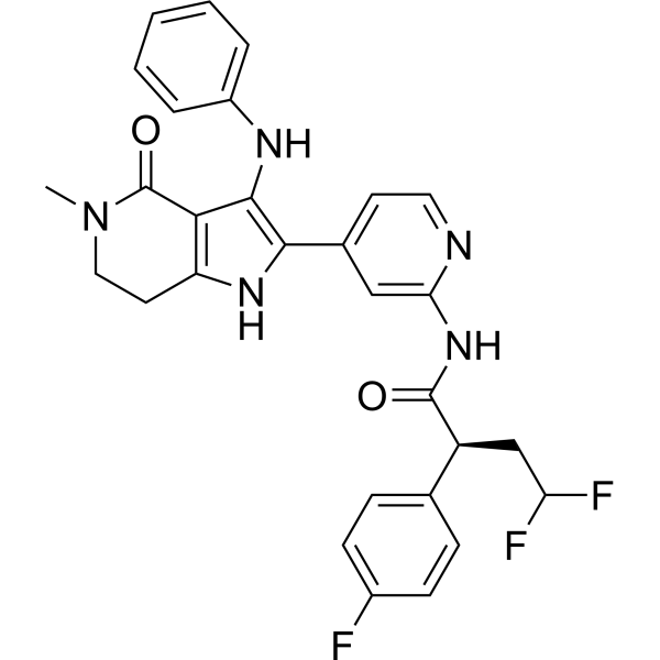


Biological Activity
Description
In Vitro
In Vivo
MedChemExpress (MCE) has not independently confirmed the accuracy of these methods. They are for reference only.


Chemical Information
-
CAS No. 2468783-75-5
-
Appearance Solid
-
Molecular Weight 483.54
-
Formula C28H26FN5O2
-
Color Light brown to yellow
-
SMILES
O=C1C2=C(NC(C3=CC=NC(NC([C@@H](C)C4=CC=C(F)C=C4)=O)=C3)=C2NC5=CC=CC=C5)CCN1C
-
Synonyms
BRD5846
-
Shipping
Room temperature in continental US; may vary elsewhere.
-
Storage
4°C, sealed storage, away from moisture and light
* In solvent : -80°C, 6 months; -20°C, 1 month (sealed storage, away from moisture and light)


Solvent & Solubility
In Vitro:
DMSO : 50 mg/mL (103.40 mM; Need ultrasonic; Hygroscopic DMSO has a significant impact on the solubility of product, please use newly opened DMSO)
Please refer to the solubility information to select the appropriate solvent. Once prepared, please aliquot and store the solution to prevent product inactivation from repeated freeze-thaw cycles.
Storage method and period of stock solution: -80°C, 6 months; -20°C, 1 month (sealed storage, away from moisture and light). When stored at -80°C, please use it within 6 months. When stored at -20°C, please use it within 1 month.
Please refer to the solubility information to select the appropriate solvent. Once prepared, please aliquot and store the solution to prevent product inactivation from repeated freeze-thaw cycles.
Storage method and period of stock solution: -80°C, 6 months; -20°C, 1 month (sealed storage, away from moisture and light). When stored at -80°C, please use it within 6 months. When stored at -20°C, please use it within 1 month.
Concentration (start) × Volume (start) = Concentration (final) × Volume (final)


Purity & Documentation
-
Data Sheet (276 KB)
-
SDS (252 KB)
- English - EN (252 KB)
- Français - FR (252 KB)
- Deutsch - DE (252 KB)
- Norwegian - NO (252 KB)
- Español - ES (252 KB)
- Swedish - SV (252 KB)
- Italian - IT (252 KB)
- Korean - KR (252 KB)
- Portuguese - PT (252 KB)
-
Handling Instructions (2659 KB)
References


Complete Stock Solution Preparation Table
Please refer to the solubility information to select the appropriate solvent. Once prepared, please aliquot and store the solution to prevent product inactivation from repeated freeze-thaw cycles.
Storage method and period of stock solution: -80°C, 6 months; -20°C, 1 month (sealed storage, away from moisture and light). When stored at -80°C, please use it within 6 months. When stored at -20°C, please use it within 1 month.
| Optional Solvent | Concentration Solvent Mass | 1 mg | 5 mg | 10 mg | 25 mg |
|---|---|---|---|---|---|
| DMSO | 1 mM | 2.0681 mL | 10.3404 mL | 20.6808 mL | 51.7020 mL |
| 5 mM | 0.4136 mL | 2.0681 mL | 4.1362 mL | 10.3404 mL | |
| 10 mM | 0.2068 mL | 1.0340 mL | 2.0681 mL | 5.1702 mL | |
| 15 mM | 0.1379 mL | 0.6894 mL | 1.3787 mL | 3.4468 mL | |
| 20 mM | 0.1034 mL | 0.5170 mL | 1.0340 mL | 2.5851 mL | |
| 25 mM | 0.0827 mL | 0.4136 mL | 0.8272 mL | 2.0681 mL | |
| 30 mM | 0.0689 mL | 0.3447 mL | 0.6894 mL | 1.7234 mL | |
| 40 mM | 0.0517 mL | 0.2585 mL | 0.5170 mL | 1.2926 mL | |
| 50 mM | 0.0414 mL | 0.2068 mL | 0.4136 mL | 1.0340 mL | |
| 60 mM | 0.0345 mL | 0.1723 mL | 0.3447 mL | 0.8617 mL | |
| 80 mM | 0.0259 mL | 0.1293 mL | 0.2585 mL | 0.6463 mL | |
| 100 mM | 0.0207 mL | 0.1034 mL | 0.2068 mL | 0.5170 mL |


BAY-888 Related Classifications
HY-145939 Related Classifications
 Powered by Bioz