



Beloranib

Based on 1 Customer Validation
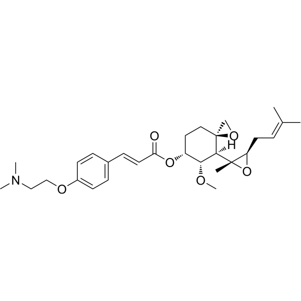
Beloranib (ZGN-440; CKD-732 free base) is a selective, irreversible inhibitor of methionine aminopeptidase MetAP2 that suppresses appetite and increases energy expenditure. Beloranib blocks the enzymatic cleavage of N-terminal methionine from nascent proteins by forming a covalent bond with MetAP2, thereby regulating fatty acid metabolism, adrenergic signaling, and hypothalamic NF-κB expression. Beloranib significantly reduces food intake, body weight, and fat accumulation, while improving glucose tolerance, insulin sensitivity, and lipid metabolism. Beloranib also elevates energy expenditure and fat oxidation levels, without affecting body temperature, spontaneous activity, or the inflammatory cytokine IL-1β. Beloranib can be used in research on obesity and hypothalamic obesity.
For research use only. We do not sell to patients.
- Purity : 89.63%
- CAS No.: 251111-30-5
- Formula: C29H41NO6
- Molecular Weight:499.64
-
Storage:
-20°C, protect from light, stored under nitrogen
* In solvent : -80°C, 6 months; -20°C, 1 month (protect from light, stored under nitrogen)

To place orders, for customer services and technical support, please contact: MedChemExpress USA
Tel: 609-228-6898 E-mail: [email protected] [email protected]


-
 Biological Activity
-
Chemical Information
- Protocols
- Purity & Documentation
- References
-
 Help & FAQs




Biological Activity
Description
IC50 & Target
|
MetAp2 |
In Vitro
Beloranib potently inhibits endothelial cell proliferation via selective inhibition of MetAP2[2].
Beloranib (24 h pre-incubation; 3 h norepinephrine treatment) enhances norepinephrine-induced lipolysis in primary rat brown adipocytes, increasing glycerol release to 4- to 5-fold relative to vehicle[3].
Beloranib (24 h pre-incubation) enhances norepinephrine-induced energy expenditure in primary rat brown adipocytes, increasing the %OCR AUC relative to vehicle[3].
Beloranib (24 h concurrent treatment) reverses norepinephrine desensitization in primary rat brown adipocytes, sustaining ucp1 gene expression at 177-fold after 24 h of norepinephrine treatment[3].
MedChemExpress (MCE) has not independently confirmed the accuracy of these methods. They are for reference only.
In Vivo
Beloranib (0.1 mg/kg; subcutaneous injection; once daily; for 14 consecutive days) induces significant weight loss and reduces food intake by 28% in young adult male MC4rKO obese rats[1].
Beloranib (0.1 mg/kg, subcutaneous injection, once daily for 21 consecutive days) induces a 10% body weight loss, reduces food intake, and improves insulin sensitivity in weight-stable aged male MC4rKO obese rats[1].
Administration of Beloranib to nude mice with xenografts inhibits angiogenesis and tumorigenesis[2].
Beloranib (1 mg/kg; subcutaneous injection; once daily; for 12 consecutive days) reduces body weight by 20-23% over 12 days in high-fat diet-induced obese male C57BL/6 mice[3].
Beloranib (1 mg/kg; subcutaneous injection; once daily; for 14 consecutive days) reduces body weight by 22-25% within 14 days in high-fat diet-induced obese C57BL/6 mice, while decreasing fat mass and increasing lean mass, but exerts minimal effects on body weight and body composition in lean mice[3].
Beloranib (0.3 mg/kg; subcutaneous injection; administered twice at 9:00 a.m. on Day 1 and Day 2) inhibits MetAP2 activity in brown adipose tissue of C57BL/6 mice with obesity induced by a high-fat diet[3].
MedChemExpress (MCE) has not independently confirmed the accuracy of these methods. They are for reference only.
-
Animal Model:Sprague Dawley (male, juvenile, 75-100 g at arrival, 246.4 ± 1.8 g at surgery, combined medial hypothalamic lesion induced)[1]
-
Dosage:0.1 mg/kg
-
Administration:subcutaneously; daily; 12 days
-
Result:Reduced mean daily body weight gain to 0.2 g/d and total body weight gain to 4.2 g over 12 days; body weight was significantly lower after 8 days of treatment.
Reduced mean 24-hour food intake by 30%, with consistent reductions across light and dark cycles.
Reduced mean 24-hour water intake by 28%, while the water-to-food intake ratio remained unchanged.
Lowered fasting insulin levels significantly; reduced fasting blood glucose significantly relative to pretreatment levels.
Increased circulating α-melanocyte stimulating hormone (α-MSH) levels significantly; increased the α-MSH-to-leptin ratio significantly.
Reduced Lee index (adiposity marker) from pretreatment levels, while vehicle-treated CMHL rats showed an increase.
Downregulated hypothalamic NF-κB mRNA expression significantly; SOCS-3 expression remained unchanged relative to vehicle.
Did not affect body temperature, locomotor activity, or serum lipid levels (cholesterol, triglycerides, HDL).
-
Animal Model:C57BL/6 (26-33 weeks old, high-fat diet-induced obesity; normal chow-fed lean)[3]
-
Dosage:1 mg/kg
-
Administration:s.c.; q.d.; 14 days
-
Result:Reduced body weight by 22-25% after 14 days in obese mice.
Achieved statistically significant weight loss versus vehicle control on days 4-14 in obese mice.
Had minimal effect on body weight in lean mice, with only a statistically significant effect on day 7.
Reduced fat mass and increased lean mass in obese mice.
Had no effect on fat mass or lean mass in lean mice.
Clinical Trial
| NCT Number | Sponsor | Condition | Start Date |
Phase

|
|---|---|---|---|---|
| NCT01329991 | 2011-05 | PHASE1 |


Chemical Information
-
CAS No. 251111-30-5
-
Appearance Liquid (Density: 1.159±0.10 g/cm3)
-
Molecular Weight 499.64
-
Formula C29H41NO6
-
Color Colorless to light yellow
-
SMILES
C[C@]1([C@H](O1)C/C=C(C)\C)[C@]2([H])[C@]3(CC[C@H]([C@H]2OC)OC(/C=C/C4=CC=C(C=C4)OCCN(C)C)=O)CO3
-
Synonyms
ZGN-440; CKD-732 free base
-
Shipping
Room temperature in continental US; may vary elsewhere.
-
Storage
-20°C, protect from light, stored under nitrogen
* In solvent : -80°C, 6 months; -20°C, 1 month (protect from light, stored under nitrogen)


Purity & Documentation
-
Data Sheet (275 KB)
-
SDS (252 KB)
- English - EN (252 KB)
- Français - FR (252 KB)
- Deutsch - DE (252 KB)
- Norwegian - NO (252 KB)
- Español - ES (252 KB)
- Swedish - SV (252 KB)
- Italian - IT (252 KB)
- Korean - KR (252 KB)
- Portuguese - PT (252 KB)
-
Handling Instructions (2659 KB)
References
[1]. Elfers CT, et al. Robust Reductions of Excess Weight and Hyperphagia by Beloranib in Rat Models of Genetic and Hypothalamic Obesity. Endocrinology. 2017;158(1):41-55. [Content Brief]
[2]. Hughes TE, et al. Ascending dose-controlled trial of beloranib, a novel obesity treatment for safety, tolerability, and weight loss in obese women. Obesity (Silver Spring). 2013;21(9):1782-1788. [Content Brief]
[3]. Huang HJ, et al. MetAP2 inhibition increases energy expenditure through direct action on brown adipocytes. J Biol Chem. 2019;294(24):9567-9575. [Content Brief]

Calculators
Concentration (start) × Volume (start) = Concentration (final) × Volume (final)


Beloranib Related Classifications
HY-14811 Related Classifications
 Powered by Bioz
Keywords
- Beloranib
- 251111-30-5
- ZGN-440
- CKD-732
- ZGN440
- ZGN 440
- CKD732
- CKD 732
- MetAP
- NF-κB
- male MC4rKO obese rats
- endothelial cell proliferation
- methionine aminopeptidase 2
- xenografted nude mice
- high-fat diet-induced obese male C57BL/6 mice
- male CMHL-induced obese Sprague Dawley rats
- obesity
- MetAP2
- primary rat brown adipocytes
- hypothalamic obesity
- Inhibitor
- inhibitor
- inhibit