



β-Amyrin

Based on 1 Customer Validation
β-Amyrin shows effectively counteract amyloid β (Aβ)-induced impairment of long-term potentiation (LTP). β-Amyrin is a promising candidate for Alzheimer's disease (AD) research. β-Amyrin exhibits anti-inflammatory effects, protective activity against pulmonary fibrosis, and notable antibacterial capabilities. β-Amyrin is an orally active natural triterpenoid compound.
For research use only. We do not sell to patients.
- Purity : 99.95%
- CAS No.: 559-70-6
- Formula: C30H50O
- Molecular Weight:426.72
-
Storage:Powder -20°C, 3 years , 4°C, 2 years ; In solvent -80°C, 6 months , -20°C, 1 month

To place orders, for customer services and technical support, please contact: MedChemExpress USA
Tel: 609-228-6898 E-mail: [email protected] [email protected]


-
 Biological Activity
-
Chemical Information
-
Solvent & Solubility
- Protocols
- Purity & Documentation
- References
-
 Help & FAQs


-
HY-N8423
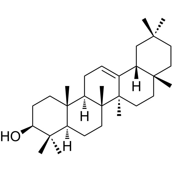
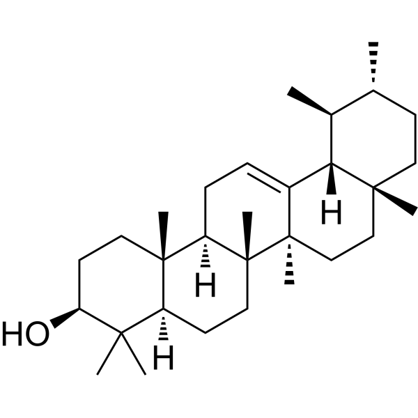
α-AmyrinPentacyclic Triterpenoid -
HY-13740
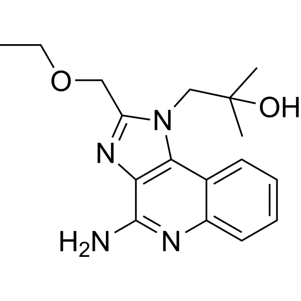
ResiquimodTLR7/8 Agonist -
HY-N1178
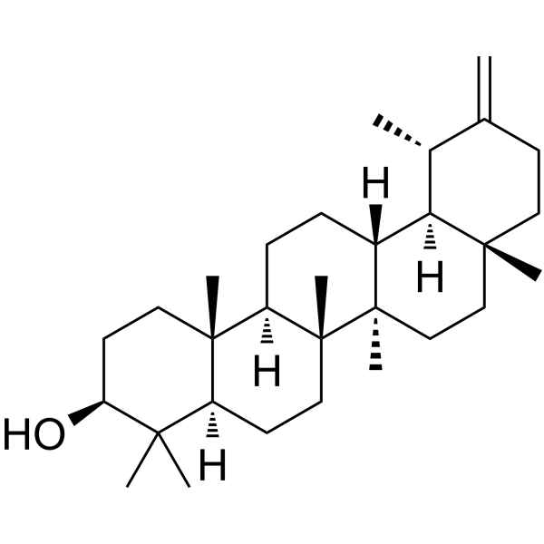
TaraxasterolAnti-Inflammatory Agent; LXRα Activator -

HY-N2922R
β-Amyrin (Standard)Anti-inflammatory compound -
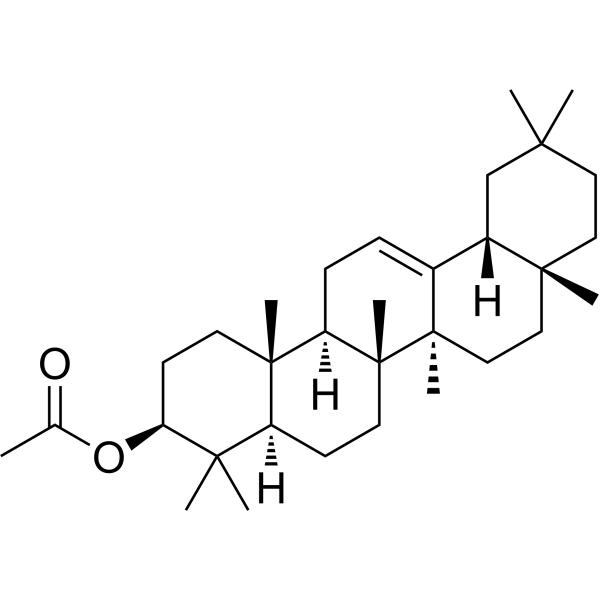
HY-N2923
β-Amyrin acetateAnti-inflammatory Agent -
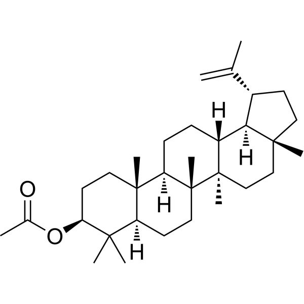
HY-126114
Lupeol acetateAnti-inflammatory Agent -
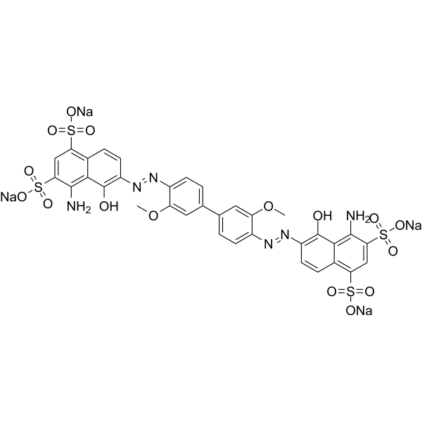
HY-D0939
Direct Blue 1VGLUT Inhibitor -
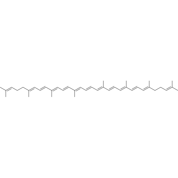
HY-N0287
LycopeneAntioxidant -
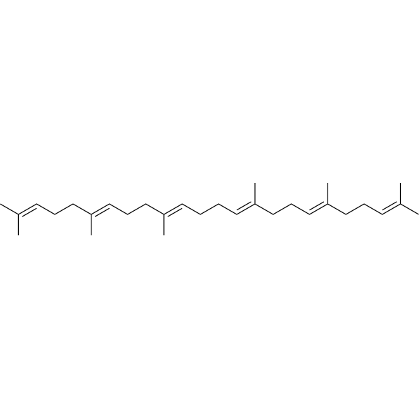
HY-N1214
SqualeneHypolipidemic Agent -

HY-N1214R
Squalene (Standard)Hypolipidemic Agent -
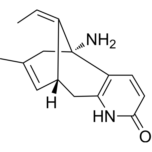
HY-17387
(-)-Huperzine AAChE/NMDA Receptor Inhibitor -
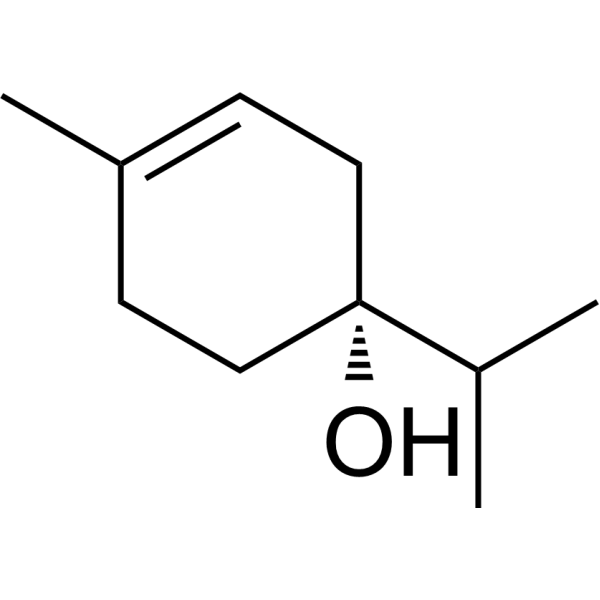
HY-N7927
(-)-Terpinen-4-olInsect Pheromones


Biological Activity
Description
Cellular Effect
|
Cell Line

|
Type | Value | Description | References |
|---|---|---|---|---|
| A549 | IC50 |
23.55 μM
Compound: 10
|
Cytotoxicity against human A549 cells assessed as reduction in cell viability after 48 hrs by MTT assay
Cytotoxicity against human A549 cells assessed as reduction in cell viability after 48 hrs by MTT assay
|
[PMID: 28482219] |
| B16 | ED50 |
>50 μM
Compound: 14
|
Induction of melanogenesis in mouse B16 2F2 cells assessed as intracellular melanin content after 3 days
Induction of melanogenesis in mouse B16 2F2 cells assessed as intracellular melanin content after 3 days
|
[PMID: 12027734] |
| B16 | IC50 |
48.8 μM
Compound: 14
|
Growth inhibition of mouse B16 2F2 cells after 3 days
Growth inhibition of mouse B16 2F2 cells after 3 days
|
[PMID: 12027734] |
| DU-145 | IC50 |
27.88 μM
Compound: 10
|
Cytotoxicity against human DU145 cells assessed as reduction in cell viability after 48 hrs by MTT assay
Cytotoxicity against human DU145 cells assessed as reduction in cell viability after 48 hrs by MTT assay
|
[PMID: 28482219] |
| HEK293 | IC50 |
94.4 μM
Compound: 10
|
Cytotoxicity against human HEK293 cells assessed as reduction in cell viability after 48 hrs by MTT assay
Cytotoxicity against human HEK293 cells assessed as reduction in cell viability after 48 hrs by MTT assay
|
[PMID: 28482219] |
| HeLa | IC50 |
>50 μM
Compound: 4
|
Cytotoxicity against human HeLa cells by MTT assay
Cytotoxicity against human HeLa cells by MTT assay
|
[PMID: 19447618] |
| HeLa | IC50 |
27.94 μM
Compound: 10
|
Cytotoxicity against human HeLa cells assessed as reduction in cell viability after 48 hrs by MTT assay
Cytotoxicity against human HeLa cells assessed as reduction in cell viability after 48 hrs by MTT assay
|
[PMID: 28482219] |
| J774 | IC50 |
>234.3 μM
Compound: 2
|
Cytotoxicity against mouse J774 cells by alamar blue assay
Cytotoxicity against mouse J774 cells by alamar blue assay
|
[PMID: 17637068] |
| MCF7 | IC50 |
>50 μM
Compound: 4
|
Cytotoxicity against human MCF7 cells by MTT assay
Cytotoxicity against human MCF7 cells by MTT assay
|
[PMID: 19447618] |
| MCF7 | IC50 |
35.31 μM
Compound: 10
|
Cytotoxicity against human MCF7 cells assessed as reduction in cell viability after 48 hrs by MTT assay
Cytotoxicity against human MCF7 cells assessed as reduction in cell viability after 48 hrs by MTT assay
|
[PMID: 28482219] |
| SK-MEL-1 | IC50 |
>50 μM
Compound: 4
|
Cytotoxicity against human SK-MEL-1 cells by MTT assay
Cytotoxicity against human SK-MEL-1 cells by MTT assay
|
[PMID: 19447618] |
In Vitro
β-Amyrin (1-100 μM, 0-80 min) blocks Aβ-induced synaptic dysfunction in the hippocampal tissues of mice[1].
β-Amyrin (1-10 μg, 24 h 15 min) shows antioxidant activity in cultured hPBMCs (human peripheral blood mononuclear cells)[2].
β-Amyrin (5-20 μg/mL, 16-24 h) significantly inhibits PGE2 (HY-101952), IL-6 secretion and NF-κB activation dose-dependently in LPS-induced hPBMCs[2].
β-Amyrin (5 μM, 2 h) exhibits antibacterial mode of action, leading promoting apoptosis-like death in E. coli by producing ROS.
MedChemExpress (MCE) has not independently confirmed the accuracy of these methods. They are for reference only. Further protocols information, click here.
-
Cell Line:E. coli (BW 25113)
-
Concentration:5 μM
-
Incubation Time:2 h
-
Result:Decreased GSH/GSSG ratio and induced membrane depolarization and DNA cleavage.
Activated caspase-like protein expression.
In Vivo
β-Amyrin (20-100 μg, i.p.) inhibits carrageenan-induced paw edema of rats[2].
β-Amyrin (20-80 mg/kg, i.g., daily, 5 days) has a potent efficacy in protecting against BLM-induced mice pulmonary fibrosis via suppressing inflammatory response and oxidative stress[3].
Pharmacokinetic parameters of β-Amyrin in rats
| β-Amyrin standard IV (group 3) | β-Amyrin standard oral dose (group 2) | ||
| Dose (mg kg−1) | 1 | 3 | 300 (equivalent of 3mg kg−1 β-amyrin) |
| V (ml kg−1) | 1317 | ||
| AUC0→24 h (min μg ml−1) | 411 | 10.6 | 47.3 |
| Cl (ml min−1 kg−1) | 2.04 | ||
| t1/2λZ (h) | 10.2 | 10.3 | 8.3 |
| MRT0→24 h (h) (h) | 8 | 8.5 | 8.7 |
| Cmax (ng ml−1) (h) (h) | 18.7 | 75 | |
| tmax (h) | - | 3 (n = 2) or 5 (n = 1) | 3 (n = 3) or 5 (n = 1) |
| F (%) | 0.86 | 3.83 |
MedChemExpress (MCE) has not independently confirmed the accuracy of these methods. They are for reference only.
-
Animal Model:Male CD-1 mice, 26-28 g, 6 weeks old. 5 μL of Aβ was acutely injected into the left lateral ventricle by hand[1].
-
Dosage:4 mg/kg
-
Administration:p.o., daily, 5 days
-
Result:Increased number of transitions between the light and dark compartments.
Increased the total exploration time on the light compartment of the apparatus and the time spent in open arms.
Ameliorated the reduction of neurogenesis.
-
Animal Model:Adult male rats of Sprague–Dawley strain, weighing 150-180 g. Carrageenan induces edema in the hind paw of the rat[2].
-
Dosage:20, 50, 100 μg/kg
-
Administration:i.p.
-
Result:Significant decreased edema by in a dose dependent manner.
-
Animal Model:Adult male rats of Sprague–Dawley strain, weighing 150-180 g. Carrageenan induces Use Bleomycin (BLM) (HY-108345), 5mg/kg, one-time intratracheally injection, in 50μL saline per mouse to induce the marine model of pulmonary fibrosis[3].
-
Dosage:20, 40, 80 mg/kg
-
Administration:i.g., daily, 5 days
-
Result:Promoted the values of SO2 and PO2 and inhibited the value of PCO2.
Significantly improved inflammatory cell infiltration, the thickened alveolar septum and the lesion collapses of the alveoli and formation of flaky lung tissue.


Chemical Information
-
CAS No. 559-70-6
-
Appearance Solid
-
Molecular Weight 426.72
-
Formula C30H50O
-
Color White to off-white
-
SMILES
CC1(C)[C@@H](O)CC[C@]2(C)[C@@]3([H])CC=C4[C@]5([H])CC(C)(C)CC[C@@](C)5CC[C@](C)4[C@@](C)3CC[C@@]12[H]
-
Structure Classification
-
Initial Source
-
Shipping
Room temperature in continental US; may vary elsewhere.
-
Storage
Powder -20°C 3 years 4°C 2 years In solvent -80°C 6 months -20°C 1 month


Solvent & Solubility
In Vitro:
Ethanol : 5 mg/mL (11.72 mM; Need ultrasonic)
Please refer to the solubility information to select the appropriate solvent. Once prepared, please aliquot and store the solution to prevent product inactivation from repeated freeze-thaw cycles.
Storage method and period of stock solution: -80°C, 6 months; -20°C, 1 month. When stored at -80°C, please use it within 6 months. When stored at -20°C, please use it within 1 month.
Please refer to the solubility information to select the appropriate solvent. Once prepared, please aliquot and store the solution to prevent product inactivation from repeated freeze-thaw cycles.
Storage method and period of stock solution: -80°C, 6 months; -20°C, 1 month. When stored at -80°C, please use it within 6 months. When stored at -20°C, please use it within 1 month.
Concentration (start) × Volume (start) = Concentration (final) × Volume (final)
In Vivo:
Select the appropriate dissolution method based on your experimental animal and administration route.
- For the following dissolution methods, please ensure to first prepare a clear stock solution using an In Vitro approach and then sequentially add co-solvents:
- To ensure reliable experimental results, the clarified stock solution can be appropriately stored based on storage conditions. As for the working solution for In Vivo experiments, it is recommended to prepare freshly and use it on the same day.
- The percentages shown for the solvents indicate their volumetric ratio in the final prepared solution. If precipitation or phase separation occurs during preparation, heat and/or sonication can be used to aid dissolution.
Add each solvent one by one: 10% EtOH 40% PEG300 5% Tween-80 45% Saline
Solubility: ≥ 0.5 mg/mL (1.17 mM); Clear solution
This protocol yields a clear solution of ≥ 0.5 mg/mL (saturation unknown).
Taking 1 mL working solution as an example, add 100 μL EtOH stock solution (5.0 mg/mL) to 400 μL PEG300, and mix evenly; then add 50 μL Tween-80 and mix evenly; then add 450 μL Saline to adjust the volume to 1 mL.
Preparation of Saline: Dissolve 0.9 g sodium chloride in ddH₂O and dilute to 100 mL to obtain a clear Saline solution.
Add each solvent one by one: 10% EtOH 90% Corn Oil
Solubility: ≥ 0.5 mg/mL (1.17 mM); Clear solution
This protocol yields a clear solution of ≥ 0.5 mg/mL (saturation unknown). If the continuous dosing period exceeds half a month, please choose this protocol carefully.
Taking 1 mL working solution as an example, add 100 μL EtOH stock solution (5.0 mg/mL) to 900 μL Corn oil, and mix evenly.
In Vivo Dissolution Calculator
Please enter the basic information of animal experiments:
-
-
-
-
Recommended: Prepare an additional quantity of animals to account for potential losses during experiments.
Working solution concentration: 0.22 mg/mL

Protocols
-
Fibrosis/Collagen Morphometry
Fibrosis and collagen morphometry is based on the quantitative visualization of fibrillar collagen deposition in tissue sections using histochemical stains such as Sirius Red (Picrosirius Red) or Masson's trichrome, followed by image-based or polarization-enhanced analysis to estimate collagen proportional area as a surrogate of extracellular matrix accumulation during fibrotic remodeling. Sirius Red combined with polarized light microscopy enhances detection of collagen fibers due to birefringence properties, enabling more specific visualization of collagen type I and III fibrils compared to conventional bright-field histology, while whole-section or region-restricted digital morphometry reduces field-selection bias in fibrosis assessment. Alternative quantitative approaches include second harmonic generation (SHG) and two-photon excited fluorescence microscopy, which enable label-free detection of fibrillar collagen and have been validated against histological staining and biochemica
-
Collagen: Sirius Red Staining
Sirius Red or picrosirius red staining is a histochemical method for visualizing collagen-rich extracellular matrix in tissue sections, and collagen fibers are detected as red-stained structures under bright-field microscopy with enhanced birefringence under polarized light. Picrosirius red is useful for assessing total collagen organization, distribution, and fibrosis burden, but polarized color should not be interpreted as a definitive collagen type I versus type III readout because color is affected by fiber orientation, thickness, and packing.
-
Research Protocol for Infectious Diseases
Infectious-disease experiments test how pathogens interact with host barriers, innate immune receptors, inflammatory signaling, pathogen replication, and tissue injury; pattern-recognition receptors such as TLRs, RIG-I-like receptors, NOD-like receptors, and inflammasomes detect microbial molecules and activate NF-κB, interferon, and cytokine responses. The central hypothesis is that infection severity reflects the balance between pathogen burden and host response: protective inflammation restricts pathogen growth, whereas excessive or mislocalized inflammation contributes to tissue damage and disease phenotype. Unresolved questions include which host pathways are protective versus pathogenic, why some infection models fail to translate to human disease, and which combined readouts best predict clinically relevant infection outcomes.
-
Connective Tissue: Masson's Trichrome/Collagen Trichrome Staining
Masson’s Trichrome (collagen/trichrome staining) is a histological technique that differentially stains tissue compartments using sequential acidic dyes to distinguish collagen from muscle and cytoplasmic components based on dye affinity and tissue permeability differences, enabling visualization of fibrosis and connective tissue architecture in histological sections. The classical formulation typically uses Weigert's iron hematoxylin for nuclear staining, Biebrich scarlet-acid fuchsin for cytoplasm and muscle, and aniline blue (or light green variants) for collagen, producing a characteristic blue/green collagen signal contrasted against red cytoplasm and dark nuclei. The staining principle relies on selective displacement of smaller dye molecules by larger anionic dyes in collagen-rich regions under controlled acidified conditions, which enhances collagen-specific dye retention. This property makes the method widely used for fibrosis assessment in organs such as heart, liver, lung, a
-
Research Protocol for Inflammation-related Diseases
The NLRP3 inflammasome is a cytosolic innate immune signaling platform that integrates priming signals and danger-signal activation to promote caspase-1 activation, maturation of IL-1β and IL-18, and gasdermin D-mediated pyroptotic cell death. The core experimental logic is to determine whether inflammatory disease phenotypes are driven by increased NLRP3 expression, ASC-containing inflammasome assembly, caspase-1 cleavage, GSDMD cleavage, and extracellular release of IL-1β/IL-18 rather than by nonspecific cell injury alone. The pathway is strongly linked to inflammation-related disease phenotypes because monosodium urate crystals activate NALP3/NLRP3 inflammasome signaling in gout-like crystal inflammation, cholesterol crystals activate NLRP3 inflammasomes in atherogenesis models, and DSS-induced intestinal inflammation has been reported to involve NLRP3 inflammasome activity. However, experimental colitis studies also show context-dependent protective effects of NLRP3 inflammasome co
-
Amyloid: Congo Red Amyloid Staining
Congo red amyloid staining is a histochemical method used to detect extracellular amyloid deposits in tissue sections based on the affinity of Congo red dye for β-pleated sheet-rich protein aggregates. When bound to amyloid, Congo red produces characteristic apple-green birefringence under polarized light microscopy, which is widely regarded as a diagnostic feature of amyloid deposition in histopathology. The diagnostic principle relies on the combination of dye binding (congophilia) and optical anisotropy under polarized illumination, which distinguishes amyloid from most non-amyloid eosinophilic extracellular deposits in routine histological evaluation. Amyloid identification by Congo red staining remains a cornerstone in diagnostic pathology despite the availability of adjunct methods such as immunohistochemistry and mass spectrometry, particularly because of its ability to localize deposits directly within tissue architecture. The specificity of Congo red-positive deposits is incre
-
Alzheimer’s Disease Modeling
Alzheimer’s Disease (AD) is a neurodegenerative disorder characterized by a progressive decline in cognitive functions and loss of specific types of neurons and synapses. Alzheimer's symptoms can be simulated in mice by injecting drugs (such as Aβ) or genetically modified.


Purity & Documentation
-
Data Sheet (285 KB)
-
SDS (393 KB)
- English - EN (393 KB)
- Français - FR (393 KB)
- Deutsch - DE (393 KB)
- Norwegian - NO (393 KB)
- Español - ES (393 KB)
- Swedish - SV (393 KB)
- Italian - IT (393 KB)
- Korean - KR (393 KB)
- Portuguese - PT (393 KB)
-
Handling Instructions (2659 KB)
References
[1]. Park HJ, et al. β-Amyrin Ameliorates Alzheimer's Disease-Like Aberrant Synaptic Plasticity in the Mouse Hippocampus. Biomol Ther (Seoul). 2019 Jul 30. [Content Brief]
[2]. Krishnan K, et al. Anti-inflammatory potential of β-amyrin, a triterpenoid isolated from Costus igneus. Inflammopharmacology. 2014 Dec;22(6):373-85. [Content Brief]
[3]. Cai Z, Liu J, et al. β-Amyrin ameliorates pulmonary fibrosis by inhibiting inflammatory response and oxidative stress in mice. Pak J Pharm Sci. 2023 Mar;36(2):431-436. [Content Brief]
[4]. Han G, et al. Antibacterial Mode of Action of β-Amyrin Promotes Apoptosis-Like Death in Escherichia coli by Producing Reactive Oxygen Species. J Microbiol Biotechnol. 2022 Dec 28;32(12):1547-1552. [Content Brief]
[5]. Ching J, et al. Quantification of α- and β-amyrin in rat plasma by gas chromatography-mass spectrometry: application to preclinical pharmacokinetic study. J Mass Spectrom. 2011 May;46(5):457-64. [Content Brief]


Complete Stock Solution Preparation Table
Please refer to the solubility information to select the appropriate solvent. Once prepared, please aliquot and store the solution to prevent product inactivation from repeated freeze-thaw cycles.
Storage method and period of stock solution: -80°C, 6 months; -20°C, 1 month. When stored at -80°C, please use it within 6 months. When stored at -20°C, please use it within 1 month.
| Optional Solvent | Concentration Solvent Mass | 1 mg | 5 mg | 10 mg | 25 mg |
|---|---|---|---|---|---|
| Ethanol | 1 mM | 2.3435 mL | 11.7173 mL | 23.4346 mL | 58.5864 mL |
| 5 mM | 0.4687 mL | 2.3435 mL | 4.6869 mL | 11.7173 mL | |
| 10 mM | 0.2343 mL | 1.1717 mL | 2.3435 mL | 5.8586 mL |


β-Amyrin Related Classifications
HY-N2922 Related Classifications
 Powered by Bioz