



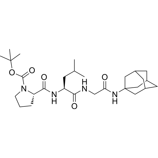
Boc-MIF-1-Am
Boc-MIF-1-Am (compound 2) is a human dopamine D2 receptor enhancer (EC50=17.82 nM) and is a bioconjugate of melanostatin (MIF-1) and amantadine. Boc-MIF-1-Am (200 μM) exhibited mild neurotoxicity in SH-SY5Y cells.
For research use only. We do not sell to patients.
- CAS No.: 3056144-84-1
- Formula: C28H46N4O5
- Molecular Weight:518.69
-
Storage:
Please store the product under the recommended conditions in the Certificate of Analysis.

To place orders, for customer services and technical support, please contact: MedChemExpress USA
Tel: 609-228-6898 E-mail: [email protected] [email protected]


-
 Biological Activity
-
Chemical Information
- Protocols
- Purity & Documentation
- References
-
 Help & FAQs


All Dopamine Receptor Isoforms
More

Biological Activity
Description
IC50 & Target
EC50: 17.82 nM (human dopamine D2 receptors, hD2R)[1]


Chemical Information
-
CAS No. 3056144-84-1
-
Molecular Weight 518.69
-
Formula C28H46N4O5
-
SMILES
O=C(N[C@@H](CC(C)C)C(NCC(NC1(C[C@H](C2)C3)C[C@H]3C[C@H]2C1)=O)=O)[C@H]4N(C(OC(C)(C)C)=O)CCC4
-
Shipping
Room temperature in continental US; may vary elsewhere.
-
Storage
Please store the product under the recommended conditions in the Certificate of Analysis.

Protocols
-
Multiplex immunofluorescence IHC
Multiplex immunofluorescence IHC detects multiple protein biomarkers in one tissue section by sequential antibody staining, HRP-mediated tyramide fluorophore deposition, heat-mediated antibody stripping, nuclear counterstaining, multispectral imaging, spectral unmixing, and digital cell phenotyping; TSA deposits fluorophore near the antigen so the fluorescence signal remains after primary and secondary antibodies are removed, enabling repeated staining cycles, including with antibodies from the same host species. Classic FFPE tumor immune-profiling applications use panels such as CD3, CD8, CD68/CD163, FOXP3, PD-1, PD-L1, pancytokeratin, Ki67, and DAPI to identify tumor cells, immune-cell subsets, checkpoint-marker expression, co-expression phenotypes, cell density, and spatial relationships in the tumor microenvironment.
-
Neurotoxicity Study
This protocol assesses in vitro neurotoxicity by combining neuronal viability, mitochondrial/metabolic activity, neurite outgrowth, and optional neuronal network function readouts. Calcein-AM or resazurin/PrestoBlue readouts estimate viable or metabolically active cells; βIII-tubulin immunofluorescence detects neuronal morphology and neurite networks; TMRE detects mitochondrial membrane potential; and MEA recordings detect functional changes in neuronal network activity.
-
Multiplex immunohistochemistry
Multiplex immunohistochemistry (mIHC), also known as tyramide dignal amplification (TSA), is an enzymatic detection method that uses horseradish peroxidase (HRP) to perform high-density in-situ labeling of target proteins or nucleic acids.


Purity & Documentation
References

Calculators
Concentration (start) × Volume (start) = Concentration (final) × Volume (final)


Boc-MIF-1-Am Related Classifications
HY-155466 Related Classifications
 Powered by Bioz