



BT173

Based on 1 Customer Validation
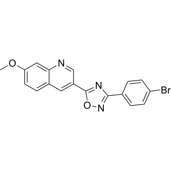
BT173 is a potent homeodomain interacting protein kinase 2 (HIPK2) inhibitor. BT173 binds to HIPK2 and does not inhibit HIPK2 kinase activity but rather, interfered allosterically with the ability of HIPK2 to associate with Smad3. BT173 attenuates renal fibrosis through suppression of the TGF-β1/Smad3 pathway. BT173 can be studied in research for kidney diseases such as renal fibrosis.
Nos produits utilisent uniquement pour la recherche. Nous ne vendons pas aux patients.
- Pureté : 98.71%
- CAS No.: 2232180-74-2
- Formule: C18H12BrN3O2
- Masse moléculaire:382.21
-
Stockage:
-20°C, protect from light
* In solvent : -80°C, 6 months; -20°C, 1 month (protect from light)

To place orders, for customer services and technical support, please contact: MedChemExpress USA
Tel: 609-228-6898 E-mail: [email protected] [email protected]


-
 Activité biologique
-
Chemical Information
-
Solvant et solubilité
- Protocole
- Pureté et documentation
- Références
-
 Help & FAQs


-
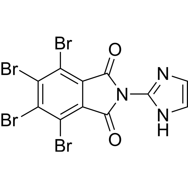
HY-100464
tBIDHIPK2 Inhibitor -
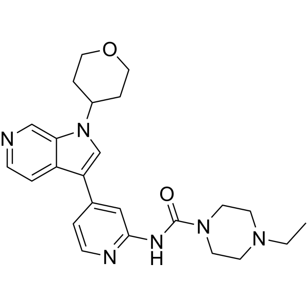
HY-142295
GNF2133DYRK1A Inhibitor -
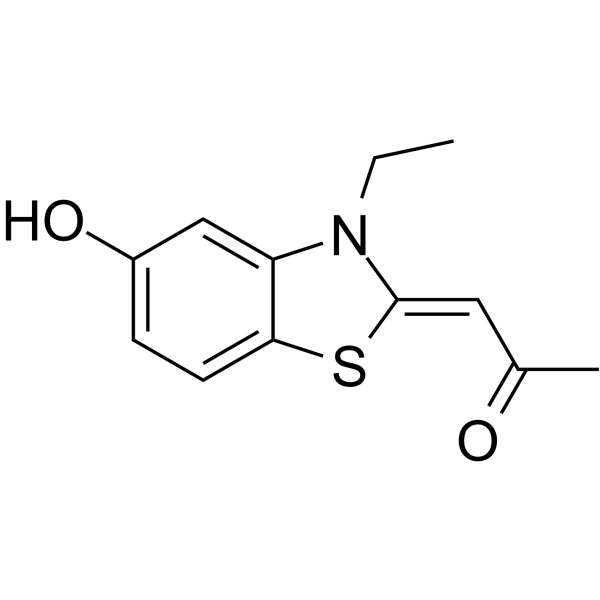
HY-108476
INDYDyrk1A And Dyrk1B Inhibitor -
HY-U00439A
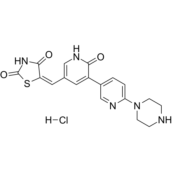
Protein kinase inhibitor 1 hydrochloride -
HY-12277
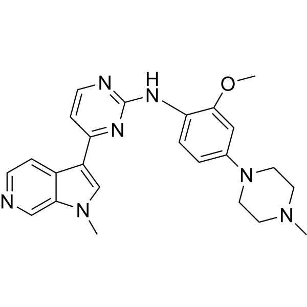
AZ191DYRK1B Inhibitor -
HY-105309
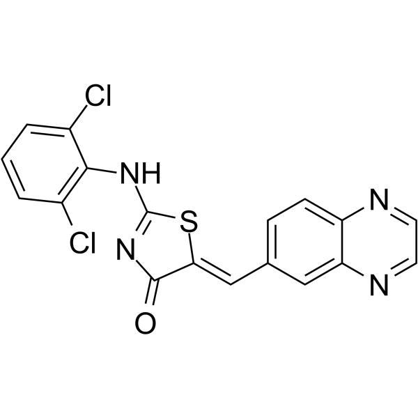
GSK-626616DYRK3 Inhibitor -
HY-15838
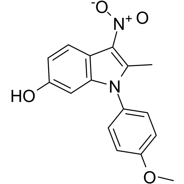
ID-8DYRK Inhibitor -
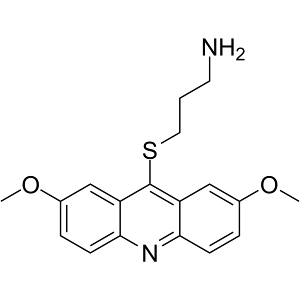
HY-13455
LDN-192960Haspin Inhibitor -
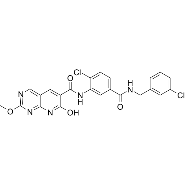
HY-12838
RO5454948Dyrk Inhibitor -
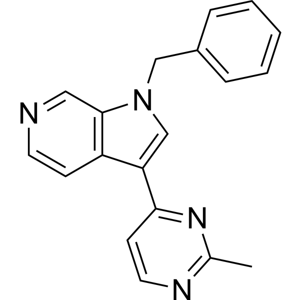
HY-117391
AZ-Dyrk1B-33Dyrk1B Kinase Inhibitor -
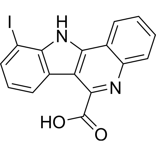
HY-146221
Dyrk1A-IN-5DYRK1A Inhibitor -
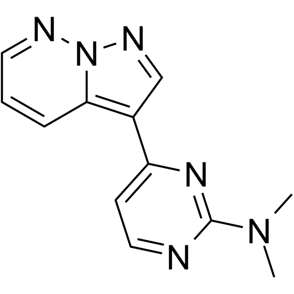
HY-132308
DYRK1-IN-1DYRK1A Inhibitor


Activité biologique
Description
In Vitro
BT173 (0-10 μM) significantly attenuates the Smad3 activity in HEK 293T cells[1].
BT173 (10 μM) has effect on HIPK2 which leads to repression of TGF-β/Smad3-dependent gene transcription in HEK 293T cells[1].
BT173 (0-100 μM) results in a partial protection of HIPK2 from prognase digestion at 30 μM and a complete protection at 100 μM in HEK 293T cells[1].
MedChemExpress (MCE) has not independently confirmed the accuracy of these methods. They are for reference only.
-
Cell Line:HEK293T
-
Concentration:0.3, 1, 3, 10, and 30 μM
-
Incubation Time:16 h
-
Result:Did not induce any significant increase of LDH level in the culture medium.
In Vivo
BT173 (20 mg/kg, p.o., 4 w) ameliorates proteinuria and kidney fibrosis in Tg26 mice[1].
MedChemExpress (MCE) has not independently confirmed the accuracy of these methods. They are for reference only.
-
Animal Model:UUO C57BL/6J renal fibrosis mouse model[1]
-
Dosage:20 mg/kg
-
Administration:Oral gavage (p.o.)
-
Result:Led to significant attenuation of fibrosis at 14 days postsurgery.
Significantly diminished the expression of genes in the Wnt/β-catenin pathways (Axin2, c-myc, and Twist).


Chemical Information
-
CAS No. 2232180-74-2
-
Appearance Solid
-
Masse moléculaire 382.21
-
Formule C18H12BrN3O2
-
Color White to light yellow
-
SMILES
COC1=CC=C2C=C(C3=NC(C4=CC=C(Br)C=C4)=NO3)C=NC2=C1
-
Livraison
Room temperature in continental US; may vary elsewhere.
-
Stockage
-20°C, protect from light
* In solvent : -80°C, 6 months; -20°C, 1 month (protect from light)


Solvant et solubilité
In Vitro:
DMSO : 2 mg/mL (5.23 mM; ultrasonic and warming and heat to 60°C; Hygroscopic DMSO has a significant impact on the solubility of product, please use newly opened DMSO)
Please refer to the solubility information to select the appropriate solvent. Once prepared, please aliquot and store the solution to prevent product inactivation from repeated freeze-thaw cycles.
Storage method and period of stock solution: -80°C, 6 months; -20°C, 1 month (protect from light). When stored at -80°C, please use it within 6 months. When stored at -20°C, please use it within 1 month.
Please refer to the solubility information to select the appropriate solvent. Once prepared, please aliquot and store the solution to prevent product inactivation from repeated freeze-thaw cycles.
Storage method and period of stock solution: -80°C, 6 months; -20°C, 1 month (protect from light). When stored at -80°C, please use it within 6 months. When stored at -20°C, please use it within 1 month.
Concentration (start) × Volume (start) = Concentration (final) × Volume (final)


Pureté et documentation
-
Fiche technique (272 KB)
-
SDS (393 KB)
- English - EN (393 KB)
- Français - FR (393 KB)
- Deutsch - DE (393 KB)
- Norwegian - NO (393 KB)
- Español - ES (393 KB)
- Swedish - SV (393 KB)
- Italian - IT (393 KB)
- Korean - KR (393 KB)
- Portuguese - PT (393 KB)
-
Instruction de manipulation (2659 KB)
Références


Complete Stock Solution Preparation Table
Please refer to the solubility information to select the appropriate solvent. Once prepared, please aliquot and store the solution to prevent product inactivation from repeated freeze-thaw cycles.
Storage method and period of stock solution: -80°C, 6 months; -20°C, 1 month (protect from light). When stored at -80°C, please use it within 6 months. When stored at -20°C, please use it within 1 month.
| Optional Solvent | Concentration Solvent Mass | 1 mg | 5 mg | 10 mg | 25 mg |
|---|---|---|---|---|---|
| DMSO | 1 mM | 2.6164 mL | 13.0818 mL | 26.1636 mL | 65.4091 mL |
| 5 mM | 0.5233 mL | 2.6164 mL | 5.2327 mL | 13.0818 mL |


BT173 Related Classifications
HY-128439 Related Classifications
 Powered by Bioz