



CA IX-IN-1
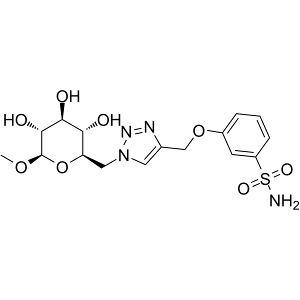
CA IX-IN-1 (compound 12g) is an effective and selective inhibitor of hCA IX with IC50 value of 7 nM. CA IX-IN-1 exhibits antitumor activity.
For research use only. We do not sell to patients.
- Formula: C16H22N4O8S
- Molecular Weight:430.43
-
Storage:
Please store the product under the recommended conditions in the Certificate of Analysis.

To place orders, for customer services and technical support, please contact: MedChemExpress USA
Tel: 609-228-6898 E-mail: [email protected] [email protected]


-
 Biological Activity
-
Chemical Information
- Protocols
- Purity & Documentation
- References
-
 Help & FAQs




Biological Activity
Description
In Vitro
CA IX-IN-1 (compound 12g) displays effective inhibition against hCA I, hCA II and hCA IX with IC50 values of 4, 0.018, 0.007 μM, respectively[1].
CA IX-IN-1 (compound 12g) (0.1 μM, 72 hours; HT-29 and MDA-MB-231 cells) exerts the potent antitumor activities against human cancer cell lines[1].
CA IX-IN-1 (compound 12g) (0-1 mM, 48 hours; HT-29 and MDA-MB-231 cells) induces highly expression level and enzyme activity of HCA IX in tumor cells with enhance the acidification of extracellular pH[1].
MedChemExpress (MCE) has not independently confirmed the accuracy of these methods. They are for reference only.
-
Cell Line:HT-29 (colon carcinoma cells) and MDA-MB-231 (breast cancer cells)
-
Concentration:0.1 mM
-
Incubation Time:72 hours
-
Result:Revealed antitumor activity against HT-29 and MDA-MB-231 (IC50 = 32 and 31 μM respectively).
-
Cell Line:MDA-MB-231 (breast cancer cells)
-
Concentration:0.1 μM
-
Incubation Time:72 hours
-
Result:Decreased (20–35%) of IC50 values were detected compared with that obtained for DOX alone.


Chemical Information
-
Molecular Weight 430.43
-
Formula C16H22N4O8S
-
SMILES
CO[C@@H]1O[C@@H]([C@H]([C@@H]([C@H]1O)O)O)CN2C=C(N=N2)COC3=CC=CC(S(N)(=O)=O)=C3
-
Shipping
Room temperature in continental US; may vary elsewhere.
-
Storage
Please store the product under the recommended conditions in the Certificate of Analysis.


Purity & Documentation
References

Calculators
Concentration (start) × Volume (start) = Concentration (final) × Volume (final)


CA IX-IN-1 Related Classifications
HY-149011 Related Classifications
 Powered by Bioz