



IO-108
IO-108 is a humanized IgG4 monoclonal antibody and a competitive inhibitor of LILRB2, with a KD value of 1.97 nM. IO-108 competitively blocks the binding of LILRB2 to its ligands including HLA-G, MHC-I, ANGPTL2 and SEMA4A, reprograms tumor-associated myeloid cells, drives the conversion of suppressive myeloid cells into a pro-inflammatory phenotype, and restores the cytotoxic activity of T cells and NK cells. IO-108 inhibits tumor growth in LILRB2 transgenic mouse models. IO-108 can be used for the research of solid tumors.
For research use only. We do not sell to patients.
-
Storage:
Please store the product under the recommended conditions in the Certificate of Analysis.


To place orders, for customer services and technical support, please contact: MedChemExpress USA
Tel: 609-228-6898 E-mail: [email protected] [email protected]


-
 Biological Activity
-
Chemical Information
- Protocols
- Purity & Documentation
- References
-
 Help & FAQs




Biological Activity
Description
Isotype
Human IgG4 kappa
Recommend Isotype Controls
Species Reactivity
Human
IC50 & Target
[1]|
LILRB2 1.97 nM (Kd) |
In Vitro
IO-108 selectively binds to LILRB2+ reporter cells, shows no cross-reactivity with other tested LILR family members or LAIR1, and binds to LILRB2 with high affinity[1].
IO-108 (0.0001-100 nM) blocks the binding of HLA-G to LILRB2 on the surface of HEK293 cells in a dose-dependent manner, while potently inhibiting the activation of LILRB2 reporter cells induced by plate-immobilized ANGPTL2 and SEMA4A[1].
IO-108 (100 nM; 3 days) enhances the production of pro-inflammatory cytokines (TNF-α, GM-CSF) and reduces the level of the anti-inflammatory cytokine IL-10 in lipopolysaccharide (LPS)-stimulated peripheral blood mononuclear cells (PBMCs)[1].
IO-108 (100 nM; 3 days) enhances the production of proinflammatory cytokines and chemokines (IFN-γ, TNF-α, IL-1β, IL-6, CXCL8) in anti-CD3-stimulated peripheral blood mononuclear cells (PBMCs)[1].
IO-108 (100 nM) enhances TNF-α production in monocyte-derived macrophages stimulated with 2’3’-cGAMP[1].
IO-108 (100 nM; 2 days) reduces the expression of tolerogenic CD209 and enhances the production of proinflammatory TNF-α in LPS-stimulated immature monocyte-derived dendritic cells[1].
IO-108 (100 nM; 6 days) enhances the pro-inflammatory effect of anti-PD-1 antibody in a co-culture system of monocyte-derived macrophages and CD4+ T cells, and increases the production level of IFN-γ[1].
IO-108 (100 nM; 6 days) promotes the differentiation of classical monocytes into CD86+ pro-inflammatory dendritic cells when co-cultured with GM-CSF and IL-4[1].
IO-108 (100 nM; 2 days) modulates the phenotype of immature monocyte-derived dendritic cells, reduces the expression of the tolerogenic CD209, and increases the expression level of the proinflammatory CD86[1].
IO-108 reverses the PMN-MDSC-mediated suppression of autologous CD8+ T cell proliferation in a cancer-derived cell co-culture system[1].
IO-108 antagonizes the tolerogenic polarization of CD33+ PBMCs induced by SK-MEL-5 melanoma cells, reduces the expression of anti-inflammatory markers, and restores the expression of pro-inflammatory markers[1].
IO-108 specifically binds to LILRB2 on the surface of human primary myeloid cells and patient-derived tumor-infiltrating myeloid cells, blocks the interactions between LILRB2 and its ligands HLA-G, SEMA4A, and ANGPTL2, enhances the inflammatory response of primary human macrophages to natural agonists, induces primary human monocyte-derived dendritic cells to develop a pro-inflammatory phenotype, and promotes T cell activation in an in vitro co-culture system with patient-derived myeloid cells[2].
MedChemExpress (MCE) has not independently confirmed the accuracy of these methods. They are for reference only. Further protocols information, click here.
-
Cell Line:monocytes
-
Concentration:100 nM
-
Incubation Time:6 days
-
Result:Promoted differentiation of healthy donor classical monocytes into CD86+ pro-inflammatory dendritic cells when cultured with GM-CSF and IL-4.
In Vivo
MedChemExpress (MCE) has not independently confirmed the accuracy of these methods. They are for reference only.
Gene ID
Accession
Target
LILRB2/ILT4
Conjugated
Unconjugated
Reconsititution
The product can be reconstituted/diluted with sterile PBS or saline.
Format
-
Product Image
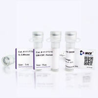
Application
ELISA, FACS, Functional assay


Chemical Information
-
Formulation
Please refer to the lot-specific COA for specific buffer information.
-
Storage
Please store the product under the recommended conditions in the Certificate of Analysis.

Protocols
-
Cell Cytotoxicity Assay
Cytotoxicity assays are usually based on the assessment of cell membrane damage, which can also be indirectly detected by measuring cell viability. Detection methods include MTT assay, CKK-8 assay, LDH assay and ATP assay, etc.
-
Patient-Derived Xenograft (PDX)
Patient-derived xenograft (PDX) models are generated by engrafting primary human tumor tissue directly into immunodeficient mice, allowing in vivo propagation of patient tumor biology without initial in vitro adaptation. These models are used to preserve key histopathological and molecular characteristics of the original tumor and enable assessment of tumor growth dynamics and therapeutic response in a living organism. The biological readout is tumor engraftment and subsequent growth in the murine host, which reflects the ability of human tumor cells to survive, vascularize, and expand in an immunocompromised microenvironment.
-
Patient-Derived Orthotopic Xenograft (PDOX)
Patient-derived orthotopic xenograft (PDOX) modeling implants fresh patient tumor tissue or patient-derived tumor cells into the anatomically corresponding organ or tissue site of immunodeficient mice, usually by surgical orthotopic implantation, to preserve patient tumor histology, local microenvironmental context, invasion, metastatic behavior, and treatment-response features better than subcutaneous implantation. PDOX readouts include tumor engraftment, orthotopic tumor growth, local invasion, metastasis, recurrence after resection, histologic similarity to the donor tumor, biomarker retention, molecular concordance, survival, and response or resistance to therapy. PDOX models are used for preclinical drug testing and individualized therapy evaluation, but engraftment success varies by tumor type and specimen quality.
-
Research Protocol for Inflammation-related Diseases
The NLRP3 inflammasome is a cytosolic innate immune signaling platform that integrates priming signals and danger-signal activation to promote caspase-1 activation, maturation of IL-1β and IL-18, and gasdermin D-mediated pyroptotic cell death. The core experimental logic is to determine whether inflammatory disease phenotypes are driven by increased NLRP3 expression, ASC-containing inflammasome assembly, caspase-1 cleavage, GSDMD cleavage, and extracellular release of IL-1β/IL-18 rather than by nonspecific cell injury alone. The pathway is strongly linked to inflammation-related disease phenotypes because monosodium urate crystals activate NALP3/NLRP3 inflammasome signaling in gout-like crystal inflammation, cholesterol crystals activate NLRP3 inflammasomes in atherogenesis models, and DSS-induced intestinal inflammation has been reported to involve NLRP3 inflammasome activity. However, experimental colitis studies also show context-dependent protective effects of NLRP3 inflammasome co
-
Subcutaneous Cell-Line-Derived Xenograft
Subcutaneous cell-line-derived xenograft (CDX) models are established by implanting cultured human cancer cell lines into immunodeficient mice, where the injected cells form localized tumors that can be monitored in vivo as a measure of tumorigenic potential, growth kinetics, and treatment response. These models are widely used in oncology research because they allow reproducible tumor formation and enable comparative assessment of tumor growth between different cell lines or genetic manipulations in a controlled in vivo microenvironment. Subcutaneous implantation of cancer cells in immunodeficient mice is a standard approach for evaluating tumor growth behavior and therapeutic response across multiple cancer types, including prostate, esophageal, pancreatic, and colon cancer models.
-
Orthotopic Cell-Line Xenograft
Orthotopic cell-line xenograft models involve implantation of human cancer cell lines into the anatomically corresponding organ of immunodeficient mice to reproduce tumor growth within a native microenvironment, enabling more clinically relevant tumor behavior compared with subcutaneous models. These models are widely used because orthotopic placement better recapitulates tumor progression, including invasion and metastatic spread, which are often underrepresented in heterotopic implantation systems. Compared with conventional xenografts, orthotopic implantation is described as more technically complex but provides improved simulation of tumor-microenvironment interactions and metastatic behavior, making it particularly valuable for translational oncology research. Surgical orthotopic implantation approaches have been emphasized as enabling faithful reproduction of clinical cancer features, including metastasis and disease progression patterns that align with the tumor’s organ of origi
-
Cell Viability Determination by MTT Colorimetric Assay
The following protocol uses the MTT colorimetric assay as a classic literature-established method for assessing cell viability/metabolic activity in cultured mammalian cells. MTT[3-(4,5-dimethylthiazol-2-yl)-2,5-diphenyltetrazolium bromide] is reduced by metabolically active cells to a colored formazan product; the amount of formazan is quantified spectrophotometrically and provides an indirect measure of metabolically active viable cells. Importantly, MTT reduction reflects cellular oxidoreductase/metabolic activity rather than an absolute direct count of living cells, so changes in cellular metabolism can alter the signal independently of cell number.


Purity & Documentation
References

Calculators
Concentration (start) × Volume (start) = Concentration (final) × Volume (final)


IO-108 Related Classifications
HY-P992388 Related Classifications
 Powered by Bioz