



SMS121
Based on 5 publication(s) in Google Scholar
SMS121 is a CD36 inhibitor with a KD values of about 5 µM. SMS121 reduces the uptake of lipids and inhibits cell viability in acute myeloid leukemia cells. SMS121 has antitumor activity.
For research use only. We do not sell to patients.
- Purity : 99.06%
- Formula: C20H21NO5
- Molecular Weight:355.38
-
Storage:Powder -20°C, 3 years , 4°C, 2 years ; In solvent -80°C, 6 months , -20°C, 1 month


To place orders, for customer services and technical support, please contact: MedChemExpress USA
Tel: 609-228-6898 E-mail: [email protected] [email protected]


-
 Biological Activity
-
Chemical Information
-
Solvent & Solubility
- Protocols
- Purity & Documentation
- References
-
 Help & FAQs


-
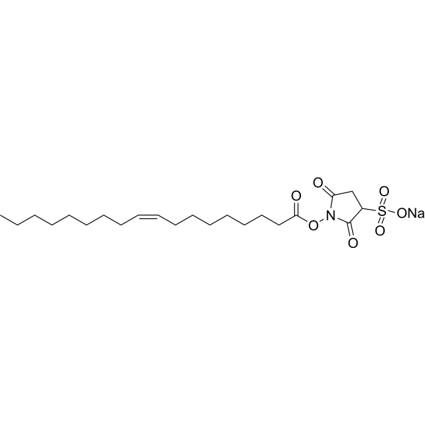
HY-112847A
Sulfosuccinimidyl oleate sodiumFatty Acid Transport Inhibitor -

HY-10071
Y-27632ROCK Inhibitor -
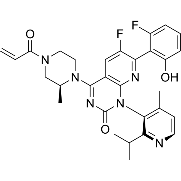
HY-114277
SotorasibKRAS G12C Inhibitor -

HY-15141
StaurosporinePKC/PKA Inhibitor -
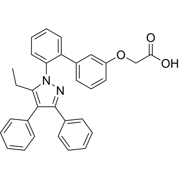
HY-101903
BMS-309403FABP4 Inhibitor -
HY-15859
AtglistatinATGL Inhibitor -

HY-18739
Phorbol 12-myristate 13-acetatePKC Activator -
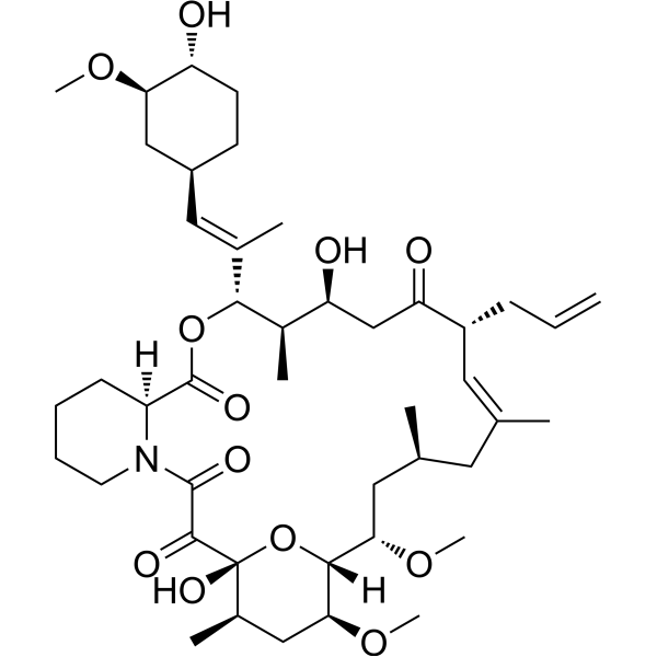
HY-13756
TacrolimusCalcineurin Inhibitor -
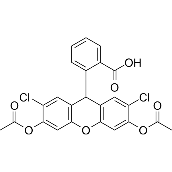
HY-D0940
H2DCFDAROS-Sensitive Probe -
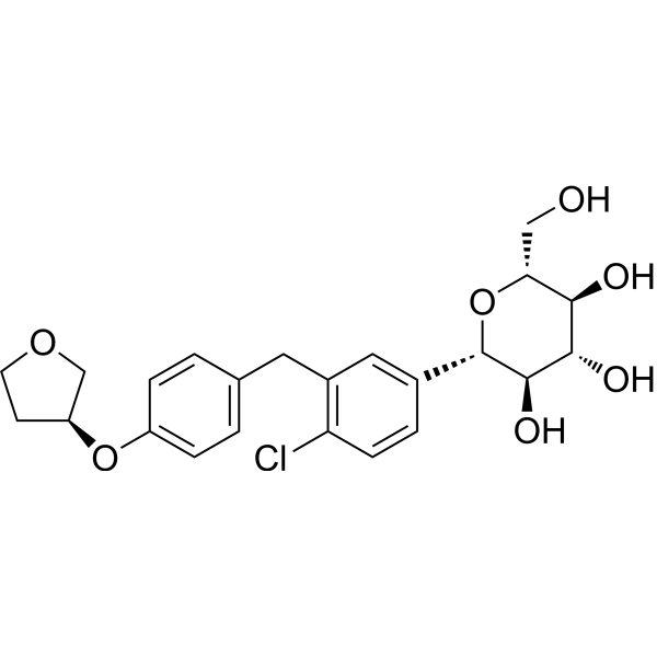
HY-15409
EmpagliflozinSGLT2 Inhibitor -
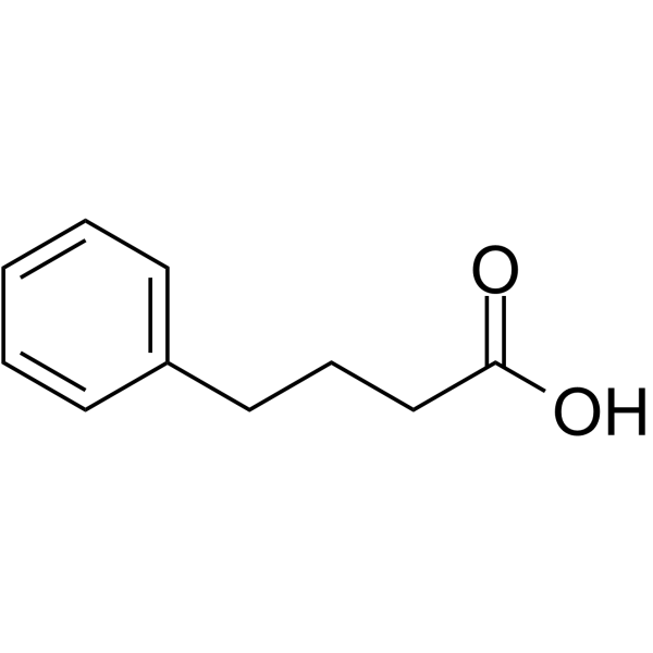
HY-A0281
4-Phenylbutyric acidHDAC Inhibitor -
HY-19312
3-MethyladenineAutophagy/PI3K Inhibitor
Publications Citing Use of MedChemExpress (MCE) SMS121
More Customer Validation & Images

-
Cell Migration/Invasion Assay


Biological Activity
Description
IC50 & Target
CD36[1]
In Vitro
SMS121 (0-500 µM; 50 min) inhibits lipid uptake in KG-1 cells with an IC50 of 164 µM[1].
SMS121 (0-400 µM; 72 h) inhibits the viability of KG-1 cells with an IC50 of 156 µM[1].
MedChemExpress (MCE) has not independently confirmed the accuracy of these methods. They are for reference only.
-
Cell Line:BODIPY fluorescent fatty acid analogue treated KG-1 cell
-
Concentration:200 µM
-
Incubation Time:50 min
-
Result:Significantly reduced green fluorescence compared to control group.


Chemical Information
-
Appearance Solid
-
Molecular Weight 355.38
-
Formula C20H21NO5
-
Color White to yellow
-
SMILES
O=C(O)COC1=CC=C(/C=C/C(C2=CC=C(N(C)C)C=C2)=O)C=C1OC
-
Shipping
Room temperature in continental US; may vary elsewhere.
-
Storage
Powder -20°C 3 years 4°C 2 years In solvent -80°C 6 months -20°C 1 month


Publications (5)
-
Journal Impact Factor
-
Most Recent
-
Cancer Cell
Macrophage-mediated brain-bone marrow crosstalk promotes chronic stress-induced glioma growth. [Abstract]2026 Jun 18:S1535-6108(26)00263-1. PMID: 42314665 -
Cancer Cell
Multiomic integration reveals tumoral heterogeneity of lipid dependence within lethal group 3 medulloblastoma. [Abstract]2026 Feb 9;44(2):383-404.e18. PMID: 41544627 -
J Lipid Res
2026 Mar 16:101023. PMID: 41850682 -
Am J Pathol
Characterizing Kupffer Cell Production of CD5 Antigen-Like and Its Function on Regulating Migration of Natural Killer T Cells. [Abstract]2025 Jul 8:S0002-9440(25)00235-4. PMID: 40639718
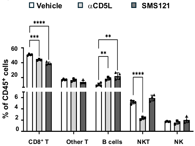
SMS121 purchased from MedChemExpress. Usage Cited in: Am J Pathol. 2025 Jul 8:S0002-9440(25)00235-4. [Abstract]
Transwell assay performed with primary liver immune cells in the insert and LMac in the bottom chamber. Treatment with or without CD5L neutralizing antibody or CD36 inhibitor SMS121 (200 μM) is compared with vehicle treatment (0.1% FBS).
-
iScience
Integrin-linked kinase promotes hepatic fat accumulation via F-actin stabilization-dependent CD36 plasma membrane localization. [Abstract]2026 Jul 14;29(8):116610. PMID: 42519009


Solvent & Solubility
In Vitro:
DMSO : 13.33 mg/mL (37.51 mM; ultrasonic and warming and heat to 60°C; Hygroscopic DMSO has a significant impact on the solubility of product, please use newly opened DMSO)
Please refer to the solubility information to select the appropriate solvent. Once prepared, please aliquot and store the solution to prevent product inactivation from repeated freeze-thaw cycles.
Storage method and period of stock solution: -80°C, 6 months; -20°C, 1 month. When stored at -80°C, please use it within 6 months. When stored at -20°C, please use it within 1 month.
Please refer to the solubility information to select the appropriate solvent. Once prepared, please aliquot and store the solution to prevent product inactivation from repeated freeze-thaw cycles.
Storage method and period of stock solution: -80°C, 6 months; -20°C, 1 month. When stored at -80°C, please use it within 6 months. When stored at -20°C, please use it within 1 month.
Concentration (start) × Volume (start) = Concentration (final) × Volume (final)
In Vivo:
Select the appropriate dissolution method based on your experimental animal and administration route.
- For the following dissolution methods, please ensure to first prepare a clear stock solution using an In Vitro approach and then sequentially add co-solvents:
- To ensure reliable experimental results, the clarified stock solution can be appropriately stored based on storage conditions. As for the working solution for In Vivo experiments, it is recommended to prepare freshly and use it on the same day.
- The percentages shown for the solvents indicate their volumetric ratio in the final prepared solution. If precipitation or phase separation occurs during preparation, heat and/or sonication can be used to aid dissolution.
Add each solvent one by one: 10% DMSO 40% PEG300 5% Tween-80 45% Saline
Solubility: ≥ 1.33 mg/mL (3.74 mM); Clear solution
This protocol yields a clear solution of ≥ 1.33 mg/mL (saturation unknown).
Taking 1 mL working solution as an example, add 100 μL DMSO stock solution (13.3 mg/mL) to 400 μL PEG300, and mix evenly; then add 50 μL Tween-80 and mix evenly; then add 450 μL Saline to adjust the volume to 1 mL.
Preparation of Saline: Dissolve 0.9 g sodium chloride in ddH₂O and dilute to 100 mL to obtain a clear Saline solution.
Add each solvent one by one: 10% DMSO 90% (20% SBE-β-CD in Saline)
Solubility: ≥ 1.33 mg/mL (3.74 mM); Clear solution
This protocol yields a clear solution of ≥ 1.33 mg/mL (saturation unknown).
Taking 1 mL working solution as an example, add 100 μL DMSO stock solution (13.3 mg/mL) to 900 μL 20% SBE-β-CD in Saline, and mix evenly.
Preparation of 20% SBE-β-CD in Saline (4°C, storage for one week): 2 g SBE-β-CD powder is dissolved in 10 mL Saline, completely dissolve until clear.
In Vivo Dissolution Calculator
Please enter the basic information of animal experiments:
-
-
-
-
Recommended: Prepare an additional quantity of animals to account for potential losses during experiments.
Please enter your animal formula composition:
-
%DMSO +
Recommended: Keep the proportion of DMSO in working solution below 2% if your animal is weak.
-
%+
-
+%Tween-80 + +
-
%Saline +
The co-solvents required include: DMSO, . All of co-solvents are available by MedChemExpress (MCE). , Tween 80. All of co-solvents are available by MedChemExpress (MCE).
Working solution concentration: 0.22 mg/mL
Method for preparing stock solution: mg drug dissolved in μL DMSO. Stock solution concentration: mg/mL.
1. Take μL DMSO stock solution;
2. Add μL .
μL , mix evenly;
3. Then add μL Tween 80, mix evenly;
4. Then add μL
Please ensure that the stock solution in the first step is dissolved to a clear state, and add co-solvents in sequence. You can use ultrasonic heating (ultrasonic cleaner, recommended frequency 20-40 kHz), vortexing, etc. to assist dissolution.


Purity & Documentation
-
Data Sheet (271 KB)
-
SDS (251 KB)
- English - EN (251 KB)
- Français - FR (251 KB)
- Deutsch - DE (251 KB)
- Norwegian - NO (251 KB)
- Español - ES (251 KB)
- Swedish - SV (251 KB)
- Italian - IT (251 KB)
- Korean - KR (251 KB)
- Portuguese - PT (251 KB)
-
Handling Instructions (2659 KB)
References


Complete Stock Solution Preparation Table
Please refer to the solubility information to select the appropriate solvent. Once prepared, please aliquot and store the solution to prevent product inactivation from repeated freeze-thaw cycles.
Storage method and period of stock solution: -80°C, 6 months; -20°C, 1 month. When stored at -80°C, please use it within 6 months. When stored at -20°C, please use it within 1 month.
| Optional Solvent | Concentration Solvent Mass | 1 mg | 5 mg | 10 mg | 25 mg |
|---|---|---|---|---|---|
| DMSO | 1 mM | 2.8139 mL | 14.0694 mL | 28.1389 mL | 70.3472 mL |
| 5 mM | 0.5628 mL | 2.8139 mL | 5.6278 mL | 14.0694 mL | |
| 10 mM | 0.2814 mL | 1.4069 mL | 2.8139 mL | 7.0347 mL | |
| 15 mM | 0.1876 mL | 0.9380 mL | 1.8759 mL | 4.6898 mL | |
| 20 mM | 0.1407 mL | 0.7035 mL | 1.4069 mL | 3.5174 mL | |
| 25 mM | 0.1126 mL | 0.5628 mL | 1.1256 mL | 2.8139 mL | |
| 30 mM | 0.0938 mL | 0.4690 mL | 0.9380 mL | 2.3449 mL |


SMS121 Related Classifications
HY-163541 Related Classifications
 Powered by Bioz