



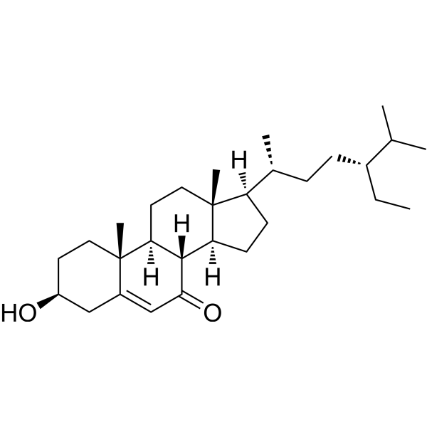
7-Ketositosterol

Based on 1 Customer Validation
7-Ketositosterol is an orally active inducer of apoptosis and ferroptosis. 7-Ketositosterol inhibits the phosphorylation of ERK1/2 and NF-κB, promotes the opening of the mitochondrial/apoptotic pathway, and induces ferroptosis in macrophages by increasing the levels of malondialdehyde, Fe2+ and ROS. 7-Ketositosterol upregulates the expression of gut microbiota-dependent PDLIM3, activates the p38MAPK/NF-κB signaling pathway, alters the composition of gut microbiota, increases the abundance of pathogenic bacteria, and exacerbates colitis in mice. 7-Ketositosterol can be used in studies related to breast cancer, liver cancer, and colitis.
Nos produits utilisent uniquement pour la recherche. Nous ne vendons pas aux patients.
- Pureté : 99.9%
- CAS No.: 2034-74-4
- Formule: C29H48O2
- Masse moléculaire:428.69
-
Stockage:Powder -20°C, 3 years ; In solvent -80°C, 6 months , -20°C, 1 month

To place orders, for customer services and technical support, please contact: MedChemExpress USA
Tel: 609-228-6898 E-mail: [email protected] [email protected]


-
 Activité biologique
-
Chemical Information
-
Solvant et solubilité
- Protocole
- Pureté et documentation
- Références
-
 Help & FAQs




Activité biologique
Description
Cellular Effect
|
Cell Line

|
Type | Value | Description | References |
|---|---|---|---|---|
| A549 | IC50 |
>20 μg/mL
Compound: 8
|
Cytotoxicity against human A549 cells
Cytotoxicity against human A549 cells
|
[PMID: 23511021] |
| BV-2 | IC50 |
>100 μM
Compound: 36
|
Antineuroinflammatory activity in mouse BV2 cells assessed as inhibition of LPS-induced NO production after 24 hrs in presence of LPS by Griess reaction based assay
Antineuroinflammatory activity in mouse BV2 cells assessed as inhibition of LPS-induced NO production after 24 hrs in presence of LPS by Griess reaction based assay
|
[PMID: 28911817] |
| Hep 3B2 | IC50 |
>20 μg/mL
Compound: 8
|
Cytotoxicity against human Hep3B cells
Cytotoxicity against human Hep3B cells
|
[PMID: 23511021] |
| HepG2 | IC50 |
>20 μg/mL
Compound: 8
|
Cytotoxicity against human HepG2 cells
Cytotoxicity against human HepG2 cells
|
[PMID: 23511021] |
| HT-29 | IC50 |
48 μM
Compound: 29
|
Cytotoxicity against human HT-29 cells after 48 hrs by Alamar blue assay
Cytotoxicity against human HT-29 cells after 48 hrs by Alamar blue assay
|
[PMID: 20931970] |
| MCF7 | IC50 |
>20 μg/mL
Compound: 8
|
Cytotoxicity against human MCF7 cells
Cytotoxicity against human MCF7 cells
|
[PMID: 23511021] |
| MDA-MB-231 | IC50 |
>20 μg/mL
Compound: 8
|
Cytotoxicity against human MDA-MB-231 cells
Cytotoxicity against human MDA-MB-231 cells
|
[PMID: 23511021] |
In Vitro
7-Ketositosterol (30 µM; 24 h) reduces viability of human breast MCF-7 and liver HepG2 cancer cells by ~30% and ~25%, respectively, but does not affect viability of non-cancerous BJ fibroblasts[1].
7-Ketositosterol (30 µM; 24 h) significantly suppresses phosphorylation of ERK1/2 and NF-κB p65 (Ser536) in human breast MCF-7 and liver HepG2 cancer cells[1].
7-Ketositosterol (30 µM; 24 h) significantly suppresses PCNA protein levels in human breast MCF-7 and liver HepG2 cancer cells, indicating reduced cell proliferation[1].
7-Ketositosterol (30 µM; 24 h) significantly increases intracellular C18-C24 ceramide levels and reduces S1P levels in human breast MCF-7 and liver HepG2 cancer cells[1].
7-Ketositosterol (30 µM; 24 h) significantly induces apoptosis in human breast MCF-7 and liver HepG2 cancer cells, as measured by TUNEL staining and annexin V-FITC/PI flow cytometry[1].
7-Ketositosterol (1 µM; 24 h) induces M1 polarization and ferroptosis in RAW 264.7 macrophages by disrupting the ALKBH5-GCLM axis, reducing glutathione biosynthesis, and increasing oxidative stress and iron accumulation[2].
7-Ketositosterol does not directly stimulate the growth of Staphylococcus lentus in cell-free bacterial cultures[3].
MedChemExpress (MCE) has not independently confirmed the accuracy of these methods. They are for reference only. Further protocols information, click here.
-
Cell Line:human breast cancer (MCF-7), human liver cancer (HepG2), human non-cancerous fibroblast (BJ) cells
-
Concentration:5 µM, 10 µM, 15 µM, 30 µM
-
Incubation Time:12 h, 18 h, 24 h
-
Result:Did not significantly affect MCF-7 cell viability at 5, 10, or 15 µM after 24 h.
Reduced MCF-7 cell viability by approximately 30% at 30 µM after 24 h.
Did not significantly affect HepG2 cell viability at 5-30 µM for 12-18 h.
Reduced HepG2 cell viability by approximately 25% at 30 µM after 24 h.
Did not significantly affect BJ fibroblast cell viability at 5-30 µM after 24 h.
-
Cell Line:human breast cancer (MCF-7), human liver cancer (HepG2) cells
-
Concentration:30 µM
-
Incubation Time:24 h
-
Result:Increased fluorescence intensity (indicating apoptotic DNA fragmentation) significantly in both MCF-7 and HepG2 cells compared to control and DMSO groups via TUNEL staining.
Increased the percentage of early and late apoptotic cells significantly in both cell lines, with a corresponding decrease in viable cell percentage via annexin V-FITC/PI flow cytometry.
In Vivo
MedChemExpress (MCE) has not independently confirmed the accuracy of these methods. They are for reference only.
-
Animal Model:C57BL/6 (female, 7 weeks old, DSS-induced ulcerative colitis)[2]
-
Dosage:20 mg/kg/day; 60 mg/kg/day; 100 mg/kg/day
-
Administration:p.o.; daily; 21 days
-
Result:Exacerbated DSS-induced colitis at 100 mg/kg/day, with body weight reduced to ~87% of initial weight by day 7 of DSS exposure (vs ~95% for DSS-only mice).
Elevated DAI scores to ~9 by day 7 of DSS exposure (vs ~6 for DSS-only mice) at 100 mg/kg/day.
Shortened colon length to ~4.8 cm (vs ~5.8 cm for DSS-only mice) at 100 mg/kg/day.
Increased histopathological injury scores to ~8 (vs ~5 for DSS-only mice) at 100 mg/kg/day.
Induced intestinal inflammation and mucosal damage in mice without DSS challenge at 100 mg/kg/day.
Recapitulated exacerbating effects at 60 mg/kg/day, significantly worsening colitis severity compared to DSS-only mice.
Showed no significant alteration of body weight, DAI scores, or colon length compared to DSS-only mice at 20 mg/kg/day.
Increased mRNA expression of M1 macrophage-associated markers (IL-1β, TNF-α, iNOS, CD80) in colon tissue at 100 mg/kg/day.
Increased proportion of CD86-positive M1 macrophages at 100 mg/kg/day.
Reduced proportion of CD206-positive M2 macrophages at 100 mg/kg/day.
Promoted CD3+ T-cell recruitment in inflamed colons relative to DSS-only mice at 100 mg/kg/day.


Chemical Information
-
CAS No. 2034-74-4
-
Appearance Solid
-
Masse moléculaire 428.69
-
Formule C29H48O2
-
Color White to off-white
-
SMILES
C[C@@]12[C@](CC[C@]2([H])[C@H](C)CC[C@@H](CC)C(C)C)([H])[C@@]3([H])[C@@](CC1)([H])[C@@]4(C(C[C@H](CC4)O)=CC3=O)C
-
Structure Classification
-
Initial Source
-
Livraison
Room temperature in continental US; may vary elsewhere.
-
Stockage
Powder -20°C 3 years In solvent -80°C 6 months -20°C 1 month


Solvant et solubilité
In Vitro:
DMF : 12 mg/mL (27.99 mM; Need ultrasonic and warming)
Please refer to the solubility information to select the appropriate solvent. Once prepared, please aliquot and store the solution to prevent product inactivation from repeated freeze-thaw cycles.
Storage method and period of stock solution: -80°C, 6 months; -20°C, 1 month. When stored at -80°C, please use it within 6 months. When stored at -20°C, please use it within 1 month.
Please refer to the solubility information to select the appropriate solvent. Once prepared, please aliquot and store the solution to prevent product inactivation from repeated freeze-thaw cycles.
Storage method and period of stock solution: -80°C, 6 months; -20°C, 1 month. When stored at -80°C, please use it within 6 months. When stored at -20°C, please use it within 1 month.
Concentration (start) × Volume (start) = Concentration (final) × Volume (final)

Protocole
-
Western Blot
Western blotting (WB) is a commonly used experimental method in molecular biology, biochemistry, and immunogenetics for identifying and quantifying target proteins. It combines gel electrophoresis with immunoassay, enabling researchers to analyze protein expression, post-translational modifications, and molecular weight.
-
RNA extraction experimental
By lysing cells, releasing RNA, and removing impurities such as proteins and DNA, high-purity RNA products are finally obtained. The commonly used traditional method is the guanidine isothiocyanate/phenol/chloroform method (Trizol), which is suitable for a variety of animal materials including animal tissues, microorganisms, cultured cells, etc., and most plant materials.
-
Annexin V plus membrane-impermeant dye apoptosis staining
Annexin V-based apoptosis assays rely on the detection of phosphatidylserine (PS) externalization from the inner leaflet of the plasma membrane to the outer leaflet, an early biochemical hallmark of apoptosis. Fluorescently labeled Annexin V binds PS in a calcium-dependent manner, enabling identification of early apoptotic cells by flow cytometry or fluorescence microscopy. When combined with a membrane-impermeant DNA-binding dye (e. g. , propidium iodide), this approach allows discrimination between viable (Annexin V−/dye−), early apoptotic (Annexin V+/dye−), and late apoptotic or necrotic (Annexin V+/dye+) cell populations by assessing membrane integrity and PS exposure.
-
ROS/oxidative-stress fluorescent staining
ROS/oxidative-stress fluorescent staining uses cell-permeant fluorogenic probes that become fluorescent after oxidation inside cells or tissues; commonly used examples include DCFH-DA/DCFDA for broad cellular oxidant detection, DHE for superoxide-related signal detection, MitoSOX for mitochondrial superoxide-related signal detection, and CellROX probes for oxidative-stress-associated fluorescence readouts. The assay detects probe oxidation rather than a single ROS species unless the probe and analysis method have been chemically validated for that species. DCFH-DA enters cells, is deacetylated by intracellular esterases to DCFH, and produces fluorescent DCF after oxidation, so the readout is used as an operational measure of total cellular oxidative stress rather than a species-specific ROS measurement. DHE and MitoSOX can report superoxide-related oxidation, but red fluorescence alone can include non-specific ethidium-like oxidation products; HPLC or optimized spectral approaches are
-
Research Protocol for Microbiome Analysis
Microbiome analysis characterizes microbial communities in biological or environmental samples by measuring community composition, diversity, taxonomic structure, functional potential, and associations with host or environmental phenotypes. 16S rRNA gene amplicon sequencing is commonly used for bacterial and archaeal taxonomic profiling, while shotgun metagenomics provides higher taxonomic resolution and direct functional information, including microbial genes, pathways, viruses, fungi, and antimicrobial-resistance genes when sequencing depth and host-DNA contamination are adequately controlled. Microbiome results are strongly affected by sample collection, storage, DNA extraction, contamination, sequencing method, reference database, and bioinformatic pipeline; therefore, standardized protocols, negative controls, mock communities, and transparent analysis workflows are required. Unresolved issues include low-biomass contamination, compositional-data bias, inconsistent species-level c
-
Research Protocol for Inflammation-related Diseases
The NLRP3 inflammasome is a cytosolic innate immune signaling platform that integrates priming signals and danger-signal activation to promote caspase-1 activation, maturation of IL-1β and IL-18, and gasdermin D-mediated pyroptotic cell death. The core experimental logic is to determine whether inflammatory disease phenotypes are driven by increased NLRP3 expression, ASC-containing inflammasome assembly, caspase-1 cleavage, GSDMD cleavage, and extracellular release of IL-1β/IL-18 rather than by nonspecific cell injury alone. The pathway is strongly linked to inflammation-related disease phenotypes because monosodium urate crystals activate NALP3/NLRP3 inflammasome signaling in gout-like crystal inflammation, cholesterol crystals activate NLRP3 inflammasomes in atherogenesis models, and DSS-induced intestinal inflammation has been reported to involve NLRP3 inflammasome activity. However, experimental colitis studies also show context-dependent protective effects of NLRP3 inflammasome co
-
Liver Cancer Modeling
Liver cancer can be classified into primary liver cancer and secondary liver cancer. Secondary liver cancer is the metastatic liver cancer. Primary liver cancer includes hepatocellular carcinoma (HCC), intrahepatic cholangiocarcinoma (ICC) and fibrolamellar HCC, of which HCC is the most common form, accounting for approximately 90% of primary liver cancers[1]. HCC mouse models include chemical agent-induced models, transplanted tumor models, and genetic engineered models.
-
Breast Cancer Modeling
Breast cancer is a heterogeneous cancer, and it has been distinguished into four subtypes: luminal A, luminal B, HER2-positive and basal-like. Molecular mutations, epigenetic alterations, hormone exposure and immune microenvironment are related to the progression of breast cancer.
-
Kinase activity and phosphorylation assays
Kinase activity assays measure the ability of kinases to transfer phosphate groups from ATP to specific substrates, while phosphorylation assays detect the presence and levels of phosphorylated proteins. Common methods include radiolabeled ATP incorporation (e. g. ,), ADP release detection via bioluminescence (e. g. ,[3]), enzyme-linked immunosorbent assays (ELISA) for phospho-specific epitopes (e. g. ,[6]), and microtiter-based formats for high-throughput screening (e. g. ,[8]). The ADP-Glo assay quantifies kinase activity by measuring ADP produced during phosphorylation using a luciferase-based system. Radiometric assays involve autoradiography or scintillation counting after incorporation of 32P-labeled ATP into substrate proteins. ELISA-based approaches rely on phospho-specific antibodies to detect activated kinases in cell lysates or purified samples.
-
Apoptosis
Apoptosis, also called programmed cell death, is generally characterized by distinct morphological characteristics.
-
TUNEL staining for apoptotic DNA fragmentation
TUNEL staining detects DNA strand breaks by using terminal deoxynucleotidyl transferase to add labeled nucleotides to exposed 3′-OH DNA termini, generating either microscopic staining in fixed cells or tissue sections, or fluorescence/cytometric signal in cell suspensions. TUNEL positivity reflects DNA fragmentation but should not be interpreted alone as definitive apoptosis, because TUNEL can also label necrotic, autolytic, mechanically damaged, or DNA-repair-associated DNA breaks.
-
DSS-Induced Colitis
Dextran sulfate sodium (DSS)-induced colitis is generated by administering DSS in mouse drinking water, producing epithelial injury, barrier disruption, weight loss, diarrhea, fecal blood, colon shortening, histologic mucosal damage, and inflammatory mediator changes; the model is mainly used to study acute or chronic intestinal inflammation resembling selected features of ulcerative colitis. DSS injury is interpreted through clinical and tissue readouts rather than a single molecular endpoint: daily body weight, stool consistency, and bleeding are combined into a disease activity index, while colon length, histology, cytokines, myeloperoxidase activity, intestinal permeability, and tight-junction markers provide complementary measures of inflammation and barrier damage.
-
TNBS-Induced Colitis
TNBS-induced colitis is produced by intrarectal delivery of 2,4,6-trinitrobenzene sulfonic acid in ethanol, where ethanol disrupts the mucosal barrier and TNBS haptenates colonic proteins, generating immune-mediated colonic inflammation with weight loss, diarrhea, ulceration, transmural injury, inflammatory-cell infiltration, and cytokine responses. The model is used as an experimental intestinal inflammation model with Crohn’s disease–like features, especially when Th1-type responses, IL-12–dependent inflammation, chronic relapsing inflammation, or fibrosis-related endpoints are studied.
-
Apoptosis Solutions
Apoptosis is a regulated, generally non-lytic cell-death pathway that removes unwanted, damaged, infected, or abnormal cells through coordinated morphological changes, caspase activation, DNA fragmentation, and membrane remodeling. The intrinsic apoptosis pathway is controlled mainly by mitochondrial outer membrane permeabilization, BCL-2 family proteins, cytochrome c release, apoptosome formation, caspase-9 activation, and downstream executioner caspase-3/7 activation. The extrinsic apoptosis pathway is initiated by death receptors such as Fas, TNFR, and TRAIL receptors, which recruit adaptor proteins and activate caspase-8 before engaging executioner caspases or mitochondrial amplification through BID cleavage. Apoptosis is linked to many phenotypes, including cancer cell killing, tissue homeostasis, immune regulation, neurodegeneration, infection response, and treatment-induced cytotoxicity; unresolved questions include how apoptosis interacts with necroptosis, pyroptosis, ferroptos
-
Ferroptosis Solutions
Ferroptosis is an iron-dependent, non-apoptotic form of regulated cell death characterized by lethal lipid peroxidation and sensitivity to suppression by iron chelators or lipophilic radical-trapping antioxidants. The core pathway links cystine uptake through system Xc−, glutathione availability, GPX4-dependent detoxification of phospholipid hydroperoxides, iron-dependent oxidative reactions, and polyunsaturated-phospholipid metabolism into a cell-death program that is biochemically and morphologically distinct from apoptosis, necrosis, and autophagy. The ferroptosis pathway is experimentally linked to phenotype through chemical and genetic perturbation. Erastin induces ferroptosis by inhibiting cystine uptake through system Xc− and weakening antioxidant defenses, while GPX4 inhibition or depletion causes lipid peroxide accumulation and ferroptotic cancer-cell death. ACSL4 and oxidizable arachidonoyl- or adrenoyl-containing phosphatidylethanolamines shape ferroptosis sensitivity by con
-
Protocol for Kinase activity and phosphorylation assays
Kinase activity assays measure transfer of phosphate from ATP to a protein or peptide substrate, generating phosphorylated substrate, ADP, or incorporated radiolabeled phosphate as the readout; phosphorylation assays measure site-specific phosphorylation in cells or tissues as a proxy for kinase-pathway activation, inhibition, or substrate regulation. Phosphorylation can be detected by phospho-specific Western blot, immunoprecipitation kinase assay, phospho-immunofluorescence, phospho-flow cytometry, luminescent ADP detection, radiolabeled ATP incorporation, or reporter-based pathway assays, and these readouts can be applied to cancer cells, primary neurons, mouse tumors, organoids, inflammatory macrophages, ferroptosis studies, and mitophagy studies when the kinase target is biologically relevant.


Pureté et documentation
-
Fiche technique (280 KB)
-
SDS (251 KB)
- English - EN (251 KB)
- Français - FR (251 KB)
- Deutsch - DE (251 KB)
- Norwegian - NO (251 KB)
- Español - ES (251 KB)
- Swedish - SV (251 KB)
- Italian - IT (251 KB)
- Korean - KR (251 KB)
- Portuguese - PT (251 KB)
-
Instruction de manipulation (2659 KB)
Références
[1]. Barut Z, et al. Antiproliferative Effect of 7-Ketositosterol in Breast and Liver Cancer Cells: Possible Impact on Ceramide, Extracellular Signal-Regulated Kinases, and Nuclear Factor Kappa B Signaling Pathways. Pharmaceuticals (Basel, Switzerland). 2024 Jul 01;17(7):860. [Content Brief]
[2]. Pang X, et al. Excessive ultra-processed foods exposure aggravates ulcerative colitis via macrophage ferroptosis. Environment international. 2025 Aug;202:109706. [Content Brief]
[3]. Yan J, et al. Ultra-processed foods sourced 7-ketositosterol aggravates colitis through gut dysbiosis induced-PDLIM3 activation. Gut microbes. 2025 Dec 31;17(1):2587980. [Content Brief]


Complete Stock Solution Preparation Table
Please refer to the solubility information to select the appropriate solvent. Once prepared, please aliquot and store the solution to prevent product inactivation from repeated freeze-thaw cycles.
Storage method and period of stock solution: -80°C, 6 months; -20°C, 1 month. When stored at -80°C, please use it within 6 months. When stored at -20°C, please use it within 1 month.
| Optional Solvent | Concentration Solvent Mass | 1 mg | 5 mg | 10 mg | 25 mg |
|---|---|---|---|---|---|
| DMF | 1 mM | 2.3327 mL | 11.6634 mL | 23.3269 mL | 58.3172 mL |
| 5 mM | 0.4665 mL | 2.3327 mL | 4.6654 mL | 11.6634 mL | |
| 10 mM | 0.2333 mL | 1.1663 mL | 2.3327 mL | 5.8317 mL | |
| 15 mM | 0.1555 mL | 0.7776 mL | 1.5551 mL | 3.8878 mL | |
| 20 mM | 0.1166 mL | 0.5832 mL | 1.1663 mL | 2.9159 mL | |
| 25 mM | 0.0933 mL | 0.4665 mL | 0.9331 mL | 2.3327 mL |


7-Ketositosterol Related Classifications
HY-N2757 Related Classifications
 Powered by Bioz