



ADPM06

Based on 1 Customer Validation
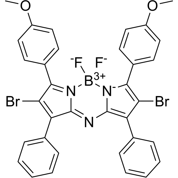
ADPM06, a lead candidate azadipyrromethene, is a novel nonporphyrin photodynamic therapeutic (PDT) agent. ADPM06 exhibits IC50 values in the micro-molar range in human tumor cells and induces apoptosis.
Nos produits utilisent uniquement pour la recherche. Nous ne vendons pas aux patients.
- Pureté : 95.0%
- CAS No.: 490035-90-0
- Formule: C34H24BBr2F2N3O2
- Masse moléculaire:715.19
-
Stockage:Powder -20°C, 3 years , 4°C, 2 years ; In solvent -80°C, 6 months , -20°C, 1 month

To place orders, for customer services and technical support, please contact: MedChemExpress USA
Tel: 609-228-6898 E-mail: [email protected] [email protected]


-
 Activité biologique
-
Chemical Information
-
Solvant et solubilité
- Protocole
- Pureté et documentation
- Références
-
 Help & FAQs




Activité biologique
Description
Cellular Effect
|
Cell Line

|
Type | Value | Description | References |
|---|---|---|---|---|
| HeLa | IC50 |
>50 μM
Compound: 1; ADPM06
|
Dark toxicity in human HeLa cells assessed as reduction in cell viability after 3 hrs by MTT assay
Dark toxicity in human HeLa cells assessed as reduction in cell viability after 3 hrs by MTT assay
|
[PMID: 32787080] |
| HeLa | IC50 |
0.13 μM
Compound: 1; ADPM06
|
Phototoxicity against human HeLa cells assessed as reduction in cell viability preincubated for 3 hrs followed by 54 J/cm2 light irradiation and measured after 24 hrs by MTT assay
Phototoxicity against human HeLa cells assessed as reduction in cell viability preincubated for 3 hrs followed by 54 J/cm2 light irradiation and measured after 24 hrs by MTT assay
|
[PMID: 32787080] |
| HeLa | IC50 |
41.7 μM
Compound: BDP-4
|
Dark cytotoxicity against human HeLa cells assessed as inhibition of cell viability incubated for 3 hrs by CCK8 assay
Dark cytotoxicity against human HeLa cells assessed as inhibition of cell viability incubated for 3 hrs by CCK8 assay
|
[PMID: 39167079] |
| HeLa | IC50 |
64.2 nM
Compound: BDP-4
|
Phototoxicity against human HeLa cells assessed as inhibition of cell viability preincubated for 3 hrs followed by photo-irradiation of 54 J/cm2 for 24 hrs by CCK8 assay
Phototoxicity against human HeLa cells assessed as inhibition of cell viability preincubated for 3 hrs followed by photo-irradiation of 54 J/cm2 for 24 hrs by CCK8 assay
|
[PMID: 39167079] |
| MCF7 | IC50 |
>50 μM
Compound: 1; ADPM06
|
Dark toxicity in human MCF7 cells assessed as reduction in cell viability after 3 hrs by MTT assay
Dark toxicity in human MCF7 cells assessed as reduction in cell viability after 3 hrs by MTT assay
|
[PMID: 32787080] |
| MCF7 | IC50 |
0.15 μM
Compound: 1; ADPM06
|
Phototoxicity against human MCF7 cells assessed as reduction in cell viability preincubated for 3 hrs followed by 54 J/cm2 light irradiation and measured after 24 hrs by MTT assay
Phototoxicity against human MCF7 cells assessed as reduction in cell viability preincubated for 3 hrs followed by 54 J/cm2 light irradiation and measured after 24 hrs by MTT assay
|
[PMID: 32787080] |
| SW480 | IC50 |
>50 μM
Compound: 1; ADPM06
|
Dark toxicity in human SW480 cells assessed as reduction in cell viability after 3 hrs by MTT assay
Dark toxicity in human SW480 cells assessed as reduction in cell viability after 3 hrs by MTT assay
|
[PMID: 32787080] |
| SW480 | IC50 |
0.16 μM
Compound: 1; ADPM06
|
Phototoxicity against human SW480 cells assessed as reduction in cell viability preincubated for 3 hrs followed by 54 J/cm2 light irradiation and measured after 24 hrs by MTT assay
Phototoxicity against human SW480 cells assessed as reduction in cell viability preincubated for 3 hrs followed by 54 J/cm2 light irradiation and measured after 24 hrs by MTT assay
|
[PMID: 32787080] |
In Vitro
The efficacy of ADPM01 is completely ablated at a 1% oxygen level in Hela and MRC5 cell lines. ADPM06 displays only a partial reduction in light-induced activity in hypoxic as compared to normoxic conditions[1].
ADPM06-PDT induces ER stress and unfolded protein response[2].
ADPM06-PDT induces apoptosis and involves caspase enzymatic activity[2].
Following ADPM06-PDT, a rapid processing of XBP1 mRNA occurs resulting in the removal of an intron from the mRNA in a spliceosome-independent manner, a post-transcriptional modification catalyzed by the action of activated inositol-requiring protein 1 (IRE1)[2].
ADPM06-PDT-induced apoptosis involves the generation of ROS[2].
MedChemExpress (MCE) has not independently confirmed the accuracy of these methods. They are for reference only. Further protocols information, click here.
-
Cell Line:Hela and MRC5 cell lines.
-
Concentration:1 nM - 100μM.
-
Incubation Time:24 h.
-
Result:Retained considerable efficacy, with EC50 values of 1.5 and 1.6 × 10⁻⁶ M for HeLa and MRC5 cells, respectively.
In Vivo
ADPM06-PDT is well tolerated in vivo and elicits impressive complete response rates in various models of cancer when a short drug-light interval is applied[2].
MedChemExpress (MCE) has not independently confirmed the accuracy of these methods. They are for reference only.
-
Animal Model:Female Balb C nu/nu mice[2].
-
Dosage:2 mg/kg in 0.3 mL solution via the lateral tail vein.
-
Administration:IV.
-
Result:Revealed a rapid reduction in tumor-specific luciferase activity as early as 1-hr post-PDT, with levels decreasing further 4-hr post-PDT.


Chemical Information
-
CAS No. 490035-90-0
-
Appearance Solid
-
Masse moléculaire 715.19
-
Formule C34H24BBr2F2N3O2
-
Color Brown to black
-
SMILES
[F-][B+3]1([N]2=C(C3=CC=C(OC)C=C3)C(Br)=C(C4=CC=CC=C4)C2=NC5=C(C(Br)=C([N-]51)C6=CC=C(OC)C=C6)C7=CC=CC=C7)[F-]
-
Livraison
Room temperature in continental US; may vary elsewhere.
-
Stockage
Powder -20°C 3 years 4°C 2 years In solvent -80°C 6 months -20°C 1 month


Solvant et solubilité
In Vitro:
DMSO : < 1 mg/mL (insoluble or slightly soluble)
In Vivo:
For the following dissolution methods, please prepare the working solution directly:
It is recommended to prepare fresh solutions and use them promptly within a short period of time.
The percentages shown for the solvents indicate their volumetric ratio in the final prepared solution. If precipitation or phase separation occurs during preparation, heat and/or sonication can be used to aid dissolution.
Add each solvent one by one: 10% Cremophor EL in PBS
Solubility: 2 mg/mL (2.80 mM); Suspended solution; Need ultrasonic

Protocole
-
Detection of Apoptosis
Apoptosis, also called programmed cell death, is generally characterized by distinct morphological characteristics.
-
TUNEL staining for apoptotic DNA fragmentation
TUNEL staining detects DNA strand breaks by using terminal deoxynucleotidyl transferase to add labeled nucleotides to exposed 3′-OH DNA termini, generating either microscopic staining in fixed cells or tissue sections, or fluorescence/cytometric signal in cell suspensions. TUNEL positivity reflects DNA fragmentation but should not be interpreted alone as definitive apoptosis, because TUNEL can also label necrotic, autolytic, mechanically damaged, or DNA-repair-associated DNA breaks.
-
Annexin V plus membrane-impermeant dye apoptosis staining
Annexin V-based apoptosis assays rely on the detection of phosphatidylserine (PS) externalization from the inner leaflet of the plasma membrane to the outer leaflet, an early biochemical hallmark of apoptosis. Fluorescently labeled Annexin V binds PS in a calcium-dependent manner, enabling identification of early apoptotic cells by flow cytometry or fluorescence microscopy. When combined with a membrane-impermeant DNA-binding dye (e. g. , propidium iodide), this approach allows discrimination between viable (Annexin V−/dye−), early apoptotic (Annexin V+/dye−), and late apoptotic or necrotic (Annexin V+/dye+) cell populations by assessing membrane integrity and PS exposure.
-
Apoptosis Solutions
Apoptosis is a regulated, generally non-lytic cell-death pathway that removes unwanted, damaged, infected, or abnormal cells through coordinated morphological changes, caspase activation, DNA fragmentation, and membrane remodeling. The intrinsic apoptosis pathway is controlled mainly by mitochondrial outer membrane permeabilization, BCL-2 family proteins, cytochrome c release, apoptosome formation, caspase-9 activation, and downstream executioner caspase-3/7 activation. The extrinsic apoptosis pathway is initiated by death receptors such as Fas, TNFR, and TRAIL receptors, which recruit adaptor proteins and activate caspase-8 before engaging executioner caspases or mitochondrial amplification through BID cleavage. Apoptosis is linked to many phenotypes, including cancer cell killing, tissue homeostasis, immune regulation, neurodegeneration, infection response, and treatment-induced cytotoxicity; unresolved questions include how apoptosis interacts with necroptosis, pyroptosis, ferroptos


Pureté et documentation
-
Fiche technique (287 KB)
-
SDS (252 KB)
- English - EN (252 KB)
- Français - FR (252 KB)
- Deutsch - DE (252 KB)
- Norwegian - NO (252 KB)
- Español - ES (252 KB)
- Swedish - SV (252 KB)
- Italian - IT (252 KB)
- Korean - KR (252 KB)
- Portuguese - PT (252 KB)
-
Instruction de manipulation (2659 KB)
Références
[1]. W M Gallagher, et al. A potent nonporphyrin class of photodynamic therapeutic agent: cellular localisation, cytotoxic potential and influence of hypoxia. Br J Cancer. 2005 May 9; 92(9): 1702-1710. [Content Brief]
[2]. Aisling E O'Connor, et al. Mechanism of cell death mediated by a BF2-chelated tetraaryl-azadipyrromethene photodynamic therapeutic: dissection of the apoptotic pathway in vitro and in vivo. Int J Cancer. 2012 Feb 1;130(3):705-15. [Content Brief]

Calculators
Concentration (start) × Volume (start) = Concentration (final) × Volume (final)


ADPM06 Related Classifications
HY-13547 Related Classifications
 Powered by Bioz