



Diethylstilbestrol
Based on 3 publication(s) in Google Scholar
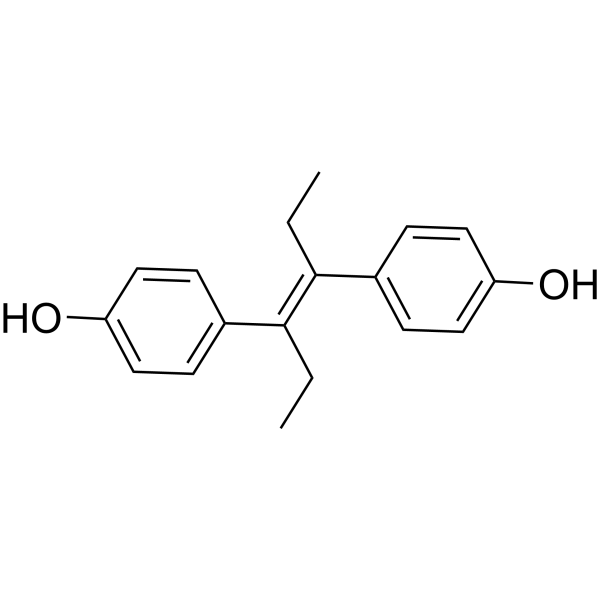
Diethylstilbestrol is a non-steroidal female hormone that has oral activity and can act on menopausal and postmenopausal disorders. Diethylstilbestrol can induce DNA oxidation and Apoptosis of spermatogonial stem cells. Diethylstilbestrol can induce thymocyte Autophagy Diethylstilbestrol is a 11β-hydroxysteroid dehydrogenase 2 (HSD11B2) inhibitor..
Nos produits utilisent uniquement pour la recherche. Nous ne vendons pas aux patients.
- Pureté : 99.62%
- CAS No.: 56-53-1
- Formule: C18H20O2
- Masse moléculaire:268.35
-
Stockage:Powder -20°C, 3 years , 4°C, 2 years ; In solvent -80°C, 2 years , -20°C, 1 year

To place orders, for customer services and technical support, please contact: MedChemExpress USA
Tel: 609-228-6898 E-mail: [email protected] [email protected]


-
 Activité biologique
-
Chemical Information
-
Solvant et solubilité
- Protocole
- Pureté et documentation
- Références
-
 Help & FAQs


-
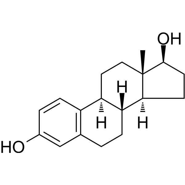
HY-B0141
EstradiolHuman Endogenous Metabolite -
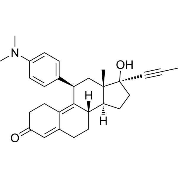
HY-13683
MifepristoneProgesterone Receptor Antagonist -
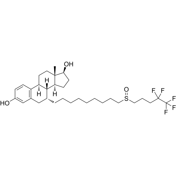
HY-13636
FulvestrantEstrogen Receptor Antagonist -
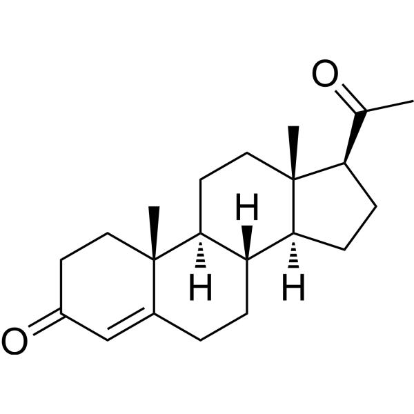
HY-N0437
ProgesteroneEndogenous Metabolite -
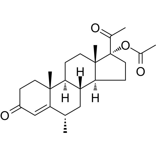
HY-B0469
Medroxyprogesterone acetateProgesterone Receptor Agonist -
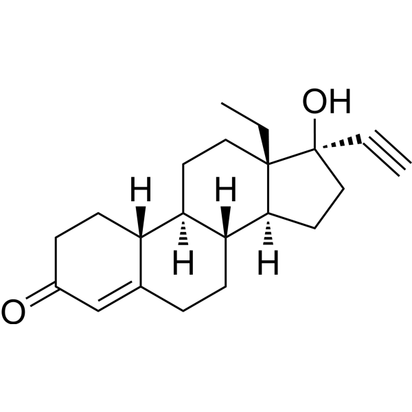
HY-B0257
LevonorgestrelProgesterone Inhibitor -
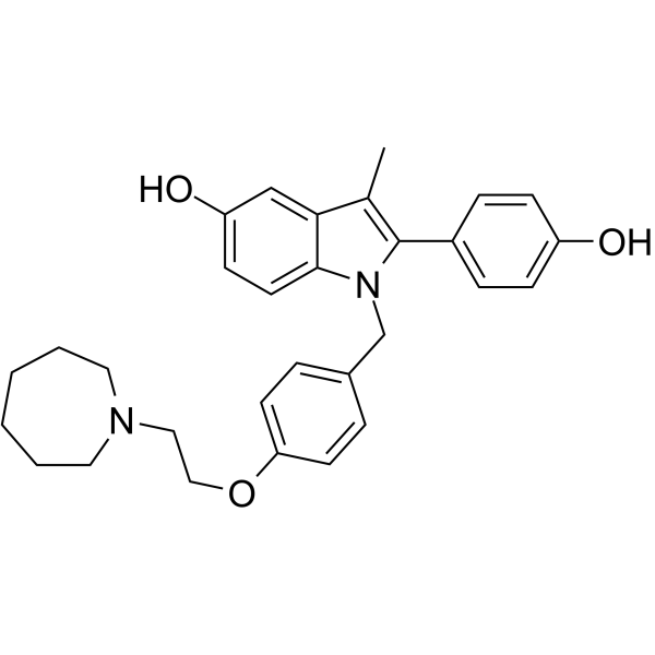
HY-A0031
BazedoxifeneSERM -
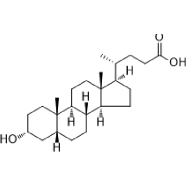
HY-B0172
Lithocholic acidAutophagy Inducer -

HY-14598R
Diethylstilbestrol (Standard)Estrogen Receptor/ERR Agonist -
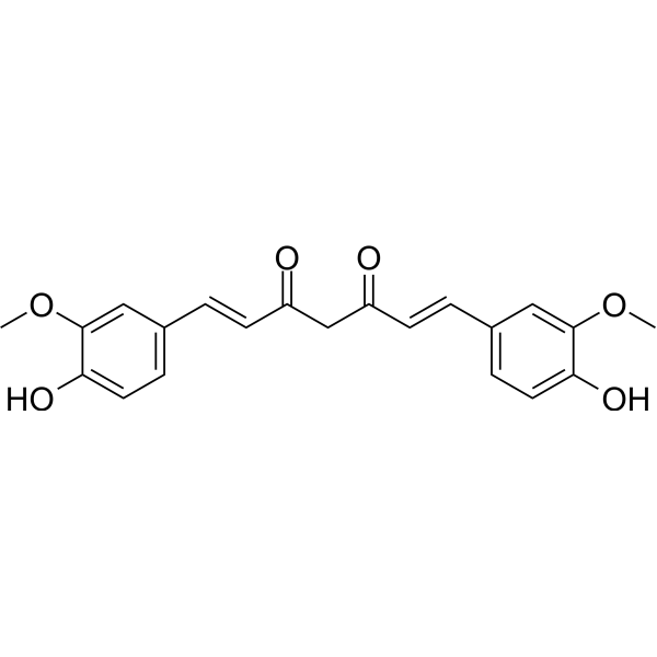
HY-N0005
Curcuminp300 Histone Acetylatransferase Inhibitor -
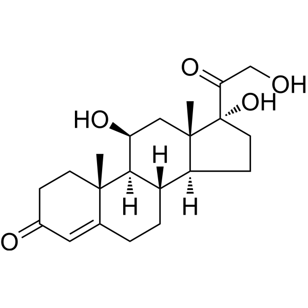
HY-N0583
HydrocortisoneGlucocorticoid Receptor Agonist -
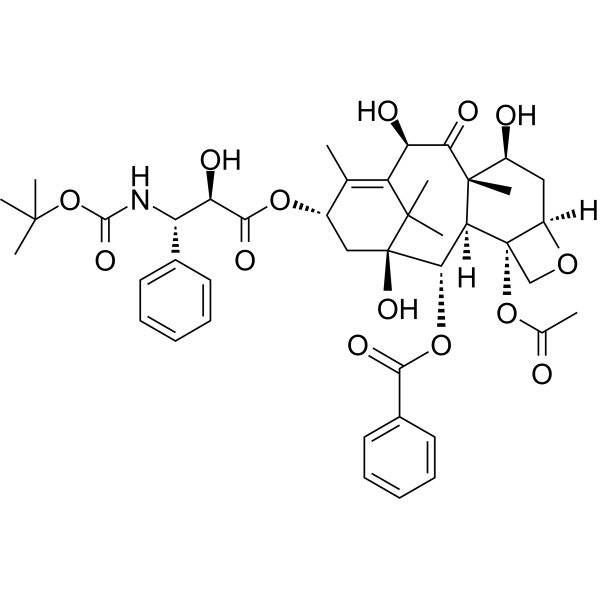
HY-B0011
DocetaxelMicrotubule Depolymerization Inhibitor
Publications Citing Use of MedChemExpress (MCE) Diethylstilbestrol
More

Activité biologique
Description
Cellular Effect
|
Cell Line

|
Type | Value | Description | References |
|---|---|---|---|---|
| ANN-1 | IC50 |
3 μM
Compound: 15b, Diethylstilbestrol
|
Cytotoxic effect on v-abl transformed murine ANN-1 cells
Cytotoxic effect on v-abl transformed murine ANN-1 cells
|
[PMID: 8201603] |
| Caco-2 | IC50 |
75 μM
Compound: 6
|
Antiproliferative activity against human Caco2 cells
Antiproliferative activity against human Caco2 cells
|
[PMID: 26539626] |
| CHO-K1 | EC50 |
8.5 μM
Compound: 3
|
Cytotoxicity against CHO-K1 cells by Alamar blue assay
Cytotoxicity against CHO-K1 cells by Alamar blue assay
|
[PMID: 16962325] |
| HEK293 | EC50 |
0.11 nM
Compound: DES
|
Selective estrogen receptor down-regulator activity at FLAG-tagged ERalpha (unknown origin) expressed in HEK293 cells assessed as induction of ERalpha degradation by luciferase reporter gene assay
Selective estrogen receptor down-regulator activity at FLAG-tagged ERalpha (unknown origin) expressed in HEK293 cells assessed as induction of ERalpha degradation by luciferase reporter gene assay
|
[PMID: 30940565] |
| HEK293 | EC50 |
0.2 nM
Compound: DES
|
Agonist activity at FLAG-tagged ERalpha (unknown origin) expressed in HEK293 cells assessed as induction of ER-alpha-mediated transcriptional activity by luciferase reporter gene assay
Agonist activity at FLAG-tagged ERalpha (unknown origin) expressed in HEK293 cells assessed as induction of ER-alpha-mediated transcriptional activity by luciferase reporter gene assay
|
[PMID: 30940565] |
| HeLa | EC50 |
0.02 nM
Compound: diethylstilbestrol
|
Activation of estrogen response element in HeLa cells stably transfected with human Estrogen receptor beta.
Activation of estrogen response element in HeLa cells stably transfected with human Estrogen receptor beta.
|
[PMID: 11906280] |
| HeLa | EC50 |
0.06 nM
Compound: diethylstilbestrol
|
Activation of estrogen response element in HeLa cells stably transfected with human Estrogen receptor alpha.
Activation of estrogen response element in HeLa cells stably transfected with human Estrogen receptor alpha.
|
[PMID: 11906280] |
| HepG2 | IC50 |
14.6 μM
Compound: 2; DES
|
Antagonist activity at full length human PXR transfected in human HepG2 cells assessed as reduction in rifaximin-induced receptor transactivation after 18 hrs by luciferase reporter assay
Antagonist activity at full length human PXR transfected in human HepG2 cells assessed as reduction in rifaximin-induced receptor transactivation after 18 hrs by luciferase reporter assay
|
[PMID: 26408814] |
| K562 | IC50 |
>0.5 μM
Compound: Diethylstilbestrol
|
TP_TRANSPORTER: drug resistance(SN-38) in BCRP-expressing K562 cells
TP_TRANSPORTER: drug resistance(SN-38) in BCRP-expressing K562 cells
|
[PMID: 12533678] |
| K562 | IC50 |
0.5 μM
Compound: Diethylstilbestrol
|
TP_TRANSPORTER: drug resistance(Mitoxantrone) in BCRP-expressing K562 cells
TP_TRANSPORTER: drug resistance(Mitoxantrone) in BCRP-expressing K562 cells
|
[PMID: 12533678] |
| MCF7 | EC50 |
0.007 nM
Compound: 3, Diethylstilbestrol
|
In vitro agonist effect on estrogen receptor alpha transcriptional activation in MCF-7 cells at 10 pM
In vitro agonist effect on estrogen receptor alpha transcriptional activation in MCF-7 cells at 10 pM
|
[PMID: 11356100] |
| MCF7 | IC50 |
0.4 nM
Compound: Diethylstilbestrol
|
Antiproliferative activity against human MCF7 cells assessed as inhibition of cell growth incubated for 6 days by SRB assay
Antiproliferative activity against human MCF7 cells assessed as inhibition of cell growth incubated for 6 days by SRB assay
|
[PMID: 33257172] |
| MCF7 | IC50 |
5 μM
Compound: 15b, Diethylstilbestrol
|
Cytotoxic effect on MCF-7 human breast carcinoma cells
Cytotoxic effect on MCF-7 human breast carcinoma cells
|
[PMID: 8201603] |
| NIH3T3 | IC50 |
2.5 μM
Compound: 15b, Diethylstilbestrol
|
Cytotoxic effect on 3T3 cells
Cytotoxic effect on 3T3 cells
|
[PMID: 8201603] |
| Sf21 | IC50 |
0.61 μM
Compound: Diethylstilbestrol
|
Displacement of [3H]-estradiol from human recombinant ERbeta expressed in Sf21 cells after 2 hrs
Displacement of [3H]-estradiol from human recombinant ERbeta expressed in Sf21 cells after 2 hrs
|
[PMID: 22652053] |
| Sf21 | IC50 |
0.77 μM
Compound: Diethylstilbestrol
|
Displacement of [3H]-estradiol from human recombinant ERalpha expressed in Sf21 cells after 2 hrs
Displacement of [3H]-estradiol from human recombinant ERalpha expressed in Sf21 cells after 2 hrs
|
[PMID: 22652053] |
| Sf9 | IC50 |
0.77 nM
Compound: diethylstilbestrol
|
Displacement of [3H]Estradiol from human recombinant estrogen receptor alpha expressed in Sf9 cells after 2 hrs
Displacement of [3H]Estradiol from human recombinant estrogen receptor alpha expressed in Sf9 cells after 2 hrs
|
[PMID: 23403082] |
In Vitro
Diethylstilbestrol (0-100 μM) activates CatSper, and promotes Ca2+ flux into human spermatozoa, and disturbs progesterone actions in human spermatozoa[5].
Diethylstilbestrol (0-10 μM, 1 h) induces oxidative DNA damage, and induces apoptosis of spermatogonial stem cells[5].
MedChemExpress (MCE) has not independently confirmed the accuracy of these methods. They are for reference only. Further protocols information, click here.
In Vivo
Diethylstilbestrol (340 μg/kg, p.o., every 2 days for 2 weeks) decreases adrenal cholesterol and corticosterone in rats[4].
Diethylstilbestrol (5 μg/kg, i.p.) reduces number of thymocytes, and induces autophagy in thymocytes of adult mice[6].
Diethylstilbestrol (0.5 mg/kg, p,o.) decreases HSD11B2 activity in rat and human placenta[8].
MedChemExpress (MCE) has not independently confirmed the accuracy of these methods. They are for reference only.
-
Animal Model:Adult mice[6]
-
Dosage:5 μg/kg
-
Administration:i.p.
-
Result:Reduced number of thymocytes, and induced autophagy in thymocytes.
Increased expression of Becn1, LC3 I and LC3 II.
Essai clinique
| NCT Number | Sponsor | Condition | Start Date |
Phase
|
|---|---|---|---|---|
| NCT01329991 | Plexxikon| | 2011-05 | PHASE1 |


Chemical Information
-
CAS No. 56-53-1
-
Appearance Solid
-
Masse moléculaire 268.35
-
Formule C18H20O2
-
Color White to off-white
-
SMILES
OC1=CC=C(/C(CC)=C(CC)/C2=CC=C(O)C=C2)C=C1
-
Livraison
Room temperature in continental US; may vary elsewhere.
-
Stockage
Powder -20°C 3 years 4°C 2 years In solvent -80°C 2 years -20°C 1 year


Publications (3)
-
Journal Impact Factor
-
Most Recent
-
Biochem Biophys Res Commun
Long non-coding RNA MIAT is estrogen-responsive and promotes estrogen-induced proliferation in ER-positive breast cancer cells. [Abstract]2018 Sep 3;503(1):45-50. PMID: 29792859 -
-
Chemosphere
Diethylstilbestrol exposure disrupts mouse oocyte meiotic maturation in vitro through affecting spindle assembly and chromosome alignment. [Abstract]2020 Jun:249:126182. PMID: 32078850


Solvant et solubilité
In Vitro:
DMSO : 50 mg/mL (186.32 mM; Need ultrasonic; Hygroscopic DMSO has a significant impact on the solubility of product, please use newly opened DMSO)
Please refer to the solubility information to select the appropriate solvent. Once prepared, please aliquot and store the solution to prevent product inactivation from repeated freeze-thaw cycles.
Storage method and period of stock solution: -80°C, 2 years; -20°C, 1 year. When stored at -80°C, please use it within 2 years. When stored at -20°C, please use it within 1 year.
Please refer to the solubility information to select the appropriate solvent. Once prepared, please aliquot and store the solution to prevent product inactivation from repeated freeze-thaw cycles.
Storage method and period of stock solution: -80°C, 2 years; -20°C, 1 year. When stored at -80°C, please use it within 2 years. When stored at -20°C, please use it within 1 year.
Concentration (start) × Volume (start) = Concentration (final) × Volume (final)
In Vivo:
Select the appropriate dissolution method based on your experimental animal and administration route.
- For the following dissolution methods, please ensure to first prepare a clear stock solution using an In Vitro approach and then sequentially add co-solvents:
- To ensure reliable experimental results, the clarified stock solution can be appropriately stored based on storage conditions. As for the working solution for In Vivo experiments, it is recommended to prepare freshly and use it on the same day.
- The percentages shown for the solvents indicate their volumetric ratio in the final prepared solution. If precipitation or phase separation occurs during preparation, heat and/or sonication can be used to aid dissolution.
Add each solvent one by one: 10% DMSO 90% (20% SBE-β-CD in Saline)
Solubility: ≥ 2.5 mg/mL (9.32 mM); Clear solution
This protocol yields a clear solution of ≥ 2.5 mg/mL (saturation unknown).
Taking 1 mL working solution as an example, add 100 μL DMSO stock solution (25.0 mg/mL) to 900 μL 20% SBE-β-CD in Saline, and mix evenly.
Preparation of 20% SBE-β-CD in Saline (4°C, storage for one week): 2 g SBE-β-CD powder is dissolved in 10 mL Saline, completely dissolve until clear.
In Vivo Dissolution Calculator
Please enter the basic information of animal experiments:
-
-
-
-
Recommended: Prepare an additional quantity of animals to account for potential losses during experiments.
Please enter your animal formula composition:
-
%DMSO +
Recommended: Keep the proportion of DMSO in working solution below 2% if your animal is weak.
-
%+
-
+%Tween-80 + +
-
%Saline +
The co-solvents required include: DMSO, . All of co-solvents are available by MedChemExpress (MCE). , Tween 80. All of co-solvents are available by MedChemExpress (MCE).
Working solution concentration: 0.22 mg/mL
Method for preparing stock solution: mg drug dissolved in μL DMSO. Stock solution concentration: mg/mL.
1. Take μL DMSO stock solution;
2. Add μL .
μL , mix evenly;
3. Then add μL Tween 80, mix evenly;
4. Then add μL
Please ensure that the stock solution in the first step is dissolved to a clear state, and add co-solvents in sequence. You can use ultrasonic heating (ultrasonic cleaner, recommended frequency 20-40 kHz), vortexing, etc. to assist dissolution.

Protocole
-
Apoptosis
Apoptosis, also called programmed cell death, is generally characterized by distinct morphological characteristics.
-
TUNEL staining for apoptotic DNA fragmentation
TUNEL staining detects DNA strand breaks by using terminal deoxynucleotidyl transferase to add labeled nucleotides to exposed 3′-OH DNA termini, generating either microscopic staining in fixed cells or tissue sections, or fluorescence/cytometric signal in cell suspensions. TUNEL positivity reflects DNA fragmentation but should not be interpreted alone as definitive apoptosis, because TUNEL can also label necrotic, autolytic, mechanically damaged, or DNA-repair-associated DNA breaks.
-
Annexin V plus membrane-impermeant dye apoptosis staining
Annexin V-based apoptosis assays rely on the detection of phosphatidylserine (PS) externalization from the inner leaflet of the plasma membrane to the outer leaflet, an early biochemical hallmark of apoptosis. Fluorescently labeled Annexin V binds PS in a calcium-dependent manner, enabling identification of early apoptotic cells by flow cytometry or fluorescence microscopy. When combined with a membrane-impermeant DNA-binding dye (e. g. , propidium iodide), this approach allows discrimination between viable (Annexin V−/dye−), early apoptotic (Annexin V+/dye−), and late apoptotic or necrotic (Annexin V+/dye+) cell populations by assessing membrane integrity and PS exposure.
-
Research Protocol for Endocrine Diseases
Endocrine diseases often arise from disrupted hormone production, hormone signaling, or target-tissue responsiveness; for diabetes-focused endocrine disease models, insulin signaling regulates glucose uptake, hepatic glucose output, lipid metabolism, and β-cell compensation. Type 2 diabetes develops through interacting defects in insulin resistance, β-cell dysfunction, adipose inflammation, hepatic glucose overproduction, altered incretin signaling, and ectopic lipid metabolism. A major unresolved question is whether endocrine dysfunction is driven primarily by target-tissue insulin resistance, intrinsic β-cell failure, immune/inflammatory stress, or combined multi-organ failure that differs by disease stage.
-
Autophagy
Autophagy is a process in which eukaryotic cells use lysosomes to degrade their own cytoplasmic proteins and damaged organelles under the regulation of autophagy related gene (Atg). Microtubule-associated proteins light chain 3 (LC3) is recognized as autophagy marker, which transfers from cytoplasmic LC3 (LC3-I) to membrane type (LC3-II). LC3-II/I ratio could be detected by Western Blot and fluorescence microscopy.
-
Apoptosis Solutions
Apoptosis is a regulated, generally non-lytic cell-death pathway that removes unwanted, damaged, infected, or abnormal cells through coordinated morphological changes, caspase activation, DNA fragmentation, and membrane remodeling. The intrinsic apoptosis pathway is controlled mainly by mitochondrial outer membrane permeabilization, BCL-2 family proteins, cytochrome c release, apoptosome formation, caspase-9 activation, and downstream executioner caspase-3/7 activation. The extrinsic apoptosis pathway is initiated by death receptors such as Fas, TNFR, and TRAIL receptors, which recruit adaptor proteins and activate caspase-8 before engaging executioner caspases or mitochondrial amplification through BID cleavage. Apoptosis is linked to many phenotypes, including cancer cell killing, tissue homeostasis, immune regulation, neurodegeneration, infection response, and treatment-induced cytotoxicity; unresolved questions include how apoptosis interacts with necroptosis, pyroptosis, ferroptos
-
Lysosome and acidic-vesicle live-cell staining
Lysosome and acidic-vesicle live-cell staining detects acidic intracellular compartments by using membrane-permeant acidotropic probes that accumulate in low-pH vesicles, including lysosomes, late endosomes, autolysosomes, and acidic phagosomes. LysoTracker staining is commonly used as an intensity-based readout of acidic lysosomal compartment abundance or enlargement, while acridine orange produces green fluorescence in less concentrated compartments and red fluorescence after concentration-dependent accumulation in acidic vesicular organelles. Loss or reduction of acridine-orange red signal can be used as a readout of lysosomal membrane permeabilization or reduced acidic-vesicle integrity. This protocol is designed for live cultured cells and can be adapted for fluorescence microscopy, high-content imaging, plate-reader readout, or flow cytometry when the selected literature supports the readout. Because these dyes report acidotropic accumulation rather than lysosome identity alone,
-
Macroautophagy Solutions
Macroautophagy is a conserved lysosome-dependent degradation pathway in which cytoplasmic material is sequestered into double-membrane autophagosomes and delivered to lysosomes for degradation and recycling. The pathway supports cellular homeostasis during nutrient limitation, organelle stress, protein-aggregate accumulation, infection, differentiation, and tissue remodeling by coupling cargo sequestration, autophagosome maturation, lysosomal fusion, and degradation of cargo-derived macromolecules. The core molecular sequence includes initiation by nutrient- and stress-regulated autophagy machinery, autophagosome nucleation, LC3/ATG8-family conjugation to autophagosomal membranes, cargo selection through receptors such as SQSTM1/p62, autophagosome-lysosome fusion, and lysosomal degradation. LC3 was identified as a mammalian homolog of yeast Atg8 that localizes to autophagosomal membranes after processing, and p62/SQSTM1 was shown to connect ubiquitinated cargo with autophagic degradati


Pureté et documentation
-
Fiche technique (275 KB)
-
SDS (761 KB)
- English - EN (761 KB)
- Français - FR (761 KB)
- Deutsch - DE (761 KB)
- Norwegian - NO (761 KB)
- Español - ES (761 KB)
- Swedish - SV (761 KB)
- Italian - IT (761 KB)
- Korean - KR (761 KB)
- Portuguese - PT (761 KB)
-
Instruction de manipulation (2659 KB)
Références
[1]. Troisi, R., et al., Medical conditions among adult offspring prenatally exposed to diethylstilbestrol. Epidemiology, 2013. 24(3): p. 430-8. [Content Brief]
[2]. Kebir, O. and M.O. Krebs, Diethylstilbestrol and risk of psychiatric disorders: a critical review and new insights. World J Biol Psychiatry, 2012. 13(2): p. 84-95. [Content Brief]
[3]. Li Y, et al. Diethylstilbestrol (DES)-stimulated hormonal toxicity is mediated by ERα alteration of target gene methylation patterns and epigenetic modifiers (DNMT3A, MBD2, and HDAC2) in the mouse seminal vesicle. Environ Health Perspect. 2014 Mar;122(3):262-8. [Content Brief]
[4]. Haeno S, et al. Diethylstilbestrol decreased adrenal cholesterol and corticosterone in rats. J Endocrinol. 2014 Apr 22;221(2):261-72. [Content Brief]
[5]. Zou QX, et al. Diethylstilbestrol activates CatSper and disturbs progesterone actions in human spermatozoa. Hum Reprod. 2017 Feb;32(2):290-298. [Content Brief]
[6]. Singh NP, et al. Diethylstilbestrol (DES) induces autophagy in thymocytes by regulating Beclin-1 expression through epigenetic modulation. Toxicology. 2018 Dec 1;410:49-58. [Content Brief]
[7]. Habas K, et al. Diethylstilbestrol induces oxidative DNA damage, resulting in apoptosis of spermatogonial stem cells in vitro. Toxicology. 2017 May 1;382:117-121. [Content Brief]
[8]. Wang Y, et al. Diethylstilbestrol inhibits human and rat 11β-hydroxysteroid dehydrogenase 2. Endocr Connect. 2019 Jul;8(7):1061-1069. [Content Brief]


Complete Stock Solution Preparation Table
Please refer to the solubility information to select the appropriate solvent. Once prepared, please aliquot and store the solution to prevent product inactivation from repeated freeze-thaw cycles.
Storage method and period of stock solution: -80°C, 2 years; -20°C, 1 year. When stored at -80°C, please use it within 2 years. When stored at -20°C, please use it within 1 year.
| Optional Solvent | Concentration Solvent Mass | 1 mg | 5 mg | 10 mg | 25 mg |
|---|---|---|---|---|---|
| DMSO | 1 mM | 3.7265 mL | 18.6324 mL | 37.2648 mL | 93.1619 mL |
| 5 mM | 0.7453 mL | 3.7265 mL | 7.4530 mL | 18.6324 mL | |
| 10 mM | 0.3726 mL | 1.8632 mL | 3.7265 mL | 9.3162 mL | |
| 15 mM | 0.2484 mL | 1.2422 mL | 2.4843 mL | 6.2108 mL | |
| 20 mM | 0.1863 mL | 0.9316 mL | 1.8632 mL | 4.6581 mL | |
| 25 mM | 0.1491 mL | 0.7453 mL | 1.4906 mL | 3.7265 mL | |
| 30 mM | 0.1242 mL | 0.6211 mL | 1.2422 mL | 3.1054 mL | |
| 40 mM | 0.0932 mL | 0.4658 mL | 0.9316 mL | 2.3290 mL | |
| 50 mM | 0.0745 mL | 0.3726 mL | 0.7453 mL | 1.8632 mL | |
| 60 mM | 0.0621 mL | 0.3105 mL | 0.6211 mL | 1.5527 mL | |
| 80 mM | 0.0466 mL | 0.2329 mL | 0.4658 mL | 1.1645 mL | |
| 100 mM | 0.0373 mL | 0.1863 mL | 0.3726 mL | 0.9316 mL |


Diethylstilbestrol Related Classifications
HY-14598 Related Classifications
 Powered by Bioz