



mHuwentoxin-IV
Based on 1 publication(s) in Google Scholar
mHuwentoxin-IV is a naturally modified Huwentoxin-IV (HY-P1220). mHuwentoxin-IV inhibits tetrodotoxin-sensitive (TTX-S) voltage-gated sodium channels of dorsal root ganglion neurons with an IC50 of 54.16 nM. mHuwentoxin-IV inhibition of tetrodotoxin-sensitive sodium channels is not reversed by strong depolarization voltages.
Nos produits utilisent uniquement pour la recherche. Nous ne vendons pas aux patients.
- Formule: C174H276N52O50S6
- Masse moléculaire:4088.76
-
Stockage:
Please store the product under the recommended conditions in the Certificate of Analysis.

To place orders, for customer services and technical support, please contact: MedChemExpress USA
Tel: 609-228-6898 E-mail: [email protected] [email protected]


-
 Activité biologique
-
Chemical Information
- Protocole
- Pureté et documentation
- Références
-
 Help & FAQs


Publications Citing Use of MedChemExpress (MCE) mHuwentoxin-IV
More

Activité biologique
Description
IC50 & Target
IC50: 54.16 nM (tetrodotoxin-sensitive sodium channel)[1]


Chemical Information
-
Masse moléculaire 4088.76
-
Formule C174H276N52O50S6
-
Sequence
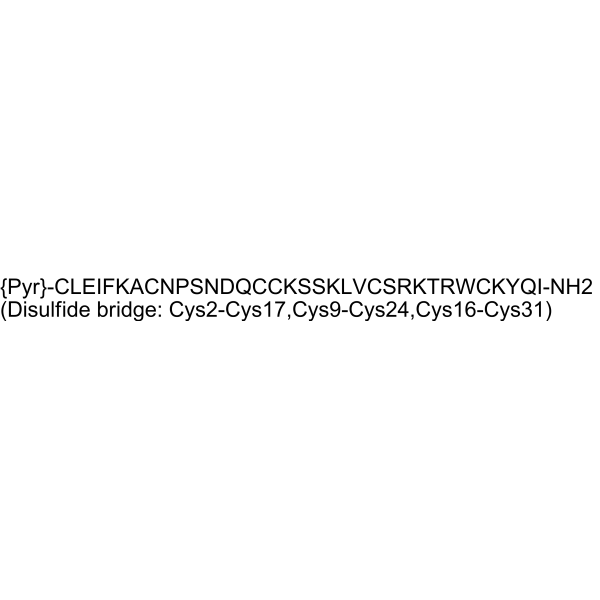
{Pyr}-Cys-Leu-Glu-Ile-Phe-Lys-Ala-Cys-Asn-Pro-Ser-Asn-Asp-Gln-Cys-Cys-Lys-Ser-Ser-Lys-Leu-Val-Cys-Ser-Arg-Lys-Thr-Arg-Trp-Cys-Lys-Tyr-Gln-Ile-NH2 (Disulfide bridge: Cys2-Cys17,Cys9-Cys24,Cys16-Cys31)
-
Sequence Shortening
{Pyr}-CLEIFKACNPSNDQCCKSSKLVCSRKTRWCKYQI-NH2 (Disulfide bridge: Cys2-Cys17,Cys9-Cys24,Cys16-Cys31)
-
Livraison
Room temperature in continental US; may vary elsewhere.
-
Stockage
Please store the product under the recommended conditions in the Certificate of Analysis.


Publications (1)
-
Journal Impact Factor
-
Most Recent
-
Cell Transplant
Bone marrow mesenchymal stem cells attenuate pain and modulate peripheral sodium channel activity in a rat model of complex regional pain syndrome type I. [Abstract]2025 Jan-Dec:34:9636897251383588. PMID: 41288153

Protocole
-
Cell Cytotoxicity Assay
Cytotoxicity assays are usually based on the assessment of cell membrane damage, which can also be indirectly detected by measuring cell viability. Detection methods include MTT assay, CKK-8 assay, LDH assay and ATP assay, etc.
-
Primary Dorsal Root Ganglion Sensory Neuron Culture
Primary dorsal root ganglion sensory neuron culture isolates DRG neuronal somata from rodent or human ganglia, dissociates tissue enzymatically and mechanically, and maintains post-mitotic sensory neurons in vitro for readouts such as neurite outgrowth, immunocytochemical marker expression, calcium imaging, electrophysiology, RNA/protein analysis, or neuropeptide release assays. The method reflects peripheral sensory neuron biology because DRG neurons are primary sensory neurons whose cell bodies reside in dorsal root ganglia and whose cultured dissociated cells can retain neuronal morphology, sensory-neuron marker expression, and stimulus-responsive properties depending on the downstream assay.


Pureté et documentation
Références

Calculators
Concentration (start) × Volume (start) = Concentration (final) × Volume (final)


mHuwentoxin-IV Related Classifications
HY-P5868 Related Classifications
 Powered by Bioz