



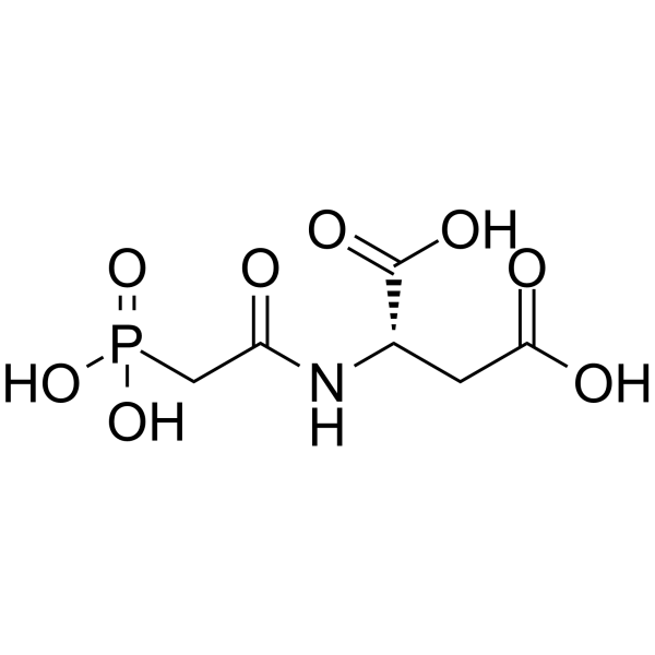
Sparfosic acid
Based on 6 publication(s) in Google Scholar
Sparfosic acid, a DNA antimetabolite agent, is a potent inhibitor of aspartate transcarbamoyl transferase, the enzyme catalyzing the second step of de novo pyrimidine biosynthesis. Sparfosic acid synergistically enhances the cytotoxicity of a combination of 5-fluorouracil (5-FU) and interferon-alpha (IFN) against human colon cancer cell lines.
Nos produits utilisent uniquement pour la recherche. Nous ne vendons pas aux patients.
- Pureté : 99.48%
- CAS No.: 51321-79-0
- Formule: C6H10NO8P
- Masse moléculaire:255.12
-
Stockage:Pure form -20°C, 3 years , 4°C, 2 years ; In solvent -80°C, 6 months , -20°C, 1 month

To place orders, for customer services and technical support, please contact: MedChemExpress USA
Tel: 609-228-6898 E-mail: [email protected] [email protected]


-
 Activité biologique
-
Chemical Information
-
Solvant et solubilité
- Protocole
- Pureté et documentation
- Références
-
 Help & FAQs


Publications Citing Use of MedChemExpress (MCE) Sparfosic acid
More Customer Validation & Images

-
Cell Proliferation/Viability Assay

-
Cell Migration/Invasion Assay

-
Cell Proliferation/Viability Assay


Activité biologique
Description
In Vitro
Sparfosic acid (N-(Phosphonacetyl)-L-aspartate, PALA) causes apoptosis in the resistant Br1 cells[1].
Sparfosic acid (300 µM) shows progressive accumulation of cells in S phase and activation of an apoptotic pathway leading to cell death[1].
MedChemExpress (MCE) has not independently confirmed the accuracy of these methods. They are for reference only. Further protocols information, click here.
-
Cell Line:Br-l and L-2 cell lines established from metastasis in nude mouse injected with the human tumor cell line MDA-MB-435
-
Concentration:300 µM
-
Incubation Time:12, 24 and 48 hours
-
Result:Cells were predominantly in S phase in both the cell lines, although slightly higher proportion of cells in S phase were noted in L-2 than Brl-3prl cells.
-
Cell Line:Br-l and L-2 cell lines
-
Concentration:300 µM
-
Incubation Time:4, 10 and 24 hours
-
Result:There was moderate difference in the level of phosphorylated Rb proteins seen in the two cell types. Marked increase in the amount of cyclin A protein was detected in the L-2 cells undergoing apoptosis with the highest level detected at 10 h post-drug treatment. In contrast, there was no increase in the level of cyclin A seen in the Brl-3prl cells. Cyclin E protein was found elevated in the L-2 cells and Brl-3prl cells compared to their respective controls.
In Vivo
MedChemExpress (MCE) has not independently confirmed the accuracy of these methods. They are for reference only.


Chemical Information
-
CAS No. 51321-79-0
-
Appearance Solid-Liquid Mixture
-
Masse moléculaire 255.12
-
Formule C6H10NO8P
-
Color Colorless to off-white
-
SMILES
O=C(O)C[C@@H](C(O)=O)NC(CP(O)(O)=O)=O
-
Livraison
Room temperature in continental US; may vary elsewhere.
-
Stockage
Pure form -20°C 3 years 4°C 2 years In solvent -80°C 6 months -20°C 1 month


Publications (6)
-
Journal Impact Factor
-
Most Recent
-
Nat Metab
Uridine-sensitized screening identifies demethoxy-coenzyme Q and NUDT5 as regulators of nucleotide synthesis. [Abstract]2025 Nov 13. PMID: 41233602
Sparfosic acid purchased from MedChemExpress. Usage Cited in: Nat Metab. 2025 Nov 13. [Abstract]
Proliferation assay of K562 cells over 5 days supplemented, as indicated, with 200 μM uridine, 200 µM CADi (Sparfosic acid trisodium), 10 μM DHODHi (brequinar) or 10 μM UMPSi (pyrazofurin).
-
Nat Commun
Hijacking of nucleotide biosynthesis and deamidation-mediated glycolysis by an oncogenic herpesvirus. [Abstract]2024 Feb 16;15(1):1442. PMID: 38365882 -
Mol Biomed
Lactate-driven pyrimidine synthesis promotes ferroptosis resistance in hepatocellular carcinoma. [Abstract]2026 Apr 17;7(1):53. PMID: 41996004 -
Life Sci
CAD drives Angiotensin II-induced vascular smooth muscle cell activation via upregulation of pyrimidine biosynthesis and rRNA synthesis. [Abstract]2026 Jun 9:401:124530. PMID: 42264140
Sparfosic acid purchased from MedChemExpress. Usage Cited in: Life Sci. 2026 Jun 9:401:124530. [Abstract]
Relative cell number in Ang II (100 nM)-treated VSMCs with or without PALA (Sparfosic acid trisodium, 150 μM).
Sparfosic acid purchased from MedChemExpress. Usage Cited in: Life Sci. 2026 Jun 9:401:124530. [Abstract]
Representative images showing the effects of PALA(Sparfosic acid trisodium, 150 μM) on VSMC migration (upper panel; scale bar = 40 μm), and quantification of migration and wound area (lower panel).
-
-


Solvant et solubilité
In Vitro:
H2O : ≥ 100 mg/mL (391.97 mM)
* "≥" means soluble, but saturation unknown.
Please refer to the solubility information to select the appropriate solvent. Once prepared, please aliquot and store the solution to prevent product inactivation from repeated freeze-thaw cycles.
Storage method and period of stock solution: -80°C, 6 months; -20°C, 1 month. When stored at -80°C, please use it within 6 months. When stored at -20°C, please use it within 1 month.
* Note: If you choose water as the stock solution, please dilute it to the working solution, then filter and sterilize it with a 0.22 μm filter before use.
Please refer to the solubility information to select the appropriate solvent. Once prepared, please aliquot and store the solution to prevent product inactivation from repeated freeze-thaw cycles.
Storage method and period of stock solution: -80°C, 6 months; -20°C, 1 month. When stored at -80°C, please use it within 6 months. When stored at -20°C, please use it within 1 month.
* Note: If you choose water as the stock solution, please dilute it to the working solution, then filter and sterilize it with a 0.22 μm filter before use.
Concentration (start) × Volume (start) = Concentration (final) × Volume (final)

Protocole
-
Cell Cytotoxicity Assay
Cytotoxicity assays are usually based on the assessment of cell membrane damage, which can also be indirectly detected by measuring cell viability. Detection methods include MTT assay, CKK-8 assay, LDH assay and ATP assay, etc.
-
Mammalian live/dead viability and cytotoxicity staining
Live/dead viability and cytotoxicity staining assays are based on the simultaneous detection of intracellular esterase activity in metabolically active (viable) cells and membrane integrity loss in non-viable cells. In commonly used dual-staining approaches, membrane-permeant fluorogenic substrates are converted by intracellular esterases into fluorescent products in live cells, while impermeant DNA-binding dyes selectively enter cells with compromised plasma membranes and label nucleic acids in dead or dying cells, enabling discrimination between viable and non-viable populations by fluorescence microscopy or flow cytometry.
-
Cell Viability Determination by MTT Colorimetric Assay
The following protocol uses the MTT colorimetric assay as a classic literature-established method for assessing cell viability/metabolic activity in cultured mammalian cells. MTT[3-(4,5-dimethylthiazol-2-yl)-2,5-diphenyltetrazolium bromide] is reduced by metabolically active cells to a colored formazan product; the amount of formazan is quantified spectrophotometrically and provides an indirect measure of metabolically active viable cells. Importantly, MTT reduction reflects cellular oxidoreductase/metabolic activity rather than an absolute direct count of living cells, so changes in cellular metabolism can alter the signal independently of cell number.


Pureté et documentation
-
Fiche technique (278 KB)
-
SDS (254 KB)
- English - EN (254 KB)
- Français - FR (254 KB)
- Deutsch - DE (254 KB)
- Norwegian - NO (254 KB)
- Español - ES (254 KB)
- Swedish - SV (254 KB)
- Italian - IT (254 KB)
- Korean - KR (254 KB)
- Portuguese - PT (254 KB)
-
Instruction de manipulation (2659 KB)
Références
[1]. Wang J, et al. Elevated cyclin A associated kinase activity promotes sensitivity of metastatic human cancer cells to DNA antimetabolite drug. Int J Oncol. 2015 Aug;47(2):782-90. [Content Brief]
[3]. Johnson RK, et al. Antitumor activity of N-(phosphonacetyl)-L-aspartic acid, a transition-state inhibitor of aspartate transcarbamylase. Cancer Res. 1976;36(8):2720-2725. [Content Brief]
[4]. Wadler S, et al. Phase II trial of N-(phosphonacetyl)-L-aspartate (PALA), 5-fluorouracil and recombinant interferon-alpha-2b in patients with advanced gastric carcinoma. Eur J Cancer. 1996;32A(7):1254-1256. [Content Brief]


Complete Stock Solution Preparation Table
Please refer to the solubility information to select the appropriate solvent. Once prepared, please aliquot and store the solution to prevent product inactivation from repeated freeze-thaw cycles.
Storage method and period of stock solution: -80°C, 6 months; -20°C, 1 month. When stored at -80°C, please use it within 6 months. When stored at -20°C, please use it within 1 month.
| Optional Solvent | Concentration Solvent Mass | 1 mg | 5 mg | 10 mg | 25 mg |
|---|---|---|---|---|---|
| H2O | 1 mM | 3.9197 mL | 19.5986 mL | 39.1972 mL | 97.9931 mL |
| 5 mM | 0.7839 mL | 3.9197 mL | 7.8394 mL | 19.5986 mL | |
| 10 mM | 0.3920 mL | 1.9599 mL | 3.9197 mL | 9.7993 mL | |
| 15 mM | 0.2613 mL | 1.3066 mL | 2.6131 mL | 6.5329 mL | |
| 20 mM | 0.1960 mL | 0.9799 mL | 1.9599 mL | 4.8997 mL | |
| 25 mM | 0.1568 mL | 0.7839 mL | 1.5679 mL | 3.9197 mL | |
| 30 mM | 0.1307 mL | 0.6533 mL | 1.3066 mL | 3.2664 mL | |
| 40 mM | 0.0980 mL | 0.4900 mL | 0.9799 mL | 2.4498 mL | |
| 50 mM | 0.0784 mL | 0.3920 mL | 0.7839 mL | 1.9599 mL | |
| 60 mM | 0.0653 mL | 0.3266 mL | 0.6533 mL | 1.6332 mL | |
| 80 mM | 0.0490 mL | 0.2450 mL | 0.4900 mL | 1.2249 mL | |
| 100 mM | 0.0392 mL | 0.1960 mL | 0.3920 mL | 0.9799 mL |
* Note: If you choose water as the stock solution, please dilute it to the working solution, then filter and sterilize it with a 0.22 μm filter before use.


Sparfosic acid Related Classifications
HY-112732 Related Classifications
 Powered by Bioz