



Catalase, Bovine Liver

Based on 1 Customer Validation
Catalase, Bovine Liver is an enzyme widely found in various organisms, including animals, plants and microorganisms. Catalase, Bovine Liver mainly exists in the peroxisome of cells and is an important antioxidant enzyme. Catalase, Bovine Liver plays an important role in removing ROS and maintaining the balance of redox state. Catalase, Bovine Liver is closely related to the occurrence and development of tumors. Catalase, Bovine Liver has the potential to be used in tumor prevention research.
For research use only. We do not sell to patients.
- CAS No.: 9001-05-2
-
Storage:
Please store the product under the recommended conditions in the Certificate of Analysis.


To place orders, for customer services and technical support, please contact: MedChemExpress USA
Tel: 609-228-6898 E-mail: [email protected] [email protected]


-
 Biological Activity
-
Chemical Information
- Protocols
- Purity & Documentation
- References
-
 Help & FAQs


-

HY-15371
ForskolinAdenylate Cyclase Activator -
HY-112879
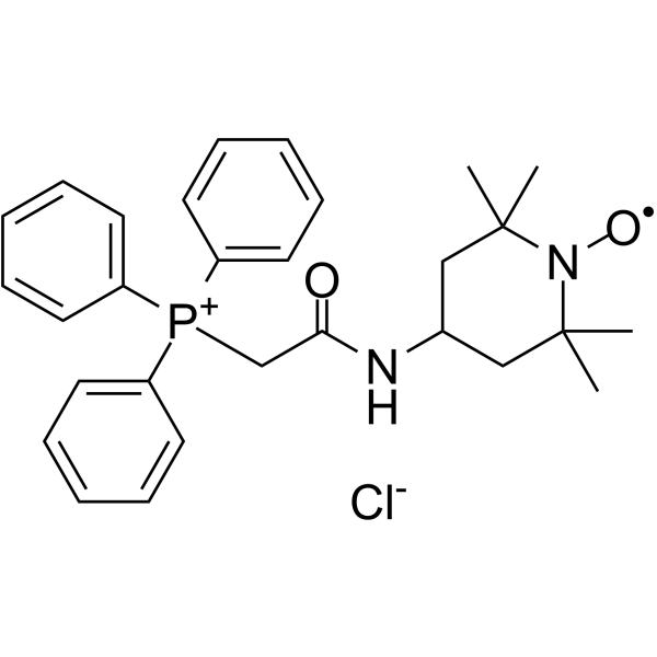
Mito-TEMPOAntioxidant -
HY-135849
Catalase, Aspergillus nigerAntioxidant Enzyme -
HY-N0390S
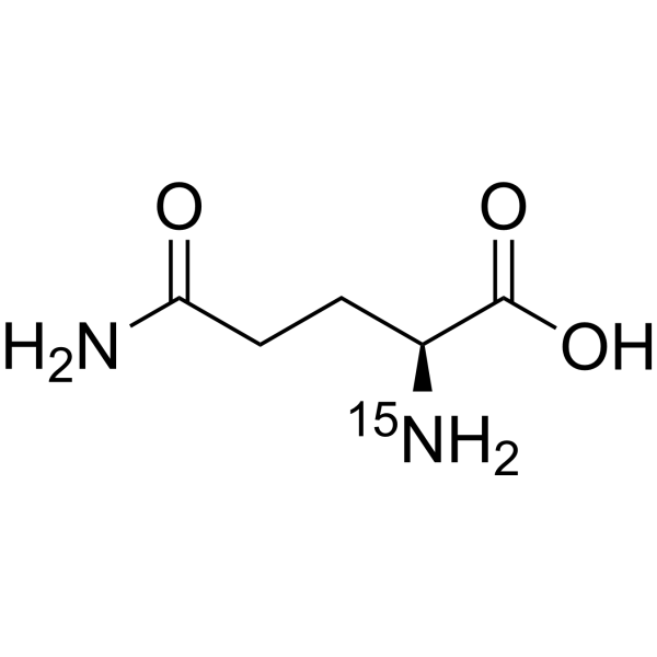
L-Glutamine-15NStable Isotope -
HY-P2733
Glycerol-3-phosphate oxidase, E. coliOxidase -

HY-135849C
Catalase, trichoderma reeseiCatalase -
HY-P2902
Glucose oxidaseOxidoreductase Enzyme -

HY-B2220
CellulaseEnzyme -
HY-P2755
Xanthine oxidase, MicroorganismROS Inducer -

HY-P2755A
Xanthine oxidase, bovine milkROS Inducer -
HY-B2220A
Cellulase R-10 from Trichoderma VrideHydrolase -
HY-125863
Glucose-6-phosphate dehydrogenase, MicroorganismEndogenous Metabolite


Biological Activity
Description
In Vitro
Product Information
Catalase is a tetramer composed of four uniform tetrahedral subunits. Each 60 kDa subunit contains a heme group and NADPH in its active center. The theoretical molecular weight is approximately 232 kDa, the optimal pH is about 7.0, and the isoelectric point is 5.4.
Activator: Sodium arsenate
Inhibitors: Cyanide, azide, hydroxylamine, aminotriazole, hydroxy ethanol, peroxide, nitroso and nitrosyl compounds, etc
Instructions
It can be dissolved in water or according to the specific experimental reference literature.
MedChemExpress (MCE) has not independently confirmed the accuracy of these methods. They are for reference only. Further protocols information, click here.


Chemical Information
-
CAS No. 9001-05-2
-
Appearance Solid
-
Color Light brown to brown
-
SMILES
[Catalase, Bovine Liver]
-
Shipping
Room temperature in continental US; may vary elsewhere.
-
Storage
Please store the product under the recommended conditions in the Certificate of Analysis.

Protocols
-
ROS/oxidative-stress fluorescent staining
ROS/oxidative-stress fluorescent staining uses cell-permeant fluorogenic probes that become fluorescent after oxidation inside cells or tissues; commonly used examples include DCFH-DA/DCFDA for broad cellular oxidant detection, DHE for superoxide-related signal detection, MitoSOX for mitochondrial superoxide-related signal detection, and CellROX probes for oxidative-stress-associated fluorescence readouts. The assay detects probe oxidation rather than a single ROS species unless the probe and analysis method have been chemically validated for that species. DCFH-DA enters cells, is deacetylated by intracellular esterases to DCFH, and produces fluorescent DCF after oxidation, so the readout is used as an operational measure of total cellular oxidative stress rather than a species-specific ROS measurement. DHE and MitoSOX can report superoxide-related oxidation, but red fluorescence alone can include non-specific ethidium-like oxidation products; HPLC or optimized spectral approaches are


Purity & Documentation
-
Data Sheet (264 KB)
-
SDS (251 KB)
- English - EN (251 KB)
- Français - FR (251 KB)
- Deutsch - DE (251 KB)
- Norwegian - NO (251 KB)
- Español - ES (251 KB)
- Swedish - SV (251 KB)
- Italian - IT (251 KB)
- Korean - KR (251 KB)
- Portuguese - PT (251 KB)
-
Handling Instructions (2659 KB)
References

Calculators
Concentration (start) × Volume (start) = Concentration (final) × Volume (final)


Catalase, Bovine Liver Related Classifications
HY-135849A Related Classifications
 Powered by Bioz