



SEN177
Based on 1 publication(s) in Google Scholar
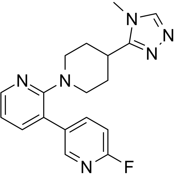
SEN177 is an orally effect glutamine cyclase (QC) inhibitor. The Ki of SEN177 for human glutamine cyclase (hQC) is 20 nM, and the IC50 is 13 nM. SEN177 interferes with the interaction between CD47 and SIRRPα, and has anti-tumor activity. SEN177 reduces aggregation and apoptosis caused by HTT mutation in Huntington model, and can be used in the study of neurodegenerative diseases.
Nur für Forschungszwecke. Wir verkaufen nicht an Patienten.
- Reinheit : 98.76%
- CAS. Nr.: 2117405-13-5
- Formel: C18H19FN6
- Molecular Weight:338.38
-
Speicherung:Powder -20°C, 3 years ; In solvent -80°C, 6 months , -20°C, 1 month

To place orders, for customer services and technical support, please contact: MedChemExpress USA
Tel: 609-228-6898 E-mail: [email protected] [email protected]


-
 Biologische Aktivität
-
Chemical Information
-
Lösungsmittel & Löslichkeit
- Protokoll
- Reinheit & Dokumentation
- Verweise
-
 Help & FAQs


Publications Citing Use of MedChemExpress (MCE) SEN177
More

Biologische Aktivität
Beschreibung
Cellular Effect
|
Cell Line

|
Type | Value | Description | References |
|---|---|---|---|---|
| Sf21 | IC50 |
16.7 nM
Compound: 4; SEN177
|
Inhibition of recombinant human N-terminal His6-tagged glutaminyl cyclase (Ala33 to Leu361 residues) expressed in baculovirus infected Sf21 insect cells using H-Gln-AMC hydrobromide as substrate measured in presence of pGAPase by fluorometric assay
Inhibition of recombinant human N-terminal His6-tagged glutaminyl cyclase (Ala33 to Leu361 residues) expressed in baculovirus infected Sf21 insect cells using H-Gln-AMC hydrobromide as substrate measured in presence of pGAPase by fluorometric assay
|
[PMID: 31411468] |
In Vitro
SEN177 (0-50 μM; 0-96 h) effectively blocks the interaction between CD47 and SIRRPα in breast cancer cell line MDA-MB-468[2].
SEN177 (10 μM; 3 days) significantly enhances the ability of EGFR antibody to induce ADCP in esophageal squamous cell carcinoma (Kyse-30) and breast cancer cell line (MDA-MB-468)[2].
SEN177 (50 μM; 24 h) decreases mutant HTT aggregate ratio and apoptosis in HeLa cells expressing HTT (Q74)[3].
MedChemExpress (MCE) has not independently confirmed the accuracy of these methods. They are for reference only.
In Vivo
MedChemExpress (MCE) has not independently confirmed the accuracy of these methods. They are for reference only.
-
Animal Model:Drosophila expressing Httex1Q46 in the eye[3]
-
Dosage:50 μM
-
Administration:Oral administration (p.o.); Change every two days
-
Result:Reduced the number of aggregates.
Rescued the number of visible rhabdomeres.
Prevented neurodegeneration.


Chemical Information
-
CAS. Nr. 2117405-13-5
-
Appearance Solid
-
Molecular Weight 338.38
-
Formel C18H19FN6
-
Color Brown to gray
-
SMILES
CN1C(C2CCN(C3=NC=CC=C3C4=CC=C(F)N=C4)CC2)=NN=C1
-
Versand
Room temperature in continental US; may vary elsewhere.
-
Speicherung
Powder -20°C 3 years In solvent -80°C 6 months -20°C 1 month


Publications (1)
-
Journal Impact Factor
-
Most Recent
-
Nat Commun
2026 Jun 9. PMID: 42265113


Lösungsmittel & Löslichkeit
In Vitro:
DMSO : 31.25 mg/mL (92.35 mM; Need ultrasonic; Hygroscopic DMSO has a significant impact on the solubility of product, please use newly opened DMSO)
Please refer to the solubility information to select the appropriate solvent. Once prepared, please aliquot and store the solution to prevent product inactivation from repeated freeze-thaw cycles.
Storage method and period of stock solution: -80°C, 6 months; -20°C, 1 month. When stored at -80°C, please use it within 6 months. When stored at -20°C, please use it within 1 month.
Please refer to the solubility information to select the appropriate solvent. Once prepared, please aliquot and store the solution to prevent product inactivation from repeated freeze-thaw cycles.
Storage method and period of stock solution: -80°C, 6 months; -20°C, 1 month. When stored at -80°C, please use it within 6 months. When stored at -20°C, please use it within 1 month.
Konzentration (Stammlösung) × Volumen (Stammlösung) = Konzentration (Ziellösung) × Volumen (Ziellösung)


Reinheit & Dokumentation
-
Data Sheet (278 KB)
-
SDS (480 KB)
- English - EN (480 KB)
- Français - FR (480 KB)
- Deutsch - DE (480 KB)
- Norwegian - NO (480 KB)
- Español - ES (480 KB)
- Swedish - SV (480 KB)
- Italian - IT (480 KB)
- Korean - KR (480 KB)
- Portuguese - PT (480 KB)
-
Handling Instructions (2659 KB)
Verweise
[1]. Cecilia Pozzi, et al. The structure of the human glutaminyl cyclase-SEN177 complex indicates routes for developing new potent inhibitors as possible agents for the treatment of neurological disorders. J Biol Inorg Chem. 2018 Dec;23(8):1219-1226. [Content Brief]
[2]. Baumann N, et al. Enhancement of epidermal growth factor receptor antibody tumor immunotherapy by glutaminyl cyclase inhibition to interfere with CD47/signal regulatory protein alpha interactions. Cancer Sci. 2021 Aug;112(8):3029-3040. [Content Brief]
[3]. Maria Jimenez-Sanchez, et al. siRNA screen identifies QPCT as a druggable target for Huntington's disease. Nat Chem Biol. 2015 May;11(5):347-354. [Content Brief]


Complete Stock Solution Preparation Table
Please refer to the solubility information to select the appropriate solvent. Once prepared, please aliquot and store the solution to prevent product inactivation from repeated freeze-thaw cycles.
Storage method and period of stock solution: -80°C, 6 months; -20°C, 1 month. When stored at -80°C, please use it within 6 months. When stored at -20°C, please use it within 1 month.
| Optional Solvent | Concentration Solvent Mass | 1 mg | 5 mg | 10 mg | 25 mg |
|---|---|---|---|---|---|
| DMSO | 1 mM | 2.9553 mL | 14.7763 mL | 29.5526 mL | 73.8814 mL |
| 5 mM | 0.5911 mL | 2.9553 mL | 5.9105 mL | 14.7763 mL | |
| 10 mM | 0.2955 mL | 1.4776 mL | 2.9553 mL | 7.3881 mL | |
| 15 mM | 0.1970 mL | 0.9851 mL | 1.9702 mL | 4.9254 mL | |
| 20 mM | 0.1478 mL | 0.7388 mL | 1.4776 mL | 3.6941 mL | |
| 25 mM | 0.1182 mL | 0.5911 mL | 1.1821 mL | 2.9553 mL | |
| 30 mM | 0.0985 mL | 0.4925 mL | 0.9851 mL | 2.4627 mL | |
| 40 mM | 0.0739 mL | 0.3694 mL | 0.7388 mL | 1.8470 mL | |
| 50 mM | 0.0591 mL | 0.2955 mL | 0.5911 mL | 1.4776 mL | |
| 60 mM | 0.0493 mL | 0.2463 mL | 0.4925 mL | 1.2314 mL | |
| 80 mM | 0.0369 mL | 0.1847 mL | 0.3694 mL | 0.9235 mL |


SEN177 Related Classifications
HY-136780 Related Classifications
 Powered by Bioz