Talacotuzumab
Based on 2 publication(s) in Google Scholar
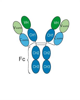
Talacotuzumab (JNJ 56022473; CSL 362) is an IgG1-type fully humanized, CD123-neutralizing monoclonal antibody containing a modified Fc structure. Talacotuzumab has KDs of 0.43 nM, 188 nM, 46 nM, 16.8 nM for CD123, CD32b/c, CD16-158F, CD16-158V, respectively. Talacotuzumab inhibits IL-3 binding to CD123, antagonizing IL-3 signaling in target cells. Talacotuzumab has mutated the Fc region to increase affinity for CD16 (FcγRIIIa), thereby enhancing antibody-dependent cell-mediated cytotoxicity (ADCC). Talacotuzumab is highly effective in vivo reducing leukemic cell growth in acute myeloid leukemia (AML) xenograft mouse models.
Nur für Forschungszwecke. Wir verkaufen nicht an Patienten.
- Reinheit : 98.82%
- CAS. Nr.: 1826831-79-1
- Molecular Weight:147.42 kDa
-
Speicherung:
Please store the product under the recommended conditions in the Certificate of Analysis.

Publications Citing Use of MedChemExpress (MCE) Talacotuzumab
More
Biologische Aktivität
Beschreibung
Isotype
Human IgG1 kappa
Recommend Isotype Controls
Species Reactivity
Human
IC50 & Target
IL-3Ra/CD123
In Vitro
Talacotuzumab (JNJ 56022473; CSL 362) strongly mediates ADCC of TF-1 cells with an ic50 of 5 ng/ml (33 pM)[1].
Talacotuzumab (1 μg/ml; pretreatment for 24 hours) inhibits TLR7-stimulated (Imiquimod; HY-B0180; 0.5 μM; for 6 days) and TLR9-stimulated (CpG C; 0.5 μM; for 6 days) IFN-α production in both SLE donors and healthy donors plasmacytoid dendritic cells (pDCs) and basophils, whereas TLR4-stimulated (LPS; HY-D1056; 10 μg/ml) production is not significantly reduced. Talacotuzumab inhibits TLR7- and TLR9-induced plasmablast expansion and proliferation by depletion of plasmacytoid dendritic cells (pDCs)[2].
MedChemExpress (MCE) has not independently confirmed the accuracy of these methods. They are for reference only. Further protocols information, click here.
In Vivo
Talacotuzumab (1, 10, 30 mg/kg; s.c.; single injection) has maximal serum concentrations at 48 hours of ~12, 190, and 380 μg/ml at doses of 1, 10, and 30 mg/kg in naive cynomolgus monkeys, respectively[2].
MedChemExpress (MCE) has not independently confirmed the accuracy of these methods. They are for reference only.
-
Animal Model:Nonobese diabetic/severe combined immunodeficiency mice injected intravenously AML xenograft cells (AML-5)[1]
-
Dosage:300 μg
-
Administration:IP; thrice weekly for 5 weeks
-
Result:Resulted in a significant delay in tumor growth compared with an isotype control.
Clinical Trial
| NCT Number | Sponsor | Condition | Start Date |
Phase
|
|---|---|---|---|---|
| NCT01329991 | Plexxikon| | 2011-05 | PHASE1 |
Gene ID
Accession
Conjugated
Unconjugated
Reconsititution
The product can be reconstituted/diluted with sterile PBS or saline.
Format
-
Human IgG1 kappa
Anwendung
ELISA, FACS, Functional assay
Verified Bioactivity
-
Immobilized IL-3R alpha/CD123 Protein, Human can bind Talacotuzumab. The EC50 for this effect is 6.855 ng/mL. -
Flow cytometric analysis of 1.5X10⁶ THP-1 cells labeling Talacotuzumab (HY-P99395, red). Cells were fixed with 4% paraformaldehyde. Then stained with the primary antibody at 1/200 dilution for an hour at 4℃. Goat Anti-Human IgG H&L (AF488) (HY-P83776) was used as the secondary antibody at 1/1,000 dilution for 30 minutes at 4℃. Human IgG1 kappa (HY-P99001, blue) was used as the isotype control, cells without incubation with primary antibody were used as the unlabeled control (black).
Chemical Information
-
CAS. Nr. 1826831-79-1
-
Appearance Liquid
-
Molecular Weight 147.42 kDa
-
Color Colorless to light yellow
-
SMILES
[Talacotuzumab]
-
Synonyms
JNJ 56022473; CSL 362
-
Versand
Shipping with dry ice.
-
Formulation
Please refer to the lot-specific COA for specific buffer information.
-
Speicherung
Please store the product under the recommended conditions in the Certificate of Analysis.
Publications (2)
-
Journal Impact Factor
-
Most Recent
-
Nat Commun
CD200R1-CD200 checkpoint inhibits phagocytosis differently from SIRPα-CD47 to suppress tumor growth. [Abstract]2025 Jun 3;16(1):5145. PMID: 40461553 -
Protokoll
-
Mammalian live/dead viability and cytotoxicity staining
Live/dead viability and cytotoxicity staining assays are based on the simultaneous detection of intracellular esterase activity in metabolically active (viable) cells and membrane integrity loss in non-viable cells. In commonly used dual-staining approaches, membrane-permeant fluorogenic substrates are converted by intracellular esterases into fluorescent products in live cells, while impermeant DNA-binding dyes selectively enter cells with compromised plasma membranes and label nucleic acids in dead or dying cells, enabling discrimination between viable and non-viable populations by fluorescence microscopy or flow cytometry.
-
Somatic Cell Culture
A method of simulating the in vivo environment in vitro to maintain the cell growth, differentation and main functions.
-
CCK-8/WST-8 Cell Proliferation Assay
The CCK-8/WST-8 assay is based on the reduction of the water-soluble tetrazolium salt WST-8 to a water-soluble formazan product by cellular dehydrogenases in metabolically active cells, where the generated formazan amount is proportional to the number of living cells and is quantified by measuring absorbance in the visible range, providing a colorimetric readout for cell viability and proliferation assessment. This class of tetrazolium-based assays improves upon earlier MTT-based systems by producing a water-soluble formazan, eliminating the need for organic solubilization steps and enabling direct spectrophotometric measurement in culture medium.
-
Cell Counting-Based Growth Curve Assay
Cell counting-based growth curve assays quantify cell proliferation by directly measuring changes in viable cell number over time using manual or automated counting methods such as hemocytometer-based counting or instrument-assisted cell enumeration, enabling construction of growth curves that reflect population expansion dynamics in response to culture conditions. A widely used approach is trypan blue exclusion with hemocytometer counting, where membrane-compromised (non-viable) cells take up the dye, allowing discrimination between viable and non-viable cells while simultaneously enabling total cell number quantification. Repeated sampling across time points allows estimation of proliferation rate, growth phases, and comparative growth kinetics between experimental conditions.
-
Cell Viability Determination by MTT Colorimetric Assay
The following protocol uses the MTT colorimetric assay as a classic literature-established method for assessing cell viability/metabolic activity in cultured mammalian cells. MTT[3-(4,5-dimethylthiazol-2-yl)-2,5-diphenyltetrazolium bromide] is reduced by metabolically active cells to a colored formazan product; the amount of formazan is quantified spectrophotometrically and provides an indirect measure of metabolically active viable cells. Importantly, MTT reduction reflects cellular oxidoreductase/metabolic activity rather than an absolute direct count of living cells, so changes in cellular metabolism can alter the signal independently of cell number.
-
Cell Cytotoxicity Assay
Cytotoxicity assays are usually based on the assessment of cell membrane damage, which can also be indirectly detected by measuring cell viability. Detection methods include MTT assay, CKK-8 assay, LDH assay and ATP assay, etc.
-
Protocol for Cell Counting and Cell Density Analysis
Cell counting and cell-density analysis estimate the number of cells in a known volume or field area. Manual hemocytometer counting uses a chamber of defined geometry to convert counted cells into cells/mL, while automated counters and image-analysis workflows detect cell objects from optical, brightfield, fluorescence, impedance, or digital-image features. Trypan blue viability counting is based on dye exclusion: viable cells with intact membranes exclude dye, while non-viable cells with compromised membranes stain blue. The readout is total cell density, viable-cell density, dead-cell density, and percent viability. Cell density can also be estimated from microscopy images by counting objects per image area, from flow cytometry using calibrated volume or reference particles, or from in situ microscopy in bioreactors after calibration against reference methods such as hemocytometer or flow cytometry.
-
Patient-Derived Xenograft (PDX)
Patient-derived xenograft (PDX) models are generated by engrafting primary human tumor tissue directly into immunodeficient mice, allowing in vivo propagation of patient tumor biology without initial in vitro adaptation. These models are used to preserve key histopathological and molecular characteristics of the original tumor and enable assessment of tumor growth dynamics and therapeutic response in a living organism. The biological readout is tumor engraftment and subsequent growth in the murine host, which reflects the ability of human tumor cells to survive, vascularize, and expand in an immunocompromised microenvironment.
-
Splenic/Portal-Vein Liver Metastasis Xenograft
Splenic and portal-vein liver metastasis xenograft models deliver tumor cells into the portal circulation so that cells reach the liver first and form hepatic metastatic lesions; splenic injection uses the spleen as an access route to the portal system, while direct portal-vein injection introduces cells into the portal vein without requiring splenectomy. The assay detects liver colonization, intrahepatic tumor growth, tumor distribution, treatment response, survival, and liver-metastasis microenvironment changes; readouts include bioluminescence or fluorescence imaging, gross liver nodule counts, liver weight or tumor burden, histology, and survival.
-
MTT Cell Proliferation Assay
The MTT assay is a colorimetric endpoint assay for estimating viable cell number, cell growth, cytotoxicity, or cell activation in cultured mammalian cells. Living cells reduce the yellow tetrazolium salt MTT into purple/blue formazan, while dead cells do not generate the same signal; the resulting color can be quantified with a multiwell spectrophotometer. MTT reduction is commonly interpreted as a readout of metabolic activity that often correlates with viable cell number, but it should not be treated as a direct cell-counting method unless the assay is optimized for the cell type and experimental condition. Studies show that MTT reduction can involve mitochondrial and non-mitochondrial reducing systems, and formazan may accumulate in intracellular lipid droplets rather than simply marking mitochondria.
-
Subcutaneous Cell-Line-Derived Xenograft
Subcutaneous cell-line-derived xenograft (CDX) models are established by implanting cultured human cancer cell lines into immunodeficient mice, where the injected cells form localized tumors that can be monitored in vivo as a measure of tumorigenic potential, growth kinetics, and treatment response. These models are widely used in oncology research because they allow reproducible tumor formation and enable comparative assessment of tumor growth between different cell lines or genetic manipulations in a controlled in vivo microenvironment. Subcutaneous implantation of cancer cells in immunodeficient mice is a standard approach for evaluating tumor growth behavior and therapeutic response across multiple cancer types, including prostate, esophageal, pancreatic, and colon cancer models.
-
Orthotopic Cell-Line Xenograft
Orthotopic cell-line xenograft models involve implantation of human cancer cell lines into the anatomically corresponding organ of immunodeficient mice to reproduce tumor growth within a native microenvironment, enabling more clinically relevant tumor behavior compared with subcutaneous models. These models are widely used because orthotopic placement better recapitulates tumor progression, including invasion and metastatic spread, which are often underrepresented in heterotopic implantation systems. Compared with conventional xenografts, orthotopic implantation is described as more technically complex but provides improved simulation of tumor-microenvironment interactions and metastatic behavior, making it particularly valuable for translational oncology research. Surgical orthotopic implantation approaches have been emphasized as enabling faithful reproduction of clinical cancer features, including metastasis and disease progression patterns that align with the tumor’s organ of origi
-
Intraperitoneal/Peritoneal Dissemination Xenograft
Intraperitoneal (IP) or peritoneal dissemination xenograft models are based on the introduction of human cancer cells into the peritoneal cavity of immunodeficient mice, where they attach to peritoneal surfaces, form multicellular aggregates or spheroids, and progressively generate disseminated tumor nodules that mimic advanced peritoneal metastatic disease. These models are widely used to study ovarian cancer progression, tumor-microenvironment interactions, and intraperitoneal therapeutic responses, often incorporating bioluminescence or fluorescence imaging to longitudinally monitor tumor burden in vivo. The biological principle relies on the capacity of tumor cells such as SKOV3 or related ovarian carcinoma lines to survive in suspension, aggregate within ascites-like fluid, adhere to mesothelial surfaces, and invade peritoneal organs, thereby recapitulating human peritoneal carcinomatosis patterns observed in advanced disease.
Reinheit & Dokumentation
-
Data Sheet (263 KB)
-
SDS (252 KB)
- English - EN (252 KB)
- Français - FR (252 KB)
- Deutsch - DE (252 KB)
- Norwegian - NO (252 KB)
- Español - ES (252 KB)
- Swedish - SV (252 KB)
- Italian - IT (252 KB)
- Korean - KR (252 KB)
- Portuguese - PT (252 KB)
-
Inhibitory Antibodies User Guide (603 KB)
Verweise
[1]. S J Busfield, et al. Targeting of acute myeloid leukemia in vitro and in vivo with an anti-CD123 mAb engineered for optimal ADCC. Leukemia. 2014 Nov;28(11):2213-21. [Content Brief]
[2]. Shereen Oon, et al. A cytotoxic anti-IL-3Rα antibody targets key cells and cytokines implicated in systemic lupus erythematosus. JCI Insight. 2016 May 5;1(6):e86131. [Content Brief]
[3]. Erwin M Lee, et al. Efficacy of an Fc-modified anti-CD123 antibody (CSL362) combined with chemotherapy in xenograft models of acute myelogenous leukemia in immunodeficient mice. Haematologica. 2015 Jul;100(7):914-26. [Content Brief]
[4]. L H Xie, et al. CD123 target validation and preclinical evaluation of ADCC activity of anti-CD123 antibody CSL362 in combination with NKs from AML patients in remission. Blood Cancer J. 2017 Jun 2;7(6):e567. [Content Brief]
Calculators
Konzentration (Stammlösung) × Volumen (Stammlösung) = Konzentration (Ziellösung) × Volumen (Ziellösung)