



Trypsin
Based on 27 publication(s) in Google Scholar
Trypsin is a serine protease enzyme, and hydrolyzes proteins at the carboxyl side of the Lysine or Arginine. Trypsin activates PAR2 and PAR4. Trypsin induces cell-to-cell membrane fusion in PDCoV infection by the interaction of S glycoprotein of PDCoV and pAPN. Trypsin also promotes cell proliferation and differentiation. Trypsin can be used in the research of wound healing and neurogenic inflammation.
Nur für Forschungszwecke. Wir verkaufen nicht an Patienten.
- Reinheit : 91.00%
- CAS. Nr.: 9002-07-7
-
Speicherung:
Please store the product under the recommended conditions in the Certificate of Analysis.


To place orders, for customer services and technical support, please contact: MedChemExpress USA
Tel: 609-228-6898 E-mail: [email protected] [email protected]


-
 Biologische Aktivität
-
Chemical Information
-
Lösungsmittel & Löslichkeit
- Protokoll
- Reinheit & Dokumentation
- Verweise
-
 Help & FAQs


-

HY-P1635
PepsinGastric Protease -
HY-108910
ChymotrypsinSerine Protease -
HY-108717
Proteinase KSerine Protease -
HY-126388
Trypsin Inhibitor, soybeanTrypsin Inhibitor -

HY-129047C
TPCK-treated TrypsinBiochemical Assay Reagent -

HY-E70005D
Collagenase IV, Clostridium histolyticaZinc peptidase, Matrix metalloproteinases (MMPs) -

HY-P1645
PapainPapain -
HY-129047A
Trypsin (MS grade)Serine Protease -
HY-108882
DNase I, Bovine pancreasEnzyme -

HY-B2118
PancreatinPancreatic Digestive Enzymes -
HY-E70005A
Collagenase, Type IZinc peptidase, Matrix metalloproteinases (MMPs) -

HY-E70005B
Collagenase, Type IIZinc peptidase, Matrix metalloproteinases (MMPs)
Publications Citing Use of MedChemExpress (MCE) Trypsin
More- Nat Aging. 2025 Jul;5(7):1295-1316. [Abstract]
- Autophagy. 2025 Oct;21(10):2168-2191. [Abstract]
- J Immunother Cancer. 2025 Feb 26;13(2):e010813. [Abstract]
- J Neuroinflammation. 2025 Nov 14;22(1):268. [Abstract]
- Int J Nanomedicine. 2026 Jan 17.
- J Med Chem. 2024 Oct 10;67(19):17124-17143. [Abstract]
- Cell Biosci. 2025 Oct 22;15(1):143. [Abstract]
- Foods. 2026 Mar 4;15(5):853. [Abstract]
- Int Immunopharmacol. 2026 Jul 3:186:117071. [Abstract]
- Oncol Res. 2025 Oct 22;33(11):3583-3603. [Abstract]
- J Neurochem. 2025 Apr;169(4):e70058. [Abstract]
- Regen Ther. 2025 Aug 5:30:491-502. [Abstract]
- BMC Genomics. 2026 Jun 13. [Abstract]
- Biochim Biophys Acta Mol Cell Biol Lipids. 2024 Aug;1869(6):159513. [Abstract]
- Toxicol Appl Pharmacol. 2026 Jun 15:514:117911. [Abstract]
- Methods. 2025 Apr:236:1-9. [Abstract]
- Adv Biol (Weinh). 2026 Apr;10(4):e00715. [Abstract]
- Biochim Biophys Acta Gen Subj. 2026 Sep;1870(9):130969. [Abstract]
- PLoS One. 2026 Jul 2;21(7):e0352422. [Abstract]
- Mol Cell Biol. 2026 Mar 19:1-18. [Abstract]
- Cytotechnology. 2026 Apr;78(2):57. [Abstract]
- Neurochirurgie. 2023 Jul 1:101465. [Abstract]
- STAR Protoc. 2025 Nov 3;6(4):104166. [Abstract]
- Gene Rep. 2024 Mar 18, 101908.
- Int J Morphol. 2023 Sep 21;41(6).
- SSRN. 2026 Jul 11.
- bioRxiv. 2025 Feb 19:2025.02.18.638829. [Abstract]
 Customer Validation & Images

-
Flow Cytometry


Biologische Aktivität
Beschreibung
IC50 & Target
|
PAR2 |
PAR4 |
In Vitro
Trypsin (5 μg/mL, 24 or 48 h) promotes porcine deltacoronavirus (PDCoV) replication in LLC-PK cells[2].
Trypsin (10 and 50 ng/mL, 12 h) enhances PDCoV cell-to-cell spread in LLC-PK cells by promoting membrane fusion in LLC-PK cells[2].
Trypsin (0.05%, 3 h) promotes C6 glioma cell proliferation in serum-free and growth factor-free medium[3].
Trypsin (20 -150 ng/mL, 5 days) potentiates PBMC differentiation[4].
MedChemExpress (MCE) has not independently confirmed the accuracy of these methods. They are for reference only.
-
Cell Line:LLC-PK cells, ST cells
-
Concentration:5 μg/mL
-
Incubation Time:24 or 48 h
-
Result:Promoted PDCoV replication in LLC-PK cells but not ST cells.
-
Cell Line:LLC-PK cells, ST cells
-
Concentration:10 and 50 ng/mL
-
Incubation Time:12 h
-
Result:Significantly increased cell-to-cell fusion activity during PDCoV infection of LLC-PK cells.
In Vivo
MedChemExpress (MCE) has not independently confirmed the accuracy of these methods. They are for reference only.
-
Animal Model:Swiss mice[5]
-
Dosage:100-500 μg per site, in saline (50 μL)
-
Administration:Intradermal injection
-
Result:Induced pruritus, and was inhibited by trypsin inhibitor.
- Research Protocol for Infectious Diseases
- BrdU Incorporation Assay
- Cell migration
- Somatic Cell Culture
- CFSE Dye Dilution Proliferation Assay
- Dye-dilution cell tracking and proliferation staining
- CCK-8/WST-8 Cell Proliferation Assay
- Protocol for Cell Cycle
- Cell Counting-Based Growth Curve Assay
- Research Protocol for Inflammation-related Diseases
- Colony Formation (Clonogenic) Assay
- Cell Viability Determination by MTT Colorimetric Assay
- EdU Incorporation Assay (Click Chemistry-Based DNA Synthesis Measurement)
- Ki-67 Immunostaining Proliferation Assay
- PCNA Immunodetection Proliferation Assay
- Protocol for Cell Counting and Cell Density Analysis
- MTT Cell Proliferation Assay
- Cell-Exclusion Zone Migration Assay
Clinical Trial
| NCT Number | Sponsor | Condition | Start Date |
Phase

|
|---|---|---|---|---|
| NCT01329991 | Plexxikon| | 2011-05 | PHASE1 |


Chemical Information
-
CAS. Nr. 9002-07-7
-
Appearance Solid
-
Color White to yellow
-
SMILES
[Trypsin]
-
Versand
Room temperature in continental US; may vary elsewhere.
-
Speicherung
Please store the product under the recommended conditions in the Certificate of Analysis.


Publications (27)
-
Journal Impact Factor
-
Most Recent
-
Nat Aging
Senescent macrophages induce ferroptosis in skeletal muscle and accelerate osteoarthritis-related muscle atrophy. [Abstract]2025 Jul;5(7):1295-1316. PMID: 40579479 -
Autophagy
METTL3-dependent m6A modification of SNAP29 induces "autophagy-mitochondrial crisis" in the ischemic microenvironment after soft tissue transplantation. [Abstract]2025 Oct;21(10):2168-2191. PMID: 40340690 -
J Immunother Cancer
Neutrophil extracellular traps impede cancer metastatic seeding via protease-activated receptor 2-mediated downregulation of phagocytic checkpoint CD24. [Abstract]2025 Feb 26;13(2):e010813. PMID: 40010762
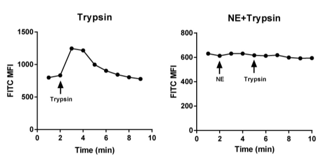
Trypsin purchased from MedChemExpress. Usage Cited in: J Immunother Cancer. 2025 Feb 26;13(2):e010813. [Abstract]
Ca2+ signals in HEK-293T cells treated with Trypsin (2 µg/mL, 8 min) and NE (flow cytometry).
-
J Neuroinflammation
Cardiac arrest triggers IL-17-mediated neuroinflammation and astrocyte polarization: insights into pathogenesis and intervention. [Abstract]2025 Nov 14;22(1):268. PMID: 41239323 -
-
J Med Chem
Peptidomimetic Analogues Act as Effective Inhibitors against SARS-CoV-2 by Blocking the Function of Cathepsin L. [Abstract]2024 Oct 10;67(19):17124-17143. PMID: 39292661 -
Cell Biosci
LARP1 acts as a key mediator in preventing angiotensin II-induced cardiac dysfunction and fibrosis. [Abstract]2025 Oct 22;15(1):143. PMID: 41126333 -
Foods
Glycosylation Remodeling and Thermal Denaturation Dictate the Functional Diversification of Protein Z. [Abstract]2026 Mar 4;15(5):853. PMID: 41829126 -
Int Immunopharmacol
USP4-mediated deubiquitination of NOTCH1 inhibits HNSCC progression through regulating oxidative stress and ferroptosis. [Abstract]2026 Jul 3:186:117071. PMID: 42398174 -
Oncol Res
MINDY1 Induces PD-L1 Deubiquitination to Promote Immune Escape in Hepatocellular Carcinoma by the Wnt/β-Catenin Pathway. [Abstract]2025 Oct 22;33(11):3583-3603. PMID: 41179305 -
J Neurochem
Demethylase FTO Regulates P2X3R Expression Contributing to the Mechanism of Hyperalgesia in Lumbar Disc Herniation. [Abstract]2025 Apr;169(4):e70058. PMID: 40170503 -
Regen Ther
Rat adipose-derived mesenchymal stem cell-derived exosomes loaded with miR-375 promote neurite outgrowth and peripheral nerve regeneration via activation of Akt by targeting EPHA4. [Abstract]2025 Aug 5:30:491-502. PMID: 41424620 -
BMC Genomics
Telomere-to-telomere genome assembly of Metarhizium acridum provides insights into Metarhizium genome evolution. [Abstract]2026 Jun 13. PMID: 42288757 -
Biochim Biophys Acta Mol Cell Biol Lipids
Proteomic and lipidomic landscape of the infrapatellar fat pad and its clinical significance in knee osteoarthritis. [Abstract]2024 Aug;1869(6):159513. PMID: 38788831 -
Toxicol Appl Pharmacol
Aureusidin protects against osteoarthritis via Caspase-3/gasdermin E-mediated pyroptosis. [Abstract]2026 Jun 15:514:117911. PMID: 42297245 -
Methods
Production of AFM wedged cantilevers for stress-relaxation experiments: Uniaxial loading of soft, spherical cells. [Abstract]2025 Apr:236:1-9. PMID: 39971021 -
Adv Biol (Weinh)
Mir-301b-3p Targets Alx4 to Suppress Cisplatin Sensitivity in Breast Cancer through DNA Damage. [Abstract]2026 Apr;10(4):e00715. PMID: 41944225 -
Biochim Biophys Acta Gen Subj
DLGAP5-induced glycolysis reprogramming reinforces macrophage M2 polarization to impact BC progression. [Abstract]2026 Sep;1870(9):130969. PMID: 42297309 -
PLoS One
ClpP deficiency attenuates contrast-induced HK-2 cell injury through changes associated with mitochondrial dynamics and apoptosis. [Abstract]2026 Jul 2;21(7):e0352422. PMID: 42391150 -
Mol Cell Biol
Paeonol Ameliorates Gastric Mucosal Enterosis and Dysplasia Associated with Chronic Atrophic Gastritis by Modulating the JAK2/STAT3 Pathway. [Abstract]2026 Mar 19:1-18. PMID: 41852098 -
Cytotechnology
METTL3/IGF2BP2 mediates m6A methylation modification of SNRPA1 to promote tumor property of non-small cell lung cancer cells. [Abstract]2026 Apr;78(2):57. PMID: 41841152 -
Neurochirurgie
Transcription factor EB (TFEB) promotes autophagy in early brain injury after subarachnoid hemorrhage in rats. [Abstract]2023 Jul 1:101465. PMID: 37400013 -
STAR Protoc
Protocol to quantify protein phosphorylation in mouse extended pluripotent stem cell-derived blastoids. [Abstract]2025 Nov 3;6(4):104166. PMID: 41191480 -
-
-
-
bioRxiv
Structural and Functional Insights into GGCX-FIX Interaction: Implications for Vitamin K-Dependent Bleeding Disorders. [Abstract]2025 Feb 19:2025.02.18.638829. PMID: 40027771


Lösungsmittel & Löslichkeit
In Vitro:
DMSO : 100 mg/mL (ultrasonic and warming and heat to 60°C; Hygroscopic DMSO has a significant impact on the solubility of product, please use newly opened DMSO)
H2O : ≥ 10 mg/mL
* "≥" means soluble, but saturation unknown.
In Vivo:
Select the appropriate dissolution method based on your experimental animal and administration route.
- For the following dissolution methods, please ensure to first prepare a clear stock solution using an In Vitro approach and then sequentially add co-solvents:
- To ensure reliable experimental results, the clarified stock solution can be appropriately stored based on storage conditions. As for the working solution for In Vivo experiments, it is recommended to prepare freshly and use it on the same day.
- The percentages shown for the solvents indicate their volumetric ratio in the final prepared solution. If precipitation or phase separation occurs during preparation, heat and/or sonication can be used to aid dissolution.
Add each solvent one by one: 10% DMSO 40% PEG300 5% Tween-80 45% Saline
Solubility: ≥ 2.5 mg/mL; Clear solution
This protocol yields a clear solution of ≥ 2.5 mg/mL (saturation unknown).
Taking 1 mL working solution as an example, add 100 μL DMSO stock solution (25.0 mg/mL) to 400 μL PEG300, and mix evenly; then add 50 μL Tween-80 and mix evenly; then add 450 μL Saline to adjust the volume to 1 mL.
Preparation of Saline: Dissolve 0.9 g sodium chloride in ddH₂O and dilute to 100 mL to obtain a clear Saline solution.
Add each solvent one by one: 10% DMSO 90% (20% SBE-β-CD in Saline)
Solubility: ≥ 2.5 mg/mL; Clear solution
This protocol yields a clear solution of ≥ 2.5 mg/mL (saturation unknown).
Taking 1 mL working solution as an example, add 100 μL DMSO stock solution (25.0 mg/mL) to 900 μL 20% SBE-β-CD in Saline, and mix evenly.
Preparation of 20% SBE-β-CD in Saline (4°C, storage for one week): 2 g SBE-β-CD powder is dissolved in 10 mL Saline, completely dissolve until clear.
For the following dissolution methods, please prepare the working solution directly:
It is recommended to prepare fresh solutions and use them promptly within a short period of time.
The percentages shown for the solvents indicate their volumetric ratio in the final prepared solution. If precipitation or phase separation occurs during preparation, heat and/or sonication can be used to aid dissolution.
Add each solvent one by one: PBS
Solubility: ≥ 25 mg/mL; Clear solution
In Vivo Dissolution Calculator
Please enter the basic information of animal experiments:
-
-
-
-
Recommended: Prepare an additional quantity of animals to account for potential losses during experiments.
Please enter your animal formula composition:
-
%DMSO +
Recommended: Keep the proportion of DMSO in working solution below 2% if your animal is weak.
-
%+
-
+%Tween-80 + +
-
%Saline +
The co-solvents required include: DMSO, . All of co-solvents are available by MedChemExpress (MCE). , Tween 80. All of co-solvents are available by MedChemExpress (MCE).
Working solution concentration: 0.22 mg/mL
Method for preparing stock solution: mg drug dissolved in μL DMSO. Stock solution concentration: mg/mL.
1. Take μL DMSO stock solution;
2. Add μL .
μL , mix evenly;
3. Then add μL Tween 80, mix evenly;
4. Then add μL
Please ensure that the stock solution in the first step is dissolved to a clear state, and add co-solvents in sequence. You can use ultrasonic heating (ultrasonic cleaner, recommended frequency 20-40 kHz), vortexing, etc. to assist dissolution.


Reinheit & Dokumentation
-
Data Sheet (279 KB)
-
SDS (557 KB)
- English - EN (557 KB)
- Français - FR (557 KB)
- Deutsch - DE (557 KB)
- Norwegian - NO (557 KB)
- Español - ES (557 KB)
- Swedish - SV (557 KB)
- Italian - IT (557 KB)
- Korean - KR (557 KB)
- Portuguese - PT (557 KB)
-
Handling Instructions (2659 KB)
Verweise
[2]. Yue-Lin Yang,et al. Trypsin promotes porcine deltacoronavirus mediating cell-to-cell fusion in a cell type-dependent manner. Emerg Microbes Infect. 2020 Feb 24;9(1):457-468. [Content Brief]
[3]. H Amano, et al. Trypsin promotes C6 glioma cell proliferation in serum- and growth factor-free medium. Neurosci Res. 1996 Jul;25(3):203-8. [Content Brief]
[5]. R Costa, et al. Evidence for the role of neurogenic inflammation components in trypsin-elicited scratching behaviour in mice. Br J Pharmacol. 2008 Jul;154(5):1094-103. [Content Brief]
[6]. F Schmidlin, et al. Protease-activated receptors: how proteases signal to cells. Curr Opin Pharmacol. 2001 Dec;1(6):575-82. [Content Brief]

Calculators
Konzentration (Stammlösung) × Volumen (Stammlösung) = Konzentration (Ziellösung) × Volumen (Ziellösung)


Trypsin Related Classifications
HY-129047 Related Classifications
 Powered by Bioz