



AP-6
Based on 1 publication(s) in Google Scholar
AP-6 is a selective inhibitor of TMEM175 with activity in modulating lysosomal function. Acute inhibition of TMEM175 by AP-6 increases lysosomal macromolecular catabolism, thereby accelerating macrophage and other digestive processes. AP-6 may be used in Parkinson's disease research.
Nos produits utilisent uniquement pour la recherche. Nous ne vendons pas aux patients.
- Pureté : 99.81%
- CAS No.: 1369963-51-8
- Formule: C16H14N4
- Masse moléculaire:262.31
-
Stockage:Powder -20°C, 3 years , 4°C, 2 years ; In solvent -80°C, 6 months , -20°C, 1 month


To place orders, for customer services and technical support, please contact: MedChemExpress USA
Tel: 609-228-6898 E-mail: [email protected] [email protected]


-
 Activité biologique
-
Chemical Information
-
Solvant et solubilité
- Protocole
- Pureté et documentation
- Références
-
 Help & FAQs



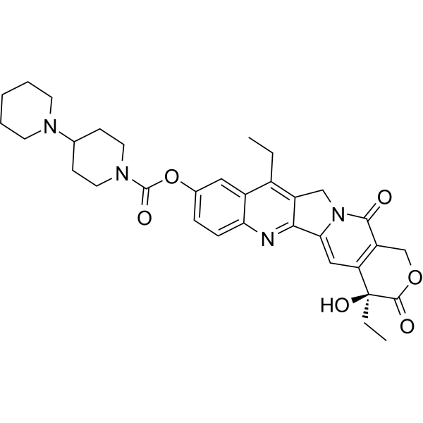
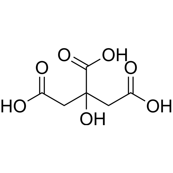
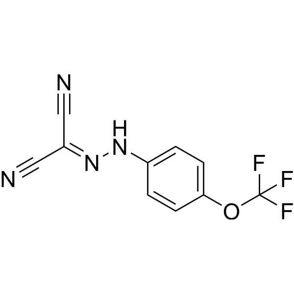
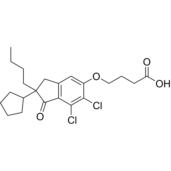

Publications Citing Use of MedChemExpress (MCE) AP-6
More

Activité biologique
Description


Chemical Information
-
CAS No. 1369963-51-8
-
Appearance Solid
-
Masse moléculaire 262.31
-
Formule C16H14N4
-
Color White to off-white
-
SMILES
NC1=CC(C2=CC=CC(C3=CC(N)=CC=N3)=C2)=NC=C1
-
Livraison
Room temperature in continental US; may vary elsewhere.
-
Stockage
Powder -20°C 3 years 4°C 2 years In solvent -80°C 6 months -20°C 1 month


Publications (1)
-
Journal Impact Factor
-
Most Recent


Solvant et solubilité
In Vitro:
DMSO : 0.93 mg/mL (3.55 mM; ultrasonic and warming and adjust pH to 3 with 1 M HCl and heat to 60°C; Hygroscopic DMSO has a significant impact on the solubility of product, please use newly opened DMSO)
H2O : < 0.1 mg/mL (insoluble)
Please refer to the solubility information to select the appropriate solvent. Once prepared, please aliquot and store the solution to prevent product inactivation from repeated freeze-thaw cycles.
Storage method and period of stock solution: -80°C, 6 months; -20°C, 1 month. When stored at -80°C, please use it within 6 months. When stored at -20°C, please use it within 1 month.
Please refer to the solubility information to select the appropriate solvent. Once prepared, please aliquot and store the solution to prevent product inactivation from repeated freeze-thaw cycles.
Storage method and period of stock solution: -80°C, 6 months; -20°C, 1 month. When stored at -80°C, please use it within 6 months. When stored at -20°C, please use it within 1 month.
Concentration (start) × Volume (start) = Concentration (final) × Volume (final)

Protocole
-
Phagocytosis Functional Assay
A phagocytosis functional assay measures the ability of phagocytic cells, such as neutrophils, macrophages, monocytes, or microglia/macrophages, to bind and internalize particulate targets including bacteria, yeast particles, beads, or myelin particles. Fluorescent flow-cytometry assays detect target uptake as fluorescence associated with gated phagocytes, while pH-sensitive dyes such as pHrodo increase signal in acidic phagosomal compartments and therefore preferentially report internalized particles rather than particles remaining outside the cell. Microscopy or high-content imaging can be used to confirm intracellular localization and, in some protocols, to follow uptake kinetics.
-
Human pluripotent stem cell midbrain dopaminergic neuron differentiation
Human pluripotent stem cells are directed toward midbrain dopaminergic neurons by first inducing a neural floor-plate-like progenitor state, then patterning cells with ventralizing SHH signaling and midbrain/WNT-FGF cues, and finally maturing progenitors into neurons expressing dopaminergic markers such as TH, NURR1/NR4A2, PITX3, DAT/SLC6A3, VMAT2/SLC18A2, GIRK2/KCNJ6, FOXA2, LMX1A, and EN1. The main readouts are loss of pluripotency, acquisition of FOXA2+/LMX1A+ midbrain floor-plate progenitors, emergence of βIII-tubulin+/MAP2+ neurons, and production of TH+ dopaminergic neurons with molecular, dopamine-release, and electrophysiological features of midbrain dopaminergic identity.
-
Lysosome and acidic-vesicle live-cell staining
Lysosome and acidic-vesicle live-cell staining detects acidic intracellular compartments by using membrane-permeant acidotropic probes that accumulate in low-pH vesicles, including lysosomes, late endosomes, autolysosomes, and acidic phagosomes. LysoTracker staining is commonly used as an intensity-based readout of acidic lysosomal compartment abundance or enlargement, while acridine orange produces green fluorescence in less concentrated compartments and red fluorescence after concentration-dependent accumulation in acidic vesicular organelles. Loss or reduction of acridine-orange red signal can be used as a readout of lysosomal membrane permeabilization or reduced acidic-vesicle integrity. This protocol is designed for live cultured cells and can be adapted for fluorescence microscopy, high-content imaging, plate-reader readout, or flow cytometry when the selected literature supports the readout. Because these dyes report acidotropic accumulation rather than lysosome identity alone,


Pureté et documentation
-
Fiche technique (272 KB)
-
SDS (251 KB)
- English - EN (251 KB)
- Français - FR (251 KB)
- Deutsch - DE (251 KB)
- Norwegian - NO (251 KB)
- Español - ES (251 KB)
- Swedish - SV (251 KB)
- Italian - IT (251 KB)
- Korean - KR (251 KB)
- Portuguese - PT (251 KB)
-
Instruction de manipulation (2659 KB)
Références


Complete Stock Solution Preparation Table
Please refer to the solubility information to select the appropriate solvent. Once prepared, please aliquot and store the solution to prevent product inactivation from repeated freeze-thaw cycles.
Storage method and period of stock solution: -80°C, 6 months; -20°C, 1 month. When stored at -80°C, please use it within 6 months. When stored at -20°C, please use it within 1 month.
| Optional Solvent | Concentration Solvent Mass | 1 mg | 5 mg | 10 mg | 25 mg |
|---|---|---|---|---|---|
| DMSO | 1 mM | 3.8123 mL | 19.0614 mL | 38.1228 mL | 95.3071 mL |


AP-6 Related Classifications
HY-161949 Related Classifications
 Powered by Bioz