



CHK1-IN-3

Based on 1 Customer Validation
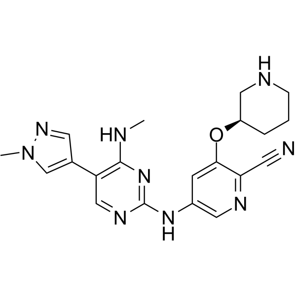
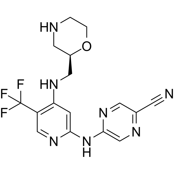
CHK1-IN-3 is a potent and selective CHK1 inhibitor with an IC50 of 0.4 nM. CHK1-IN-3 effectively inhibits the growth of malignant hematopathy cell lines and displays low affinity for hERG (IC50 > 40 μM). CHK1-IN-3 significantly suppresses the tumor growth in vivo. CHK1-IN-3 can be used for the study of hematologic malignancies.
For research use only. We do not sell to patients.
- Purity : 99.79%
- CAS No.: 2097252-39-4
- Formula: C20H23N9O
- Molecular Weight:405.46
-
Storage:Powder -20°C, 3 years ; In solvent -80°C, 6 months , -20°C, 1 month

To place orders, for customer services and technical support, please contact: MedChemExpress USA
Tel: 609-228-6898 E-mail: [email protected] [email protected]


-
 Biological Activity
-
Chemical Information
-
Solvent & Solubility
- Protocols
- Purity & Documentation
- References
-
 Help & FAQs


-
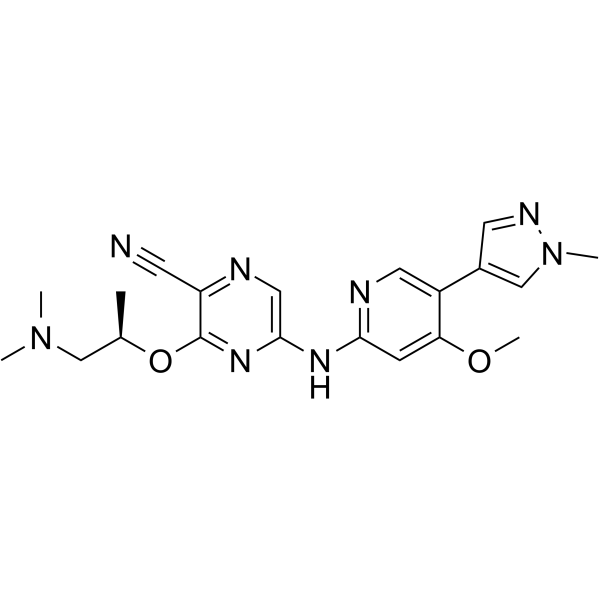
HY-18175
CCT244747 -
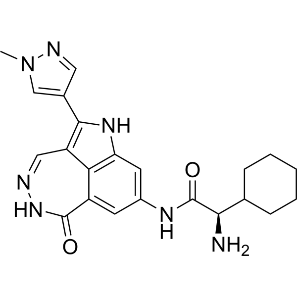
HY-10032
PF 477736Chk1 Inhibitor -
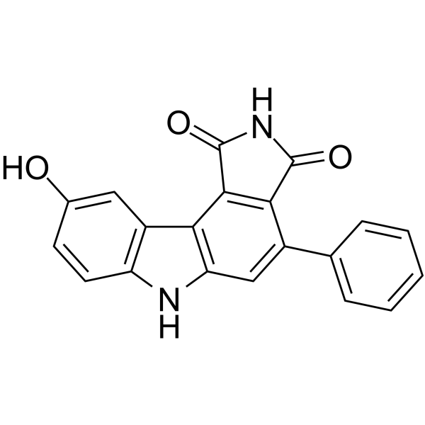
HY-18961
PD 407824Chk1/Wee1 Inhibitor -
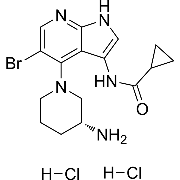
HY-112167A
GDC-0575 dihydrochlorideCHK1 Inhibitor -
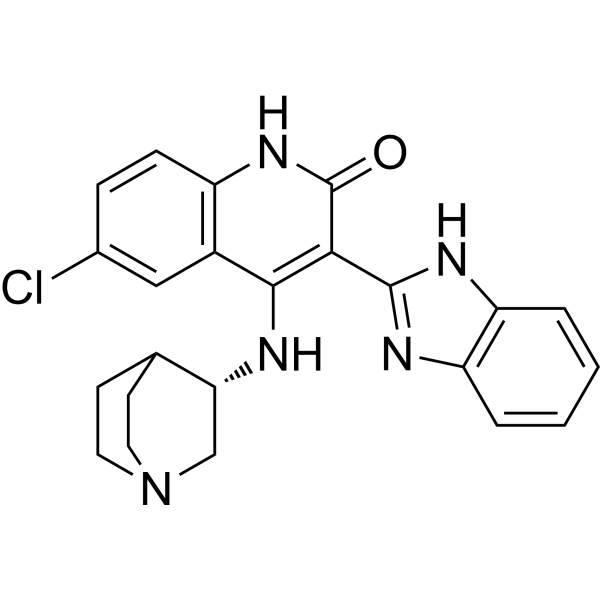
HY-13263
CHIR-124Checkpoint Kinase (Chk) Inhibitor -
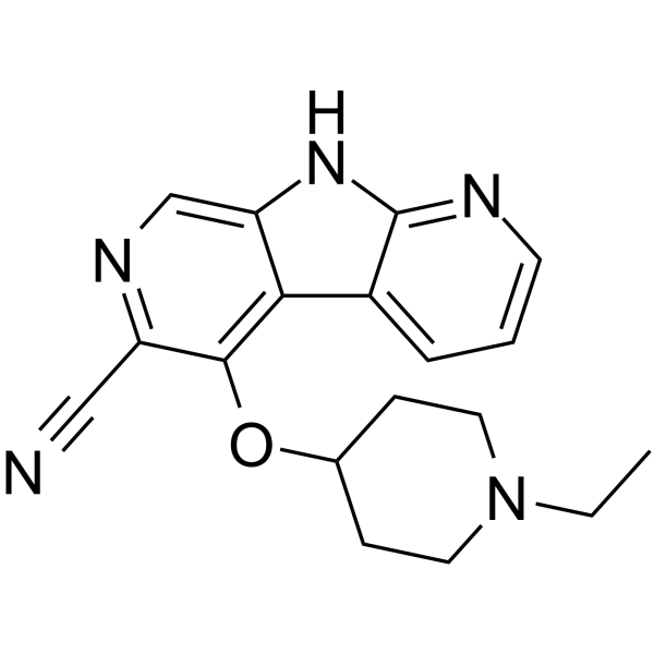
HY-19926
GDC-0425ChK1 Inhibitor -
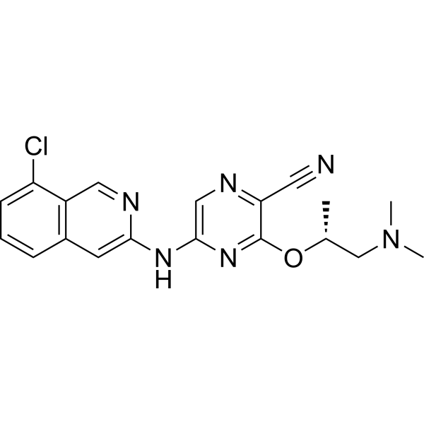
HY-100195
SAR-020106CHK1 Inhibitor -
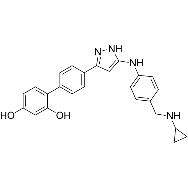
HY-103367
CHK1-IN-7CHK1 Inhibitor -
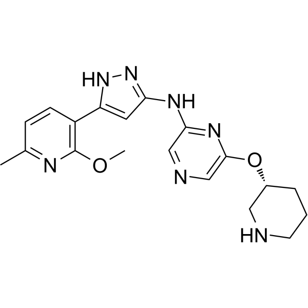
HY-148962
LY2880070CHK1 Inhibitor -
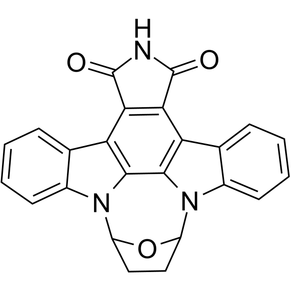
HY-107407
SB-218078Chk1 Inhibitor -
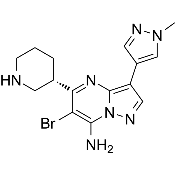
HY-15532B
SCH900776 (S-isomer)CHK1 Serine/Threonine Kinase Inhibitor -
HY-18958
CCT245737CHK1 Inhibitor


Biological Activity
Description
IC50 & Target
[1]|
Chk1 0.4 nM (IC50) |
Cellular Effect
|
Cell Line

|
Type | Value | Description | References |
|---|---|---|---|---|
| CHO | IC50 |
>40 μM
Compound: (R)-17
|
Inhibition of human ERG expressed in CHO cells at -80 mV holding potential by automated patch clamp assay
Inhibition of human ERG expressed in CHO cells at -80 mV holding potential by automated patch clamp assay
|
[PMID: 30986571] |
| JeKo-1 | IC50 |
0.036 μM
Compound: (R)-17
|
Inhibition of human JeKo1 cells incubated for 72 hrs by cellTiter 96 aqueous one solution reagent based assay
Inhibition of human JeKo1 cells incubated for 72 hrs by cellTiter 96 aqueous one solution reagent based assay
|
[PMID: 30986571] |
| MV4-11 | IC50 |
0.039 μM
Compound: (R)-17
|
Inhibition of human MV411 cells incubated for 72 hrs by cellTiter 96 aqueous one solution reagent based assay
Inhibition of human MV411 cells incubated for 72 hrs by cellTiter 96 aqueous one solution reagent based assay
|
[PMID: 30986571] |
| Ramos | IC50 |
0.095 μM
Compound: (R)-17
|
Inhibition of human Ramos cells incubated for 72 hrs by cellTiter 96 aqueous one solution reagent based assay
Inhibition of human Ramos cells incubated for 72 hrs by cellTiter 96 aqueous one solution reagent based assay
|
[PMID: 30986571] |
| RPMI-8226 | IC50 |
1.991 μM
Compound: (R)-17
|
Inhibition of human RPMI8226 cells incubated for 72 hrs by cellTiter 96 aqueous one solution reagent based assay
Inhibition of human RPMI8226 cells incubated for 72 hrs by cellTiter 96 aqueous one solution reagent based assay
|
[PMID: 30986571] |
| Z-138 | IC50 |
0.012 μM
Compound: (R)-17
|
Inhibition of human Z138 cells incubated for 72 hrs by cellTiter 96 aqueous one solution reagent based assay
Inhibition of human Z138 cells incubated for 72 hrs by cellTiter 96 aqueous one solution reagent based assay
|
[PMID: 30986571] |
In Vitro
CHK1-IN-3 (Compound (R)-17) exhibits kinase inhibition against CHK2, AMPKα2β1γ1, MPKα1β1γ1, PIM1, PIM3 with IC50s of 1729, 91.11, 107.5, 511.8 and 735.53 nM[1].
CHK1-IN-3 shows strong inhibitory effects on hematological malignancy cell lines: Mino (IC50 = 155 nM), Jeko-1 (IC50 = 36 nM), MV4-11 (IC50 = 39 nM) and Z-138 (IC50 = 13 nM)[1].
MedChemExpress (MCE) has not independently confirmed the accuracy of these methods. They are for reference only.
Parmacokinetics
| Species | Dose | Route | T1/2 | Cmax | AUC0-t | AUC0-∞ | Vss | MRT | CL |
|---|---|---|---|---|---|---|---|---|---|
| Mice[1] | 15 mg/mL | i.v. | 2.33 h | 849 ng/mL | 363 μg/L·h | 370 μg/L·h | 6.42 L/kg | 0.80 h | 135 mL/min/kg |
In Vivo
MedChemExpress (MCE) has not independently confirmed the accuracy of these methods. They are for reference only.
-
Animal Model:Z-138 cell inoculated xenograft model established in female nu/nu mice[1]
-
Dosage:10 and 20 mg/kg
-
Administration:Intravenous infection (i.v.), 5 times every week for 3 weeks
-
Result:Observed tumor growth inhibitions of 78.64% and 90.29% at doses of 10 and 20 mg/kg.
No significant body weight loss and no adverse effects were observed.


Chemical Information
-
CAS No. 2097252-39-4
-
Appearance Solid
-
Molecular Weight 405.46
-
Formula C20H23N9O
-
Color White to light yellow
-
SMILES
CNC1=C(C2=CN(C)N=C2)C=NC(NC3=CN=C(C#N)C(O[C@H]4CNCCC4)=C3)=N1
-
Shipping
Room temperature in continental US; may vary elsewhere.
-
Storage
Powder -20°C 3 years In solvent -80°C 6 months -20°C 1 month


Solvent & Solubility
In Vitro:
DMSO : ≥ 125 mg/mL (308.29 mM; Hygroscopic DMSO has a significant impact on the solubility of product, please use newly opened DMSO)
* "≥" means soluble, but saturation unknown.
Please refer to the solubility information to select the appropriate solvent. Once prepared, please aliquot and store the solution to prevent product inactivation from repeated freeze-thaw cycles.
Storage method and period of stock solution: -80°C, 6 months; -20°C, 1 month. When stored at -80°C, please use it within 6 months. When stored at -20°C, please use it within 1 month.
Please refer to the solubility information to select the appropriate solvent. Once prepared, please aliquot and store the solution to prevent product inactivation from repeated freeze-thaw cycles.
Storage method and period of stock solution: -80°C, 6 months; -20°C, 1 month. When stored at -80°C, please use it within 6 months. When stored at -20°C, please use it within 1 month.
Concentration (start) × Volume (start) = Concentration (final) × Volume (final)


Purity & Documentation
-
Data Sheet (275 KB)
-
SDS (251 KB)
- English - EN (251 KB)
- Français - FR (251 KB)
- Deutsch - DE (251 KB)
- Norwegian - NO (251 KB)
- Español - ES (251 KB)
- Swedish - SV (251 KB)
- Italian - IT (251 KB)
- Korean - KR (251 KB)
- Portuguese - PT (251 KB)
-
Handling Instructions (2659 KB)
References


Complete Stock Solution Preparation Table
Please refer to the solubility information to select the appropriate solvent. Once prepared, please aliquot and store the solution to prevent product inactivation from repeated freeze-thaw cycles.
Storage method and period of stock solution: -80°C, 6 months; -20°C, 1 month. When stored at -80°C, please use it within 6 months. When stored at -20°C, please use it within 1 month.
| Optional Solvent | Concentration Solvent Mass | 1 mg | 5 mg | 10 mg | 25 mg |
|---|---|---|---|---|---|
| DMSO | 1 mM | 2.4663 mL | 12.3317 mL | 24.6633 mL | 61.6584 mL |
| 5 mM | 0.4933 mL | 2.4663 mL | 4.9327 mL | 12.3317 mL | |
| 10 mM | 0.2466 mL | 1.2332 mL | 2.4663 mL | 6.1658 mL | |
| 15 mM | 0.1644 mL | 0.8221 mL | 1.6442 mL | 4.1106 mL | |
| 20 mM | 0.1233 mL | 0.6166 mL | 1.2332 mL | 3.0829 mL | |
| 25 mM | 0.0987 mL | 0.4933 mL | 0.9865 mL | 2.4663 mL | |
| 30 mM | 0.0822 mL | 0.4111 mL | 0.8221 mL | 2.0553 mL | |
| 40 mM | 0.0617 mL | 0.3083 mL | 0.6166 mL | 1.5415 mL | |
| 50 mM | 0.0493 mL | 0.2466 mL | 0.4933 mL | 1.2332 mL | |
| 60 mM | 0.0411 mL | 0.2055 mL | 0.4111 mL | 1.0276 mL | |
| 80 mM | 0.0308 mL | 0.1541 mL | 0.3083 mL | 0.7707 mL | |
| 100 mM | 0.0247 mL | 0.1233 mL | 0.2466 mL | 0.6166 mL |


CHK1-IN-3 Related Classifications
HY-128601 Related Classifications
 Powered by Bioz