



XL888
Based on 6 publication(s) in Google Scholar
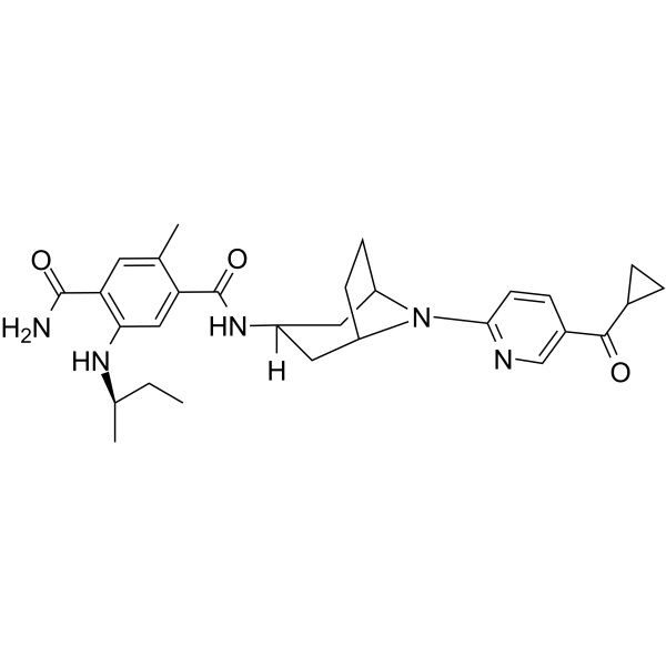
XL888 is a potent and orally active HSP90 inhibitor with an IC50 value of 24 nM. XL888 shows anti-proliferation activity and induces Apoptosis. XL888 shows anti-tumor activity.
For research use only. We do not sell to patients.
- Purity : 99.41%
- CAS No.: 1149705-71-4
- Formula: C29H37N5O3
- Molecular Weight:503.64
-
Storage:Powder -20°C, 3 years ; In solvent -80°C, 6 months , -20°C, 1 month

To place orders, for customer services and technical support, please contact: MedChemExpress USA
Tel: 609-228-6898 E-mail: [email protected] [email protected]


-
 Biological Activity
-
Chemical Information
-
Solvent & Solubility
- Protocols
- Purity & Documentation
- References
-
 Help & FAQs


-
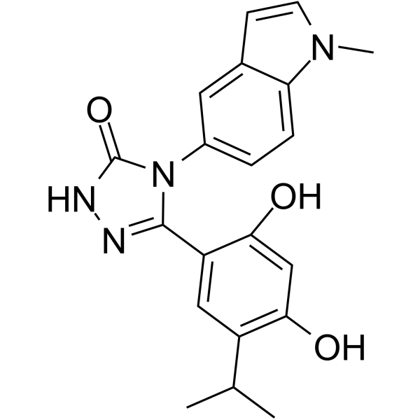
HY-15205
GanetespibHSP90 Inhibitor -
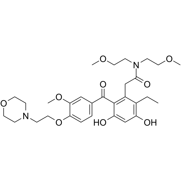
HY-13468
KW-2478 -
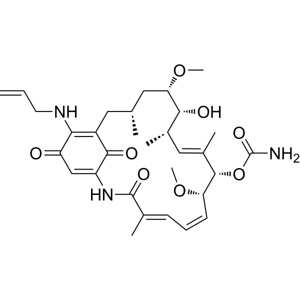
HY-10211
TanespimycinHSP90 Inhibitor -
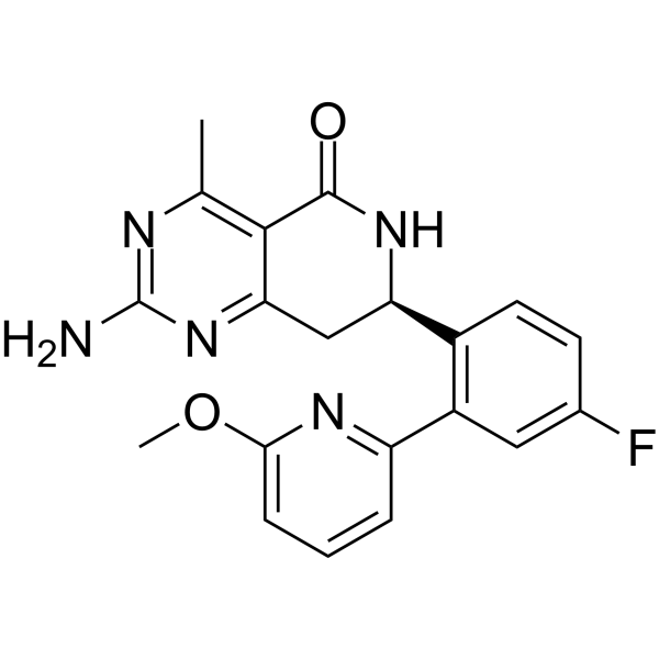
HY-15190
NVP-HSP990Apoptosis Inducer -
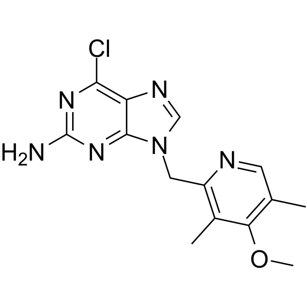
HY-10212
BIIB021HSP90 Inhibitor -
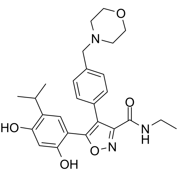
HY-10215
LuminespibHSP90 Inhibitor -
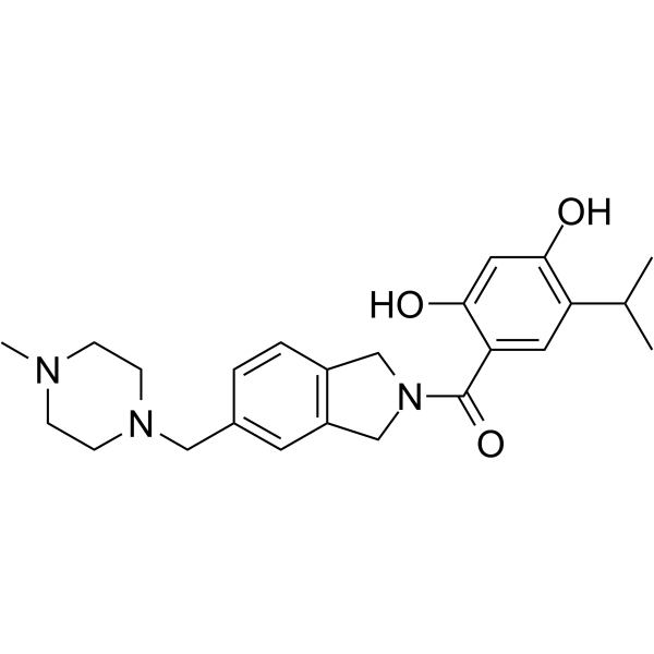
HY-14463
OnalespibHsp90 Inhibitor -
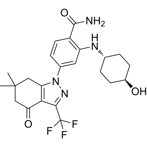
HY-10214
SNX-2112 -
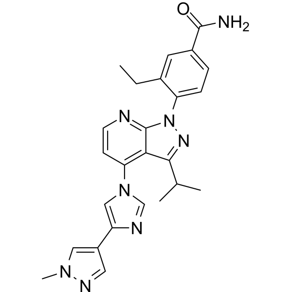
HY-15785
PimitespibHSP90α/HSP90β Inhibitor -
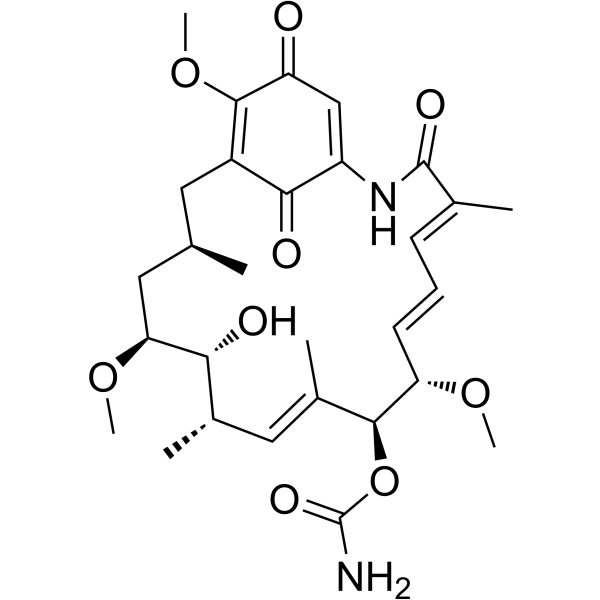
HY-15230
GeldanamycinHsp90 Inhibitor -
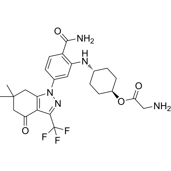
HY-10213
SNX-5422 -
HY-11038
ZelavespibHsp90 Inhibitor
Publications Citing Use of MedChemExpress (MCE) XL888
More Customer Validation & Images

-
Flow Cytometry
-
IF
-
In Vivo Efficacy Study


Biological Activity
Description
IC50 & Target
[3]|
HSP90 24 nM (IC50) |
Cellular Effect
|
Cell Line

|
Type | Value | Description | References |
|---|---|---|---|---|
| A549 | IC50 |
4.3 nM
Compound: 12i
|
Antiproliferative activity against human A549 cells overexpressing Kras after 48 hrs by BrdU labeling assay
Antiproliferative activity against human A549 cells overexpressing Kras after 48 hrs by BrdU labeling assay
|
[PMID: 22877636] |
| BT-474 | IC50 |
0.1 nM
Compound: 12i
|
Antiproliferative activity against human BT474 cells overexpressing Her2 after 48 hrs by BrdU labeling assay
Antiproliferative activity against human BT474 cells overexpressing Her2 after 48 hrs by BrdU labeling assay
|
[PMID: 22877636] |
| COLO 205 | IC50 |
11.6 nM
Compound: 12i
|
Antiproliferative activity against human COLO205 cells overexpressing Braf after 48 hrs by BrdU labeling assay
Antiproliferative activity against human COLO205 cells overexpressing Braf after 48 hrs by BrdU labeling assay
|
[PMID: 22877636] |
| HN5 | IC50 |
5.5 nM
Compound: 12i
|
Antiproliferative activity against human HN5 cells overexpressing EGFR after 48 hrs by BrdU labeling assay
Antiproliferative activity against human HN5 cells overexpressing EGFR after 48 hrs by BrdU labeling assay
|
[PMID: 22877636] |
| MCF7 | IC50 |
4.1 nM
Compound: 12i
|
Antiproliferative activity against human MCF7 cells overexpressing PI3K after 48 hrs by BrdU labeling assay
Antiproliferative activity against human MCF7 cells overexpressing PI3K after 48 hrs by BrdU labeling assay
|
[PMID: 22877636] |
| MDA-MB-453 | IC50 |
16 nM
Compound: 12i
|
Antiproliferative activity against human MDA-MB-453 cells overexpressing Her2 after 48 hrs by BrdU labeling assay
Antiproliferative activity against human MDA-MB-453 cells overexpressing Her2 after 48 hrs by BrdU labeling assay
|
[PMID: 22877636] |
| MKN-45 | IC50 |
45.5 nM
Compound: 12i
|
Antiproliferative activity against human MKN45 cells overexpressing MET after 48 hrs by BrdU labeling assay
Antiproliferative activity against human MKN45 cells overexpressing MET after 48 hrs by BrdU labeling assay
|
[PMID: 22877636] |
| NCI-H1975 | IC50 |
0.7 nM
Compound: 12i
|
Antiproliferative activity against human NCI-H1975 cells overexpressing EGFR after 48 hrs by BrdU labeling assay
Antiproliferative activity against human NCI-H1975 cells overexpressing EGFR after 48 hrs by BrdU labeling assay
|
[PMID: 22877636] |
| NCI-N87 | IC50 |
21.8 nM
Compound: 12i
|
Antiproliferative activity against human NCI-N87 cells overexpressing Her2 after 48 hrs by BrdU labeling assay
Antiproliferative activity against human NCI-N87 cells overexpressing Her2 after 48 hrs by BrdU labeling assay
|
[PMID: 22877636] |
| NCI-N87 | IC50 |
56 nM
Compound: 12i
|
Inhibition of Hsp90 in human NCI-N87 cells assessed as Her2 degradation after 24 hrs by Western blot analysis
Inhibition of Hsp90 in human NCI-N87 cells assessed as Her2 degradation after 24 hrs by Western blot analysis
|
[PMID: 22877636] |
| SK-MEL-28 | IC50 |
0.3 nM
Compound: 12i
|
Antiproliferative activity against human SK-MEL-28 cells overexpressing Braf after 48 hrs by BrdU labeling assay
Antiproliferative activity against human SK-MEL-28 cells overexpressing Braf after 48 hrs by BrdU labeling assay
|
[PMID: 22877636] |
In Vitro
XL888 (300 nM; 24, 72, 144 h) blocks the growth and survival of melanoma cell lines with diverse mechanisms of vemurafenib resistance and induces cycle arrest at G1-phase or G2/M phase, induces apoptosis[1].
XL888 (300 nM; 72h) increases the expression of BIM and decreases the expression of Mcl-1[1].
MedChemExpress (MCE) has not independently confirmed the accuracy of these methods. They are for reference only.
-
Cell Line:WM164, M229, M229R, M249, M249R, 1205Lu, WM39, WM164R, 1205LuR, RPMI 7951 cells
-
Concentration:300 nM
-
Incubation Time:24 h
-
Result:Induced cell cycle arrest at G1-phase in WM164, M229, M229R, M249, M249R, 1205Lu, WM39 cells, G2/M phase cell cycle arrest for WM164R, 1205LuR, RPMI 795 cells.
-
Cell Line:WM164R, M229R, WM39 cells
-
Concentration:300 nM
-
Incubation Time:72, 144 h
-
Result:Induced cell apoptosis.
-
Cell Line:M229R, 1205LuR, RPMI7951, WM39, WM164R, M249R cells
-
Concentration:300 nM
-
Incubation Time:72 h
-
Result:Increased the expression of BIM-EL, BIM-L and BIM-S expression in the M229R, 1205LuR, RPMI7951 and WM39 cell lines, induced expression of BIM-L and BIM-S in the WM164R cell line and BIM-EL in the M249R cell line.
-
Cell Line:NCI-N87, BT-474, MDA-MB-453, MKN45, Colo-205, SK-MEL-28, HN5, NCI-H1975, MCF7, A549 cells
-
Concentration:0-50 nM
-
Incubation Time:48 h
-
Result:Showed anti-proliferation activity with IC50s of 21.8, 0.1, 16.0, 45.5, 11.6, 0.3, 5.5, 0.7, 4.1, 4.3 nM for NCI-N87, BT-474, MDA-MB-453, MKN45, Colo-205, SK-MEL-28, HN5, NCI-H1975, MCF7, A549 cells, respectively.
In Vivo
MedChemExpress (MCE) has not independently confirmed the accuracy of these methods. They are for reference only.
-
Animal Model:BALB SCID mice (M229R and 1205LuR xenografts model)[1]
-
Dosage:100 mg/kg
-
Administration:P.o.; thrice per week for 15 days
-
Result:Inhibited the tumor growth and increases the expression of intratumoral HSP70.
Clinical Trial
| NCT Number | Sponsor | Condition | Start Date |
Phase
|
|---|---|---|---|---|
| NCT01329991 | 2011-05 | PHASE1 |


Chemical Information
-
CAS No. 1149705-71-4
-
Appearance Solid
-
Molecular Weight 503.64
-
Formula C29H37N5O3
-
Color Off-white to light yellow
-
SMILES
CC1=CC(C(N)=O)=C(N[C@H](C)CC)C=C1C(N[C@]2([H])C[C@H](CC3)N(C4=NC=C(C(C5CC5)=O)C=C4)[C@H]3C2)=O
-
Shipping
Room temperature in continental US; may vary elsewhere.
-
Storage
Powder -20°C 3 years In solvent -80°C 6 months -20°C 1 month


Publications (6)
-
Journal Impact Factor
-
Most Recent
-
Cell Stem Cell
Senolytic-sensitive p16Ink4a+ fibroblasts in the tumor stroma rewire lung cancer metabolism and plasticity. [Abstract]2025 Dec 4;32(12):1869-1885.e8. PMID: 41187746
XL888 purchased from MedChemExpress. Usage Cited in: Cell Stem Cell. 2025 Dec 4;32(12):1869-1885.e8. [Abstract]
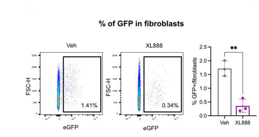
Flow cytometry analysis of GFP+ fibroblasts in PCLS treated with XL888 (1 μM).
XL888 purchased from MedChemExpress. Usage Cited in: Cell Stem Cell. 2025 Dec 4;32(12):1869-1885.e8. [Abstract]
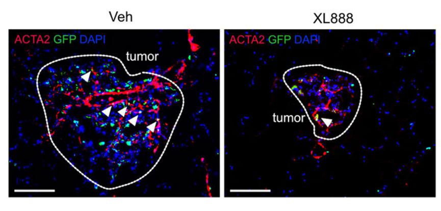
Representative image of GFP and ACTA2 immunofluorescence in PCLS treated with vehicle or XL888.
XL888 purchased from MedChemExpress. Usage Cited in: Cell Stem Cell. 2025 Dec 4;32(12):1869-1885.e8. [Abstract]
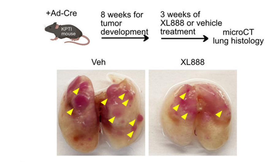
Experimental design to test tumor suppressive effect of XL888 (62.5 mg/kg, p.o.) in KPTI mouse (top), alongside representative macroscopic lung images from vehicle- and XL888-treated groups (bottom).
-
Theranostics
Inhibition of HSP90β Improves Lipid Disorders by Promoting Mature SREBPs Degradation via the Ubiquitin-proteasome System. [Abstract]2019 Aug 12;9(20):5769-5783. PMID: 31534518 -
Life Sci
The HSP90 inhibitor, XL888, enhanced cell apoptosis via downregulating STAT3 after insufficient radiofrequency ablation in hepatocellular carcinoma. [Abstract]2021 Oct 1:282:119762. PMID: 34186047 -
Transl Oncol
Integrated bioinformatics and experimental validation identifies CLIC6 as a novel tumor suppressor regulating NF-κB signaling and immune microenvironment in nasopharyngeal carcinoma. [Abstract]2026 Jul:69:102802. PMID: 42092278 -
-
bioRxiv
2024 Jul 30:2024.07.29.605645. PMID: 39131266


Solvent & Solubility
In Vitro:
DMSO : 50 mg/mL (99.28 mM; Need ultrasonic; Hygroscopic DMSO has a significant impact on the solubility of product, please use newly opened DMSO)
Please refer to the solubility information to select the appropriate solvent. Once prepared, please aliquot and store the solution to prevent product inactivation from repeated freeze-thaw cycles.
Storage method and period of stock solution: -80°C, 6 months; -20°C, 1 month. When stored at -80°C, please use it within 6 months. When stored at -20°C, please use it within 1 month.
Please refer to the solubility information to select the appropriate solvent. Once prepared, please aliquot and store the solution to prevent product inactivation from repeated freeze-thaw cycles.
Storage method and period of stock solution: -80°C, 6 months; -20°C, 1 month. When stored at -80°C, please use it within 6 months. When stored at -20°C, please use it within 1 month.
Concentration (start) × Volume (start) = Concentration (final) × Volume (final)
In Vivo:
Select the appropriate dissolution method based on your experimental animal and administration route.
- For the following dissolution methods, please ensure to first prepare a clear stock solution using an In Vitro approach and then sequentially add co-solvents:
- To ensure reliable experimental results, the clarified stock solution can be appropriately stored based on storage conditions. As for the working solution for In Vivo experiments, it is recommended to prepare freshly and use it on the same day.
- The percentages shown for the solvents indicate their volumetric ratio in the final prepared solution. If precipitation or phase separation occurs during preparation, heat and/or sonication can be used to aid dissolution.
Add each solvent one by one: 10% DMSO 40% PEG300 5% Tween-80 45% Saline
Solubility: ≥ 1.25 mg/mL (2.48 mM); Clear solution
This protocol yields a clear solution of ≥ 1.25 mg/mL (saturation unknown).
Taking 1 mL working solution as an example, add 100 μL DMSO stock solution (12.5 mg/mL) to 400 μL PEG300, and mix evenly; then add 50 μL Tween-80 and mix evenly; then add 450 μL Saline to adjust the volume to 1 mL.
Preparation of Saline: Dissolve 0.9 g sodium chloride in ddH₂O and dilute to 100 mL to obtain a clear Saline solution.
Add each solvent one by one: 10% DMSO 90% (20% SBE-β-CD in Saline)
Solubility: ≥ 1.25 mg/mL (2.48 mM); Clear solution
This protocol yields a clear solution of ≥ 1.25 mg/mL (saturation unknown).
Taking 1 mL working solution as an example, add 100 μL DMSO stock solution (12.5 mg/mL) to 900 μL 20% SBE-β-CD in Saline, and mix evenly.
Preparation of 20% SBE-β-CD in Saline (4°C, storage for one week): 2 g SBE-β-CD powder is dissolved in 10 mL Saline, completely dissolve until clear.
In Vivo Dissolution Calculator
Please enter the basic information of animal experiments:
-
-
-
-
Recommended: Prepare an additional quantity of animals to account for potential losses during experiments.
Please enter your animal formula composition:
-
%DMSO +
Recommended: Keep the proportion of DMSO in working solution below 2% if your animal is weak.
-
%+
-
+%Tween-80 + +
-
%Saline +
The co-solvents required include: DMSO, . All of co-solvents are available by MedChemExpress (MCE). , Tween 80. All of co-solvents are available by MedChemExpress (MCE).
Working solution concentration: 0.22 mg/mL
Method for preparing stock solution: mg drug dissolved in μL DMSO. Stock solution concentration: mg/mL.
1. Take μL DMSO stock solution;
2. Add μL .
μL , mix evenly;
3. Then add μL Tween 80, mix evenly;
4. Then add μL
Please ensure that the stock solution in the first step is dissolved to a clear state, and add co-solvents in sequence. You can use ultrasonic heating (ultrasonic cleaner, recommended frequency 20-40 kHz), vortexing, etc. to assist dissolution.


Purity & Documentation
-
Data Sheet (277 KB)
-
SDS (393 KB)
- English - EN (393 KB)
- Français - FR (393 KB)
- Deutsch - DE (393 KB)
- Norwegian - NO (393 KB)
- Español - ES (393 KB)
- Swedish - SV (393 KB)
- Italian - IT (393 KB)
- Korean - KR (393 KB)
- Portuguese - PT (393 KB)
-
Handling Instructions (2659 KB)
References
[1]. Paraiso KH, et al. The HSP90 inhibitor XL888 overcomes BRAF inhibitor resistance mediated through diverse mechanisms. Clin Cancer Res. 2012 May 1;18(9):2502-14. [Content Brief]
[2]. Bussenius J, et al. Discovery of XL888: a novel tropane-derived small molecule inhibitor of HSP90. Bioorg Med Chem Lett. 2012 Sep 1;22(17):5396-404. [Content Brief]


Complete Stock Solution Preparation Table
Please refer to the solubility information to select the appropriate solvent. Once prepared, please aliquot and store the solution to prevent product inactivation from repeated freeze-thaw cycles.
Storage method and period of stock solution: -80°C, 6 months; -20°C, 1 month. When stored at -80°C, please use it within 6 months. When stored at -20°C, please use it within 1 month.
| Optional Solvent | Concentration Solvent Mass | 1 mg | 5 mg | 10 mg | 25 mg |
|---|---|---|---|---|---|
| DMSO | 1 mM | 1.9855 mL | 9.9277 mL | 19.8555 mL | 49.6386 mL |
| 5 mM | 0.3971 mL | 1.9855 mL | 3.9711 mL | 9.9277 mL | |
| 10 mM | 0.1986 mL | 0.9928 mL | 1.9855 mL | 4.9639 mL | |
| 15 mM | 0.1324 mL | 0.6618 mL | 1.3237 mL | 3.3092 mL | |
| 20 mM | 0.0993 mL | 0.4964 mL | 0.9928 mL | 2.4819 mL | |
| 25 mM | 0.0794 mL | 0.3971 mL | 0.7942 mL | 1.9855 mL | |
| 30 mM | 0.0662 mL | 0.3309 mL | 0.6618 mL | 1.6546 mL | |
| 40 mM | 0.0496 mL | 0.2482 mL | 0.4964 mL | 1.2410 mL | |
| 50 mM | 0.0397 mL | 0.1986 mL | 0.3971 mL | 0.9928 mL | |
| 60 mM | 0.0331 mL | 0.1655 mL | 0.3309 mL | 0.8273 mL | |
| 80 mM | 0.0248 mL | 0.1241 mL | 0.2482 mL | 0.6205 mL |


XL888 Related Classifications
HY-13313 Related Classifications
 Powered by Bioz