



Dermatan sulphate sodium
Based on 1 publication(s) in Google Scholar
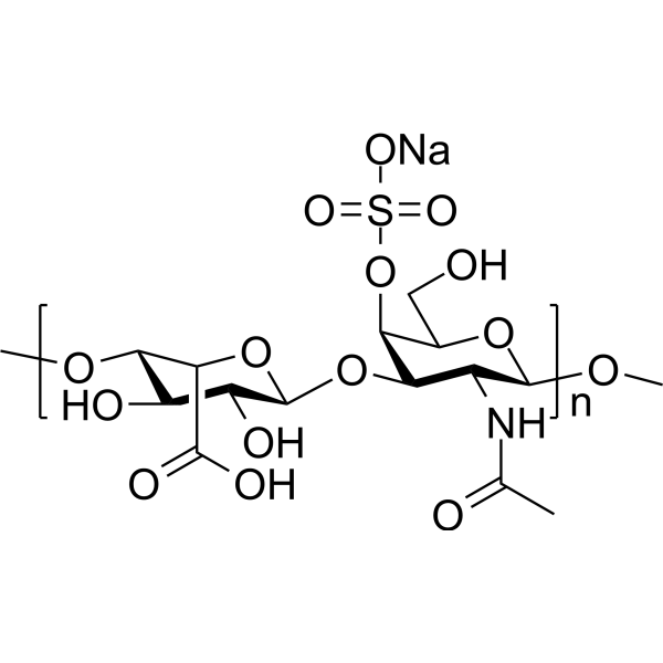
Dermatan sulphate sodium is an oral active glycosaminoglycan and thrombin inactivator with antithrombotic activity. It selectively catalyzes the heparin cofactor II-mediated inactivation of thrombin without interacting with antithrombin III. Dermatan sulphate sodium promotes stem cell differentiation, exhibits high bioavailability, and alleviates pulmonary fibrosis injury induced by Bleomycin (HY-108345). Dermatan sulphate sodium can be used in research related to cardiovascular diseases, inflammation, and stem cell differentiation.
For research use only. We do not sell to patients.
- Purity : 95.4%
- CAS No.: 54328-33-5
-
Storage:
4°C, sealed storage, away from moisture and light
* In solvent : -80°C, 6 months; -20°C, 1 month (sealed storage, away from moisture and light)

To place orders, for customer services and technical support, please contact: MedChemExpress USA
Tel: 609-228-6898 E-mail: [email protected] [email protected]


-
 Biological Activity
-
Chemical Information
-
Solvent & Solubility
- Protocols
- Purity & Documentation
- References
-
 Help & FAQs


-
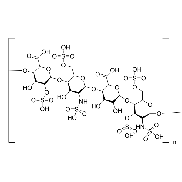
HY-101916
Heparan SulfateLinear Polysaccharide -
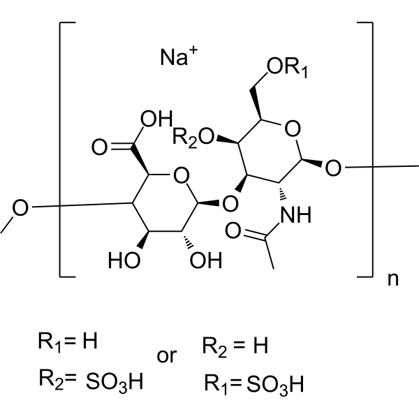
HY-B2162A
Chondroitin sulfate sodium (from shark cartilage)Glycosaminoglycan -
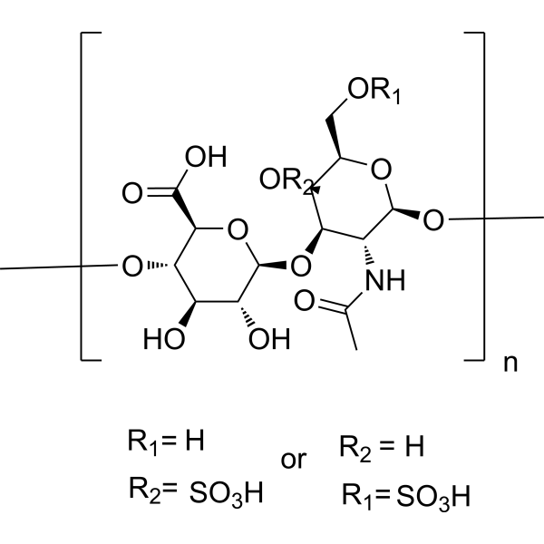
HY-B2162
Chondroitin sulfateGlycosaminoglycan -
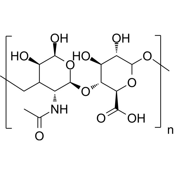
HY-B0633A
Hyaluronic acidGlycosaminoglycan -
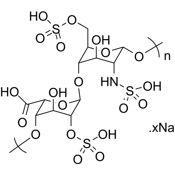
HY-17567A
Heparin sodium saltAnticoagulant -
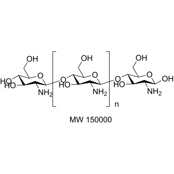
HY-B2144A
Chitosan (MW 150000) (≥85% deacetylated)Bacterial Inhibitor -
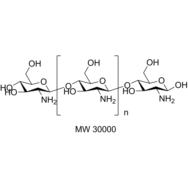
HY-B2144B
Chitosan (MW 30000) (≥85% deacetylated)Bacterial Inhibitor -

HY-B2144K
Chitosan (≥85% deacetylated, viscosity>90 mPa.s, MW 15000)Polysaccharid -
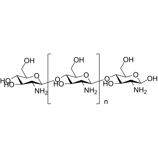
HY-B2144E
Chitosan (≥90% deacetylated, Low viscosity,<200mPa.s)Biochemical Assay Reagent -
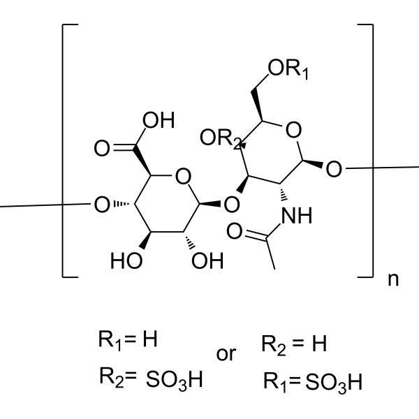
HY-B2162B
Chondroitin sulfate sodium (from bovine)Sulfated Linear Polysaccharide -
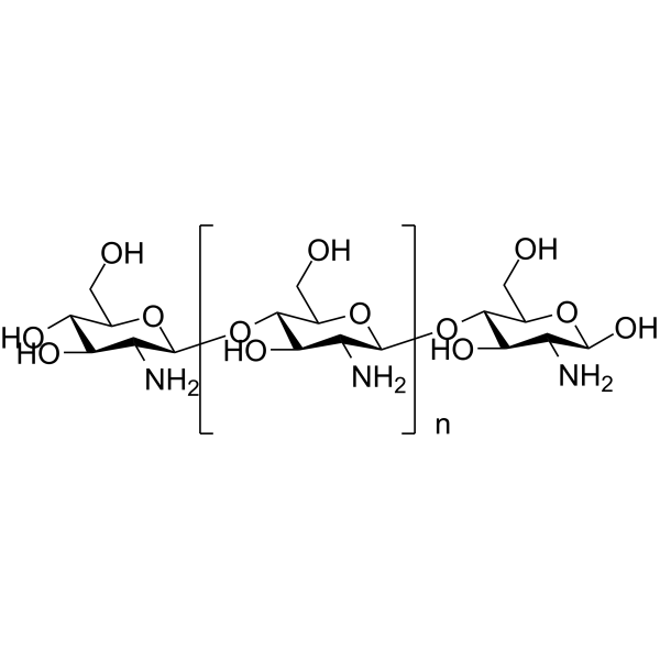
HY-B2144F
Chitosan (≥80% deacetylated, Medium viscosity,200-400mPa.s)Biochemical Assay Reagent -
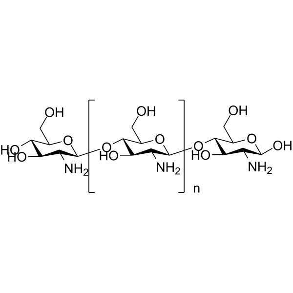
HY-B2144C
Chitosan (≥95% deacetylated,viscosity 100-200 mPa.s)Biochemical Assay Reagent
Publications Citing Use of MedChemExpress (MCE) Dermatan sulphate sodium
More

Biological Activity
Description
In Vitro
Dermatan sulphate (10 μg/mL, 7 days) sodium promoted neuronal differentiation and migration, but did not affect neurite outgrowth in hNSCs[3]. Dermatan sulphate (100 μg/mL, 12 days) sodium promoted neuronal differentiation and enhanced neurite outgrowth in mESCs[3].
MedChemExpress (MCE) has not independently confirmed the accuracy of these methods. They are for reference only. Further protocols information, click here.
-
Cell Line:Human neural stem cells (hNSCs, from 14-week human fetal cerebral cortex)
-
Concentration:10 μg/mL
-
Incubation Time:7 days
-
Result:Significantly increased expression of neuronal markers (DCX, β-III-Tubulin).
-
Cell Line:R1 mouse embryonic stem cells (mESCs, mouse, undifferentiated)
-
Concentration:100 μg/mL
-
Incubation Time:12 days
-
Result:Significantly enhanced p-Erk1/2 expression.
In Vivo
MedChemExpress (MCE) has not independently confirmed the accuracy of these methods. They are for reference only.
-
Animal Model:Bleomycin (HY-108345)-induced pulmonary fibrosis model in C57 mice[2]
-
Dosage:5.3 mg/kg
-
Administration:Oral gavage (p.o.), once daily for 28 days
-
Result:Significantly reduced lung index, alleviated pulmonary edema, inflammatory infiltration, alveolar damage and collagen deposition; Reduced lung hydroxyproline and plasma fibrinogen levels; Increased plasma tPA levels.


Chemical Information
-
CAS No. 54328-33-5
-
Appearance Solid
-
Color White to off-white
-
SMILES
N/A
-
Structure Classification
-
Initial Source
mammals and bacteria
-
Shipping
Room temperature in continental US; may vary elsewhere.
-
Storage
4°C, sealed storage, away from moisture and light
* In solvent : -80°C, 6 months; -20°C, 1 month (sealed storage, away from moisture and light)


Publications (1)
-
Journal Impact Factor
-
Most Recent
-
Biochemistry
The Crystal Structure of Acanthamoeba Polyphaga Mimivirus R655 and Its Potential Glycosaminoglycan Glycosyltransferase Activity. [Abstract]2025 Jul 1;64(13):2737-2744. PMID: 40545624


Solvent & Solubility
In Vitro:
H2O : 100 mg/mL (Need ultrasonic)
DMSO : < 1 mg/mL (insoluble or slightly soluble)

Protocols
-
Fibrosis/Collagen Morphometry
Fibrosis and collagen morphometry is based on the quantitative visualization of fibrillar collagen deposition in tissue sections using histochemical stains such as Sirius Red (Picrosirius Red) or Masson's trichrome, followed by image-based or polarization-enhanced analysis to estimate collagen proportional area as a surrogate of extracellular matrix accumulation during fibrotic remodeling. Sirius Red combined with polarized light microscopy enhances detection of collagen fibers due to birefringence properties, enabling more specific visualization of collagen type I and III fibrils compared to conventional bright-field histology, while whole-section or region-restricted digital morphometry reduces field-selection bias in fibrosis assessment. Alternative quantitative approaches include second harmonic generation (SHG) and two-photon excited fluorescence microscopy, which enable label-free detection of fibrillar collagen and have been validated against histological staining and biochemica
-
Collagen: Sirius Red Staining
Sirius Red or picrosirius red staining is a histochemical method for visualizing collagen-rich extracellular matrix in tissue sections, and collagen fibers are detected as red-stained structures under bright-field microscopy with enhanced birefringence under polarized light. Picrosirius red is useful for assessing total collagen organization, distribution, and fibrosis burden, but polarized color should not be interpreted as a definitive collagen type I versus type III readout because color is affected by fiber orientation, thickness, and packing.
-
Connective Tissue: Masson's Trichrome/Collagen Trichrome Staining
Masson’s Trichrome (collagen/trichrome staining) is a histological technique that differentially stains tissue compartments using sequential acidic dyes to distinguish collagen from muscle and cytoplasmic components based on dye affinity and tissue permeability differences, enabling visualization of fibrosis and connective tissue architecture in histological sections. The classical formulation typically uses Weigert's iron hematoxylin for nuclear staining, Biebrich scarlet-acid fuchsin for cytoplasm and muscle, and aniline blue (or light green variants) for collagen, producing a characteristic blue/green collagen signal contrasted against red cytoplasm and dark nuclei. The staining principle relies on selective displacement of smaller dye molecules by larger anionic dyes in collagen-rich regions under controlled acidified conditions, which enhances collagen-specific dye retention. This property makes the method widely used for fibrosis assessment in organs such as heart, liver, lung, a
-
Protocol for Pharmacokinetic Study
Pharmacokinetic studies quantify how an organism handles a drug over time through absorption, distribution, metabolism, and excretion, and the core experimental readout is the concentration-time profile of parent drug and, when relevant, metabolites in biological matrices such as plasma, whole blood, urine, bile, or tissue. Pharmacokinetic analysis links dose, route, exposure, clearance, half-life, distribution, bioavailability, and systemic exposure to drug efficacy and toxicity hypotheses rather than measuring a signaling pathway directly. The literature links pharmacokinetics to drug-development phenotypes by showing that drug metabolism and pharmacokinetics influence compound progression, exposure-response interpretation, safety margins, dosing strategy, and failure risk during discovery and development. DMPK science contributes to compound optimization by integrating physicochemical properties, in vitro metabolism, transporter behavior, in vivo exposure, and pharmacodynamic contex
-
Carbohydrates and Mucins: Alcian Blue/Alcian Blue-PAS Staining
Alcian Blue (AB) staining is a cationic copper phthalocyanine dye-based histochemical method that binds electrostatically to negatively charged acidic mucopolysaccharides (glycosaminoglycans and sialomucins), enabling visualization of acidic carbohydrate-rich structures such as epithelial mucins, cartilage matrix, and mast cell granules. Periodic Acid-Schiff (PAS) reaction detects neutral mucopolysaccharides and glycoconjugates by oxidizing vicinal diols to aldehydes, which subsequently react with Schiff reagent to produce a magenta signal. The combined Alcian Blue-PAS (AB-PAS) method allows simultaneous differentiation of acidic (blue) and neutral (magenta) mucins in the same tissue section, enabling mucin subtype discrimination in epithelial tissues and pathological lesions.
-
Research Protocol for Inflammation-related Diseases
The NLRP3 inflammasome is a cytosolic innate immune signaling platform that integrates priming signals and danger-signal activation to promote caspase-1 activation, maturation of IL-1β and IL-18, and gasdermin D-mediated pyroptotic cell death. The core experimental logic is to determine whether inflammatory disease phenotypes are driven by increased NLRP3 expression, ASC-containing inflammasome assembly, caspase-1 cleavage, GSDMD cleavage, and extracellular release of IL-1β/IL-18 rather than by nonspecific cell injury alone. The pathway is strongly linked to inflammation-related disease phenotypes because monosodium urate crystals activate NALP3/NLRP3 inflammasome signaling in gout-like crystal inflammation, cholesterol crystals activate NLRP3 inflammasomes in atherogenesis models, and DSS-induced intestinal inflammation has been reported to involve NLRP3 inflammasome activity. However, experimental colitis studies also show context-dependent protective effects of NLRP3 inflammasome co
-
iPSC cell differentiation
Induced pluripotent stem cells (iPSCs) are a type of cell that has similar properties to embryonic stem cells through somatic cell reprogramming.


Purity & Documentation
-
Data Sheet (279 KB)
-
SDS (252 KB)
- English - EN (252 KB)
- Français - FR (252 KB)
- Deutsch - DE (252 KB)
- Norwegian - NO (252 KB)
- Español - ES (252 KB)
- Swedish - SV (252 KB)
- Italian - IT (252 KB)
- Korean - KR (252 KB)
- Portuguese - PT (252 KB)
-
Handling Instructions (2659 KB)
References
[1]. Nenci GG. Dermatan sulphate as an antithrombotic drug. Pathophysiol Haemost Thromb. 2002 Sep-Dec;32(5-6):303-7. [Content Brief]
[2]. Xu J, et al. Effect of dermatan sulphate on a C57-mouse model of pulmonary fibrosis. J Int Med Res. 2019 Jun;47(6):2655-2665. [Content Brief]
[3]. Ogura C, et al. Dermatan sulphate promotes neuronal differentiation in mouse and human stem cells. J Biochem. 2021 Feb 6;169(1):55-64. [Content Brief]

Calculators
Concentration (start) × Volume (start) = Concentration (final) × Volume (final)


Dermatan sulphate sodium Related Classifications
HY-N8278 Related Classifications
 Powered by Bioz