



(2E)-OBAA
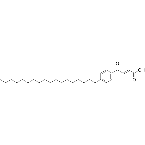
(2E)-OBAA is a potent phospholipase A2 (PLA2) inhibitor, with an IC50 of 70 nM. (2E)-OBAA induces apoptosis of HUVEC cells. (2E)-OBAA blocks Melittin-induced Ca2+ influx in Trypanosoma brucei, with an IC50 of 0.4 μM.
For research use only. We do not sell to patients.
- CAS No.: 221632-26-4
- Formula: C28H44O3
- Molecular Weight:428.66
-
Storage:
Please store the product under the recommended conditions in the Certificate of Analysis.

To place orders, for customer services and technical support, please contact: MedChemExpress USA
Tel: 609-228-6898 E-mail: [email protected] [email protected]


-
 Biological Activity
-
Chemical Information
- Protocols
- Purity & Documentation
- References
-
 Help & FAQs


-
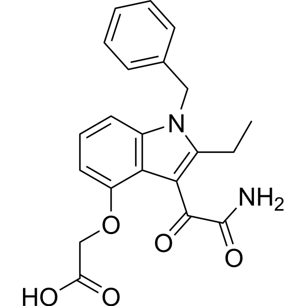
HY-13402
VarespladibsPLA2 Inhibitor -
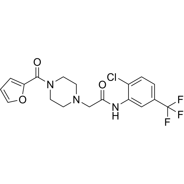
HY-100736
ML348APT1/LYPLA1 Inhibitor -
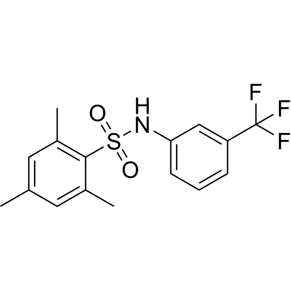
HY-19619
m-3M3FBSPhospholipase C Activator -
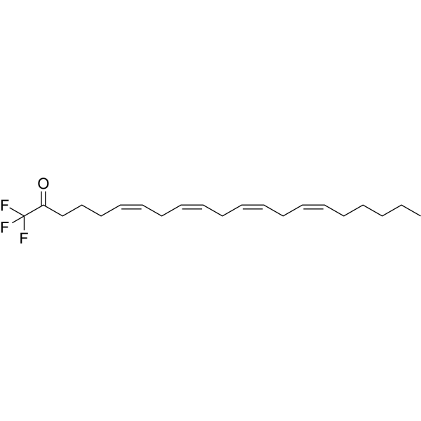
HY-108611
AACOCF3cPLA2 Inhibitor -
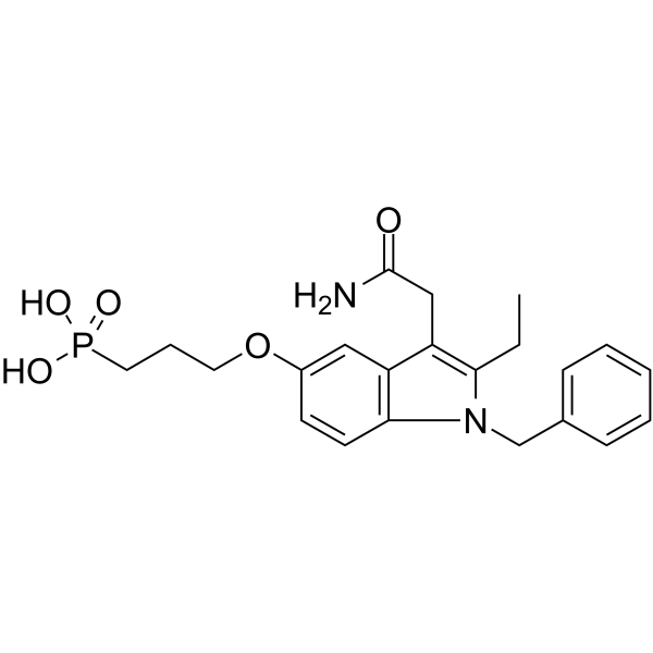
HY-107393
LY-311727sPLA2 Inhibitor -
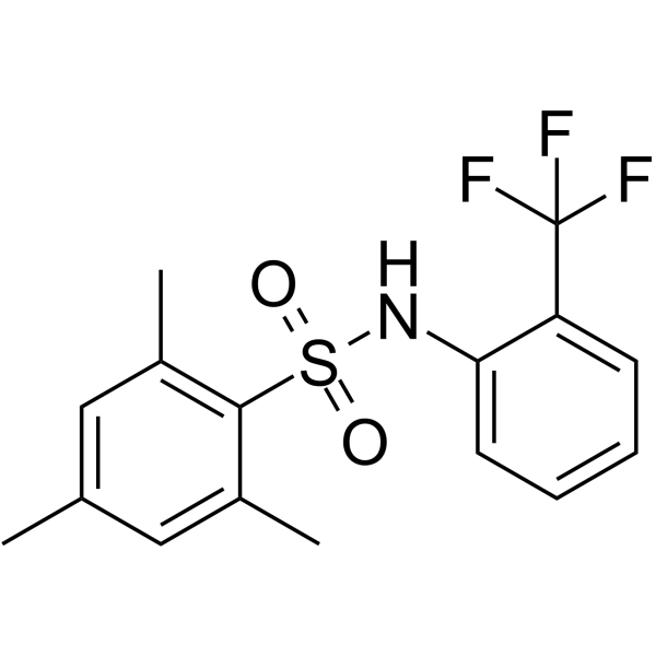
HY-108608
o-3M3FBSActive Compound -
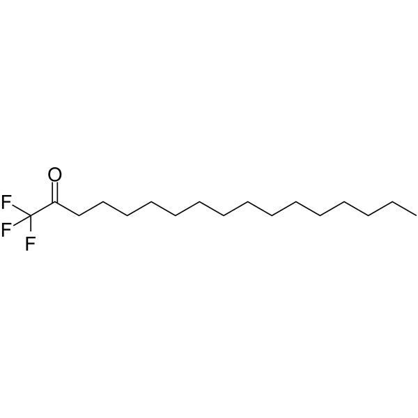
HY-108607
PACOCF3Phospholipase A2 Inhibitor -
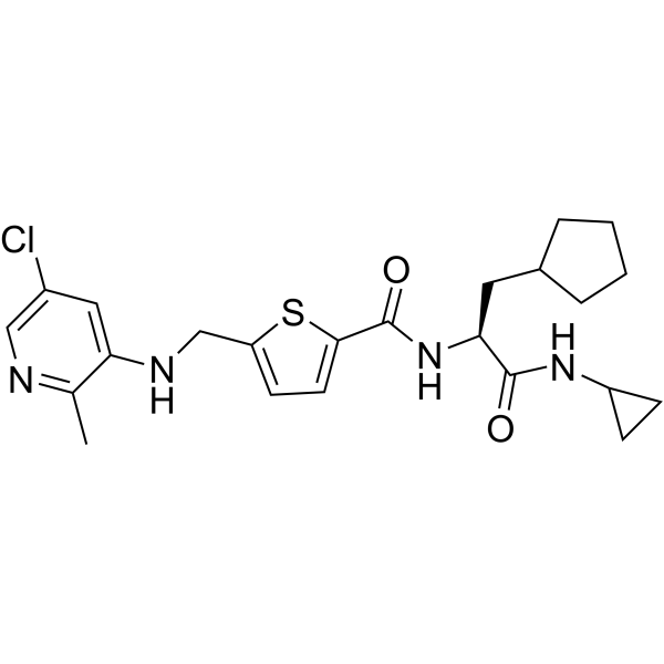
HY-15832
GSK 2830371Wip1 Phosphatase Inhibitor -
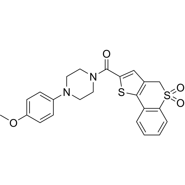
HY-100737
ML349APT2/LYPLA2 Inhibitor -
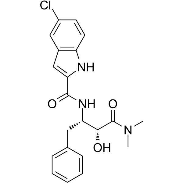
HY-13525
CP-91149Glycogen Phosphorylase Inhibitor -
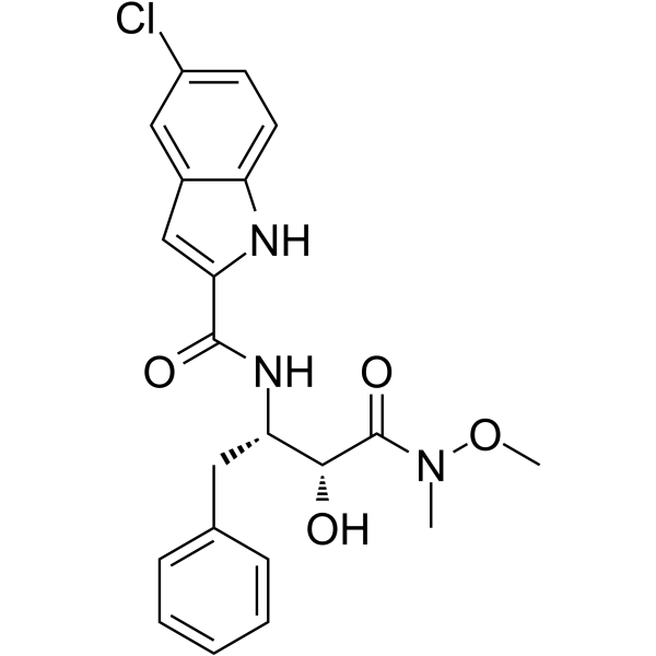
HY-108615
CP-316819Glycogen Phosphorylase Inhibitor
All Phospholipase Isoforms
More

Biological Activity
Description
IC50 & Target
|
PLA2 70 nM (IC50) |
In Vitro
(2E)-OBAA (5.7 μM) induces apoptotic cell death of human umbilical vein endothelial cells (HUVEC). After 16 h of treatment, almost all of the cells has disintegrated into apoptotic bodies[1].
MedChemExpress (MCE) has not independently confirmed the accuracy of these methods. They are for reference only.
In Vivo
MedChemExpress (MCE) has not independently confirmed the accuracy of these methods. They are for reference only.


Chemical Information
-
CAS No. 221632-26-4
-
Molecular Weight 428.66
-
Formula C28H44O3
-
SMILES
O=C(O)/C=C/C(C1=CC=C(CCCCCCCCCCCCCCCCCC)C=C1)=O
-
Shipping
Room temperature in continental US; may vary elsewhere.
-
Storage
Please store the product under the recommended conditions in the Certificate of Analysis.


Purity & Documentation
References
[1]. J Y Miao, et al. Inhibitors of phospholipase promote apoptosis of human endothelial cells. J Biochem. 1997 Mar;121(3):612-8. [Content Brief]
[2]. J Eintracht, et al. Calcium entry in Trypanosoma brucei is regulated by phospholipase A2 and arachidonic acid. Biochem J. 1998 Dec 15;336 ( Pt 3)(Pt 3):659-66. [Content Brief]
[3]. T Köhler, et al. Phospholipase A2 inhibition by alkylbenzoylacrylic acids. Biochem Pharmacol. 1992 Aug 18;44(4):805-13. [Content Brief]
[4]. Kethineedi VR, et al. Quantum dot-NBD-liposome luminescent probes for monitoring phospholipase A2 activity. Anal Bioanal Chem. 2013 Dec;405(30):9729-37. [Content Brief]

Calculators
Concentration (start) × Volume (start) = Concentration (final) × Volume (final)


(2E)-OBAA Related Classifications
HY-101015 Related Classifications
 Powered by Bioz