



DX3-213B

Based on 1 Customer Validation
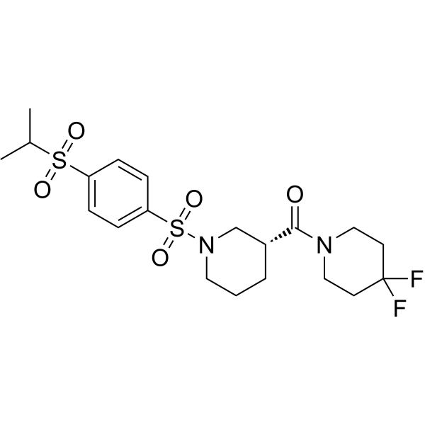
DX3-213B is a highly potent, orally active oxidative phosphorylation (OXPHOS) complex I inhibitor (IC50=3.6 nM). DX3-213B impairs ATP generation (IC50=11 nM), and blocks MIA PaCa-2 cell growth (GI50=11 nM). DX3-213B is used for the research of the pancreatic cancer.
For research use only. We do not sell to patients.
- Purity : 99.68%
- CAS No.: 2749555-66-4
- Formula: C20H28F2N2O5S2
- Molecular Weight:478.57
-
Storage:Powder -20°C, 3 years , 4°C, 2 years ; In solvent -80°C, 6 months , -20°C, 1 month

To place orders, for customer services and technical support, please contact: MedChemExpress USA
Tel: 609-228-6898 E-mail: [email protected] [email protected]


-
 Biological Activity
-
Chemical Information
-
Solvent & Solubility
- Protocols
- Purity & Documentation
- References
-
 Help & FAQs


-
HY-112037
IACS-010759OXPHOS Inhibitor -
HY-110284
BAM 15OXPHOS Uncoupler -
HY-145707
BAY-179Complex I Inhibitor -
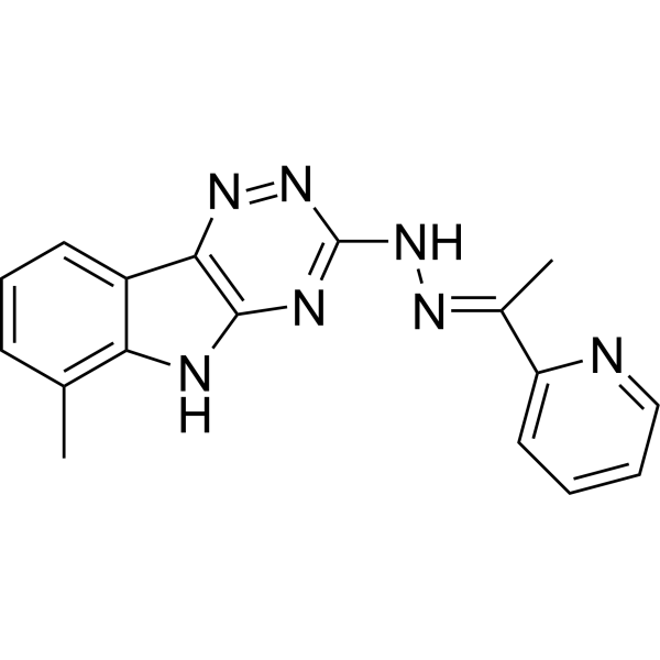
HY-12406
VLX600OXPHOS Inhibitor/Iron Chelator -
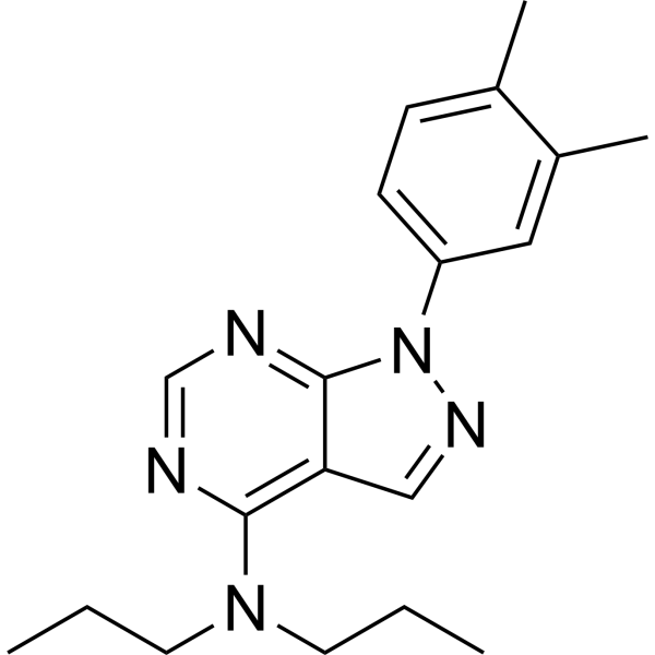
HY-110282
S3QEL-2Site IIIQo Superoxide Production Suppressor -
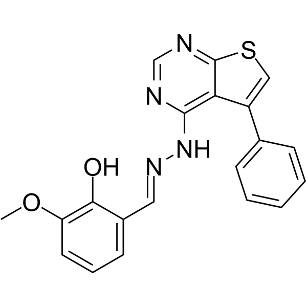
HY-144906
THP104cMitochondrial Fission Inhibitor -
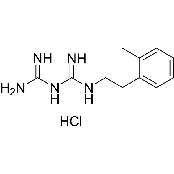
HY-125283
IM176OUT05Stem Cell Metabolism Activator -
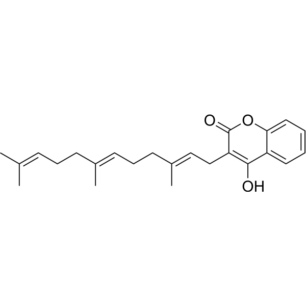
HY-129605
FerulenolMitochondrial Metabolism Inhibitor -
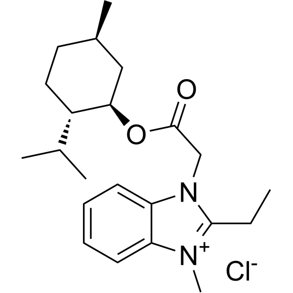
HY-111651
GboxinOXPHOS Inhibitor -
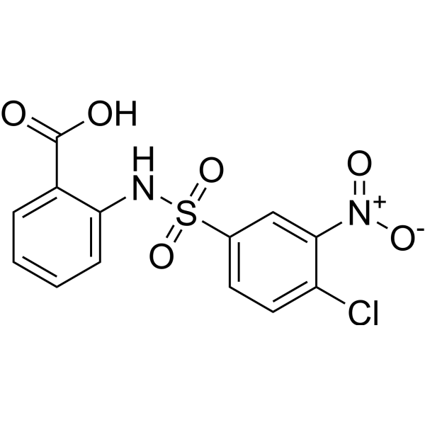
HY-123986
CTPI-2SLC25A1 Inhibitor -
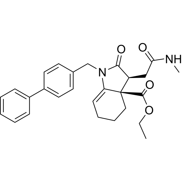
HY-100004
Fumarate hydratase-IN-1Fumarate Hydratase Inhibitor -
HY-134539
IMT1POLRMT Inhibitor


Biological Activity
Description
Cellular Effect
|
Cell Line

|
Type | Value | Description | References |
|---|---|---|---|---|
| BXPC-3 | IC50 |
0.05 μM
Compound: 23; DX3-213B
|
Cytotoxicity against human BXPC-3 cells assessed as inhibition of cell growth incubated for overnight in glucose-containing medium followed by compound addition and measured after 7 days by MTT assay
Cytotoxicity against human BXPC-3 cells assessed as inhibition of cell growth incubated for overnight in glucose-containing medium followed by compound addition and measured after 7 days by MTT assay
|
[PMID: 35167303] |
| MIA PaCa-2 | IC50 |
>3000 nM
Compound: 23; DX3-213B
|
Inhibition of oxidative phosphorylation in human MIA PaCa-2 cells assessed as reduction in ATP production incubated for 24 hrs in glucose-containing medium by celltiter-glo luminescence cell viability assay
Inhibition of oxidative phosphorylation in human MIA PaCa-2 cells assessed as reduction in ATP production incubated for 24 hrs in glucose-containing medium by celltiter-glo luminescence cell viability assay
|
[PMID: 35167303] |
| MIA PaCa-2 | IC50 |
0.011 μM
Compound: 23; DX3-213B
|
Cytotoxicity against human MIA PaCa-2 cells assessed as inhibition of cell growth incubated for overnight in glucose-containing medium followed by compound addition and measured after 7 days by MTT assay
Cytotoxicity against human MIA PaCa-2 cells assessed as inhibition of cell growth incubated for overnight in glucose-containing medium followed by compound addition and measured after 7 days by MTT assay
|
[PMID: 35167303] |
| MIA PaCa-2 | IC50 |
11 nM
Compound: 23; DX3-213B
|
Inhibition of oxidative phosphorylation in human MIA PaCa-2 cells assessed as reduction in ATP production incubated for 24 hrs in galactose-containing medium by celltiter-glo luminescence cell viability assay
Inhibition of oxidative phosphorylation in human MIA PaCa-2 cells assessed as reduction in ATP production incubated for 24 hrs in galactose-containing medium by celltiter-glo luminescence cell viability assay
|
[PMID: 35167303] |
| MIA PaCa-2 | IC50 |
9.1 nM
Compound: 23; DX3-213B
|
Cytotoxicity against human MIA PaCa-2 cells assessed as inhibition of cell growth incubated for overnight in galactose-containing medium followed by compound addition and measured after 3 days by MTT assay
Cytotoxicity against human MIA PaCa-2 cells assessed as inhibition of cell growth incubated for overnight in galactose-containing medium followed by compound addition and measured after 3 days by MTT assay
|
[PMID: 35167303] |
| MIA PaCa-2 | IC50 |
>3000 nM
Compound: 23; DX3-213B
|
Cytotoxicity against human MIA PaCa-2 cells assessed as inhibition of cell growth incubated for overnight in glucose-containing medium followed by compound addition and measured after 3 days by MTT assay
Cytotoxicity against human MIA PaCa-2 cells assessed as inhibition of cell growth incubated for overnight in glucose-containing medium followed by compound addition and measured after 3 days by MTT assay
|
[PMID: 35167303] |


Chemical Information
-
CAS No. 2749555-66-4
-
Appearance Solid
-
Molecular Weight 478.57
-
Formula C20H28F2N2O5S2
-
Color White to off-white
-
SMILES
O=S(N1C[C@@H](CCC1)C(N2CCC(F)(CC2)F)=O)(C3=CC=C(C=C3)S(C(C)C)(=O)=O)=O
-
Shipping
Room temperature in continental US; may vary elsewhere.
-
Storage
Powder -20°C 3 years 4°C 2 years In solvent -80°C 6 months -20°C 1 month


Solvent & Solubility
In Vitro:
DMSO : 125 mg/mL (261.19 mM; ultrasonic and warming and heat to 60°C; Hygroscopic DMSO has a significant impact on the solubility of product, please use newly opened DMSO)
Please refer to the solubility information to select the appropriate solvent. Once prepared, please aliquot and store the solution to prevent product inactivation from repeated freeze-thaw cycles.
Storage method and period of stock solution: -80°C, 6 months; -20°C, 1 month. When stored at -80°C, please use it within 6 months. When stored at -20°C, please use it within 1 month.
Please refer to the solubility information to select the appropriate solvent. Once prepared, please aliquot and store the solution to prevent product inactivation from repeated freeze-thaw cycles.
Storage method and period of stock solution: -80°C, 6 months; -20°C, 1 month. When stored at -80°C, please use it within 6 months. When stored at -20°C, please use it within 1 month.
Concentration (start) × Volume (start) = Concentration (final) × Volume (final)
In Vivo:
Select the appropriate dissolution method based on your experimental animal and administration route.
- For the following dissolution methods, please ensure to first prepare a clear stock solution using an In Vitro approach and then sequentially add co-solvents:
- To ensure reliable experimental results, the clarified stock solution can be appropriately stored based on storage conditions. As for the working solution for In Vivo experiments, it is recommended to prepare freshly and use it on the same day.
- The percentages shown for the solvents indicate their volumetric ratio in the final prepared solution. If precipitation or phase separation occurs during preparation, heat and/or sonication can be used to aid dissolution.
Add each solvent one by one: 10% DMSO 40% PEG300 5% Tween-80 45% Saline
Solubility: ≥ 2.08 mg/mL (4.35 mM); Clear solution
This protocol yields a clear solution of ≥ 2.08 mg/mL (saturation unknown).
Taking 1 mL working solution as an example, add 100 μL DMSO stock solution (20.8 mg/mL) to 400 μL PEG300, and mix evenly; then add 50 μL Tween-80 and mix evenly; then add 450 μL Saline to adjust the volume to 1 mL.
Preparation of Saline: Dissolve 0.9 g sodium chloride in ddH₂O and dilute to 100 mL to obtain a clear Saline solution.
Add each solvent one by one: 10% DMSO 90% (20% SBE-β-CD in Saline)
Solubility: ≥ 2.08 mg/mL (4.35 mM); Clear solution
This protocol yields a clear solution of ≥ 2.08 mg/mL (saturation unknown).
Taking 1 mL working solution as an example, add 100 μL DMSO stock solution (20.8 mg/mL) to 900 μL 20% SBE-β-CD in Saline, and mix evenly.
Preparation of 20% SBE-β-CD in Saline (4°C, storage for one week): 2 g SBE-β-CD powder is dissolved in 10 mL Saline, completely dissolve until clear.
In Vivo Dissolution Calculator
Please enter the basic information of animal experiments:
-
-
-
-
Recommended: Prepare an additional quantity of animals to account for potential losses during experiments.
Please enter your animal formula composition:
-
%DMSO +
Recommended: Keep the proportion of DMSO in working solution below 2% if your animal is weak.
-
%+
-
+%Tween-80 + +
-
%Saline +
The co-solvents required include: DMSO, . All of co-solvents are available by MedChemExpress (MCE). , Tween 80. All of co-solvents are available by MedChemExpress (MCE).
Working solution concentration: 0.22 mg/mL
Method for preparing stock solution: mg drug dissolved in μL DMSO. Stock solution concentration: mg/mL.
1. Take μL DMSO stock solution;
2. Add μL .
μL , mix evenly;
3. Then add μL Tween 80, mix evenly;
4. Then add μL
Please ensure that the stock solution in the first step is dissolved to a clear state, and add co-solvents in sequence. You can use ultrasonic heating (ultrasonic cleaner, recommended frequency 20-40 kHz), vortexing, etc. to assist dissolution.

Protocols
-
Kinase activity and phosphorylation assays
Kinase activity assays measure the ability of kinases to transfer phosphate groups from ATP to specific substrates, while phosphorylation assays detect the presence and levels of phosphorylated proteins. Common methods include radiolabeled ATP incorporation (e. g. ,), ADP release detection via bioluminescence (e. g. ,[3]), enzyme-linked immunosorbent assays (ELISA) for phospho-specific epitopes (e. g. ,[6]), and microtiter-based formats for high-throughput screening (e. g. ,[8]). The ADP-Glo assay quantifies kinase activity by measuring ADP produced during phosphorylation using a luciferase-based system. Radiometric assays involve autoradiography or scintillation counting after incorporation of 32P-labeled ATP into substrate proteins. ELISA-based approaches rely on phospho-specific antibodies to detect activated kinases in cell lysates or purified samples.
-
Western Blot
Western blotting (WB) is a commonly used experimental method in molecular biology, biochemistry, and immunogenetics for identifying and quantifying target proteins. It combines gel electrophoresis with immunoassay, enabling researchers to analyze protein expression, post-translational modifications, and molecular weight.
-
Cell Cytotoxicity Assay
Cytotoxicity assays are usually based on the assessment of cell membrane damage, which can also be indirectly detected by measuring cell viability. Detection methods include MTT assay, CKK-8 assay, LDH assay and ATP assay, etc.
-
Protocol for Cell Counting and Cell Density Analysis
Cell counting and cell-density analysis estimate the number of cells in a known volume or field area. Manual hemocytometer counting uses a chamber of defined geometry to convert counted cells into cells/mL, while automated counters and image-analysis workflows detect cell objects from optical, brightfield, fluorescence, impedance, or digital-image features. Trypan blue viability counting is based on dye exclusion: viable cells with intact membranes exclude dye, while non-viable cells with compromised membranes stain blue. The readout is total cell density, viable-cell density, dead-cell density, and percent viability. Cell density can also be estimated from microscopy images by counting objects per image area, from flow cytometry using calibrated volume or reference particles, or from in situ microscopy in bioreactors after calibration against reference methods such as hemocytometer or flow cytometry.
-
Somatic Cell Culture
A method of simulating the in vivo environment in vitro to maintain the cell growth, differentation and main functions.
-
CCK-8/WST-8 Cell Proliferation Assay
The CCK-8/WST-8 assay is based on the reduction of the water-soluble tetrazolium salt WST-8 to a water-soluble formazan product by cellular dehydrogenases in metabolically active cells, where the generated formazan amount is proportional to the number of living cells and is quantified by measuring absorbance in the visible range, providing a colorimetric readout for cell viability and proliferation assessment. This class of tetrazolium-based assays improves upon earlier MTT-based systems by producing a water-soluble formazan, eliminating the need for organic solubilization steps and enabling direct spectrophotometric measurement in culture medium.
-
Protocol for Kinase activity and phosphorylation assays
Kinase activity assays measure transfer of phosphate from ATP to a protein or peptide substrate, generating phosphorylated substrate, ADP, or incorporated radiolabeled phosphate as the readout; phosphorylation assays measure site-specific phosphorylation in cells or tissues as a proxy for kinase-pathway activation, inhibition, or substrate regulation. Phosphorylation can be detected by phospho-specific Western blot, immunoprecipitation kinase assay, phospho-immunofluorescence, phospho-flow cytometry, luminescent ADP detection, radiolabeled ATP incorporation, or reporter-based pathway assays, and these readouts can be applied to cancer cells, primary neurons, mouse tumors, organoids, inflammatory macrophages, ferroptosis studies, and mitophagy studies when the kinase target is biologically relevant.
-
Cell Counting-Based Growth Curve Assay
Cell counting-based growth curve assays quantify cell proliferation by directly measuring changes in viable cell number over time using manual or automated counting methods such as hemocytometer-based counting or instrument-assisted cell enumeration, enabling construction of growth curves that reflect population expansion dynamics in response to culture conditions. A widely used approach is trypan blue exclusion with hemocytometer counting, where membrane-compromised (non-viable) cells take up the dye, allowing discrimination between viable and non-viable cells while simultaneously enabling total cell number quantification. Repeated sampling across time points allows estimation of proliferation rate, growth phases, and comparative growth kinetics between experimental conditions.
-
MTT Cell Proliferation Assay
The MTT assay is a colorimetric endpoint assay for estimating viable cell number, cell growth, cytotoxicity, or cell activation in cultured mammalian cells. Living cells reduce the yellow tetrazolium salt MTT into purple/blue formazan, while dead cells do not generate the same signal; the resulting color can be quantified with a multiwell spectrophotometer. MTT reduction is commonly interpreted as a readout of metabolic activity that often correlates with viable cell number, but it should not be treated as a direct cell-counting method unless the assay is optimized for the cell type and experimental condition. Studies show that MTT reduction can involve mitochondrial and non-mitochondrial reducing systems, and formazan may accumulate in intracellular lipid droplets rather than simply marking mitochondria.
-
Cell Viability Determination by MTT Colorimetric Assay
The following protocol uses the MTT colorimetric assay as a classic literature-established method for assessing cell viability/metabolic activity in cultured mammalian cells. MTT[3-(4,5-dimethylthiazol-2-yl)-2,5-diphenyltetrazolium bromide] is reduced by metabolically active cells to a colored formazan product; the amount of formazan is quantified spectrophotometrically and provides an indirect measure of metabolically active viable cells. Importantly, MTT reduction reflects cellular oxidoreductase/metabolic activity rather than an absolute direct count of living cells, so changes in cellular metabolism can alter the signal independently of cell number.


Purity & Documentation
-
Data Sheet (270 KB)
-
SDS (251 KB)
- English - EN (251 KB)
- Français - FR (251 KB)
- Deutsch - DE (251 KB)
- Norwegian - NO (251 KB)
- Español - ES (251 KB)
- Swedish - SV (251 KB)
- Italian - IT (251 KB)
- Korean - KR (251 KB)
- Portuguese - PT (251 KB)
-
Handling Instructions (2659 KB)
References


Complete Stock Solution Preparation Table
Please refer to the solubility information to select the appropriate solvent. Once prepared, please aliquot and store the solution to prevent product inactivation from repeated freeze-thaw cycles.
Storage method and period of stock solution: -80°C, 6 months; -20°C, 1 month. When stored at -80°C, please use it within 6 months. When stored at -20°C, please use it within 1 month.
| Optional Solvent | Concentration Solvent Mass | 1 mg | 5 mg | 10 mg | 25 mg |
|---|---|---|---|---|---|
| DMSO | 1 mM | 2.0896 mL | 10.4478 mL | 20.8956 mL | 52.2390 mL |
| 5 mM | 0.4179 mL | 2.0896 mL | 4.1791 mL | 10.4478 mL | |
| 10 mM | 0.2090 mL | 1.0448 mL | 2.0896 mL | 5.2239 mL | |
| 15 mM | 0.1393 mL | 0.6965 mL | 1.3930 mL | 3.4826 mL | |
| 20 mM | 0.1045 mL | 0.5224 mL | 1.0448 mL | 2.6119 mL | |
| 25 mM | 0.0836 mL | 0.4179 mL | 0.8358 mL | 2.0896 mL | |
| 30 mM | 0.0697 mL | 0.3483 mL | 0.6965 mL | 1.7413 mL | |
| 40 mM | 0.0522 mL | 0.2612 mL | 0.5224 mL | 1.3060 mL | |
| 50 mM | 0.0418 mL | 0.2090 mL | 0.4179 mL | 1.0448 mL | |
| 60 mM | 0.0348 mL | 0.1741 mL | 0.3483 mL | 0.8706 mL | |
| 80 mM | 0.0261 mL | 0.1306 mL | 0.2612 mL | 0.6530 mL | |
| 100 mM | 0.0209 mL | 0.1045 mL | 0.2090 mL | 0.5224 mL |


DX3-213B Related Classifications
HY-144310 Related Classifications
 Powered by Bioz