



EG00229
Based on 12 publication(s) in Google Scholar
EG00229 is an inhibitor for VEGF-A and NRP1 interaction with an IC50 of 8 μM. EG00229 inhibits the migration of HUVECS and the phosphorylation of VEGFR2 in endothelial cells. EG00229 exhibits cytotoxicity in cancer cell A549.
연구목적의 판매만을 진행합니다. 환자를 대상으로 한 판매는 하지 않습니다.
- CAS No.: 1018927-63-3
- 화학식: C17H19N7O5S3
- 분자량:497.57
-
보관:
Please store the product under the recommended conditions in the Certificate of Analysis.


To place orders, for customer services and technical support, please contact: MedChemExpress USA
Tel: 609-228-6898 E-mail: [email protected] [email protected]


-
 Biological Activity
-
Chemical Information
- Protocol
- 순도&문서
- References
-
 Help & FAQs


Publications Citing Use of MedChemExpress (MCE) EG00229
More- EBioMedicine. 2019 May:43:525-536. [Abstract]
- Acta Pharmacol Sin. 2026 Feb 19. [Abstract]
- Proc Natl Acad Sci U S A. 2026 Mar 31;123(13):e2510265123. [Abstract]
- Mol Med. 2025 Dec 16;31(1):336. [Abstract]
- Fluids Barriers CNS. 2024 Apr 8;21(1):32. [Abstract]
- Biochem Pharmacol. 2026 Jun 12;251(Pt 2):118163. [Abstract]
- Biochem Pharmacol. 2025 Nov:241:117183. [Abstract]
- Biochem Pharmacol. 2022 May;199:115030. [Abstract]
- Bioorg Med Chem. 2020 Jan 1;28(1):115183. [Abstract]
- J Cancer. 2021 Aug 24;12(20):6105-6117. [Abstract]
- Cell Physiol Biochem. 2017;44(3):1251-1262. [Abstract]
- bioRxiv. 2024 Oct 21:2023.12.06.570398. [Abstract]
 Customer Validation & Images

-
WB
-
Cell Imaging/Staining
-
In Vivo Efficacy Study
-
Cell Proliferation/Viability Assay
-
RT-PCR
All VEGFR Isoforms
More

Biological Activity
제품 설명
Cellular Effect
|
Cell Line

|
Type | Value | Description | References |
|---|---|---|---|---|
| A549 | IC50 |
7 μM
Compound: 2, EG00229
|
Displacement of [125I]VEGF-A from human NRP1 expressed in human A549 cells
Displacement of [125I]VEGF-A from human NRP1 expressed in human A549 cells
|
[PMID: 20151671] |
| DU-145 | IC50 |
3 μM
Compound: 2, EG00229
|
Displacement of biotinylated-VEGF-A125 from human NRP1 expressed in human DU145 cells
Displacement of biotinylated-VEGF-A125 from human NRP1 expressed in human DU145 cells
|
[PMID: 20151671] |
| HEK293 | IC50 |
11.1 μM
Compound: EG00229
|
Inhibition of recombinant human biotinylated VEGF-165A expressed in HEK293 cells binding to immobilized recombinant full length human FC-tagged NRP1 (chimera active) expressed in HEK293 cells using TMB as substrate incubated for 2 hrs followed by substrat
Inhibition of recombinant human biotinylated VEGF-165A expressed in HEK293 cells binding to immobilized recombinant full length human FC-tagged NRP1 (chimera active) expressed in HEK293 cells using TMB as substrate incubated for 2 hrs followed by substrat
|
[PMID: 31744780] |
| HUVEC | IC50 |
23 μM
Compound: 2, EG00229
|
Displacement of biotinylated-VEGF-A125 from human NRP1 expressed in HUVEC
Displacement of biotinylated-VEGF-A125 from human NRP1 expressed in HUVEC
|
[PMID: 20151671] |


Chemical Information
-
CAS No. 1018927-63-3
-
분자량 497.57
-
화학식 C17H19N7O5S3
-
SMILES
N=C(N)NCCC[C@@H](C(O)=O)NC(C1=C(NS(=O)(C2=CC=CC3=NSN=C32)=O)C=CS1)=O
-
선적
Room temperature in continental US; may vary elsewhere.
-
보관
Please store the product under the recommended conditions in the Certificate of Analysis.


Publications (12)
-
Journal Impact Factor
-
Most Recent
-
EBioMedicine
Neuropilin-1 aggravates liver cirrhosis by promoting angiogenesis via VEGFR2-dependent PI3K/Akt pathway in hepatic sinusoidal endothelial cells. [Abstract]2019 May:43:525-536. PMID: 31060904
EG00229 purchased from MedChemExpress. Usage Cited in: EBioMedicine. 2019 May:43:525-536. [Abstract]
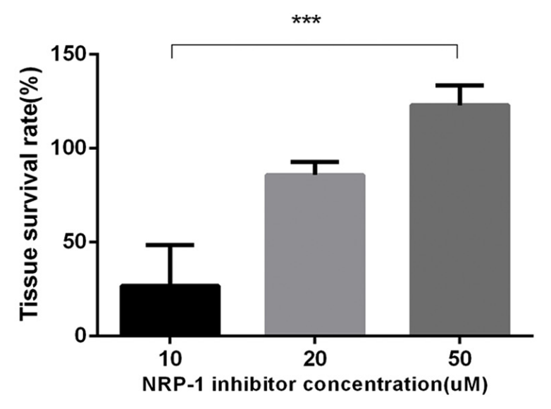
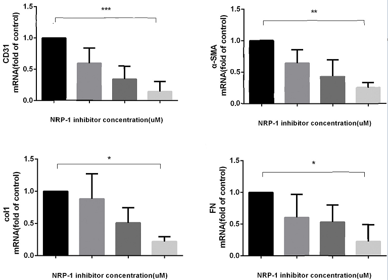
EG00229 trifluoroacetate (NRP-1 inhibitor) (10-50 μM; once daily for 7 d) concentration-dependently increased the survival rate of fibrotic liver tissues from patients.
EG00229 purchased from MedChemExpress. Usage Cited in: EBioMedicine. 2019 May:43:525-536. [Abstract]
EG00229 trifluoroacetate (NRP-1 inhibitor) (10-50 μM; once daily for 7 d) significantly decreased the mRNA levels of CD31, α-SMA, collagen1α I, and FN in fibrotic liver tissues from patients.
-
Acta Pharmacol Sin
Epigenetic regulation of NDGA and its synergistic inhibition with EZH2 inhibitors in prostate cancer via NRP1. [Abstract]2026 Feb 19. PMID: 41714725 -
Proc Natl Acad Sci U S A
Targeting the ANGPTL4/NRP1/ABL1/RAD51 axis reverses cisplatin resistance by impairing DNA damage repair in head and neck cancer. [Abstract]2026 Mar 31;123(13):e2510265123. PMID: 41886374
EG00229 purchased from MedChemExpress. Usage Cited in: Proc Natl Acad Sci U S A. 2026 Mar 31;123(13):e2510265123. [Abstract]
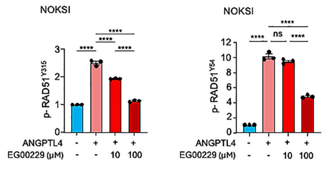
NRP1 inhibitor EG00229 (10-100 μM) blocked ANGPTL4-induced RAD51 phosphorylation in Tyr315 and Tyr54 of NOKSI cells.
EG00229 purchased from MedChemExpress. Usage Cited in: Proc Natl Acad Sci U S A. 2026 Mar 31;123(13):e2510265123. [Abstract]
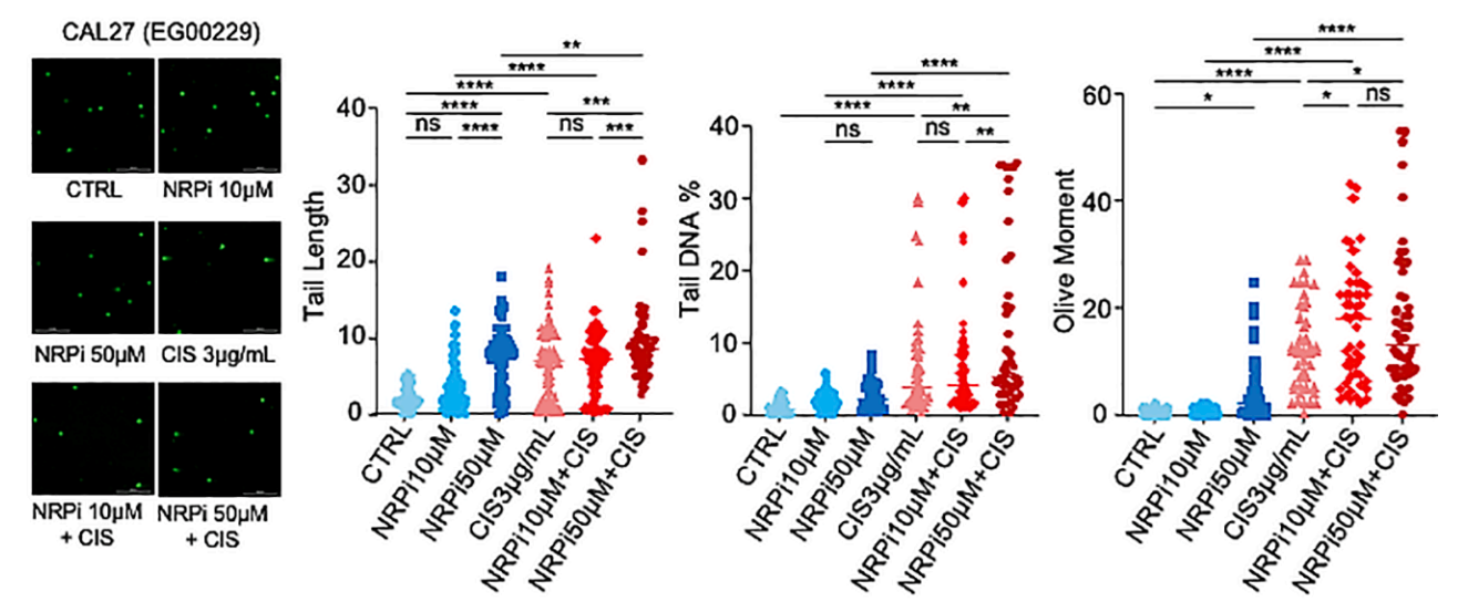
EG00229 trifluoroacetate (NRPi) (10-50 µM; 48 h) led to an increase in the DDR in CAL27 cells.
EG00229 purchased from MedChemExpress. Usage Cited in: Proc Natl Acad Sci U S A. 2026 Mar 31;123(13):e2510265123. [Abstract]
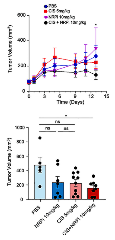
EG00229 trifluoroacetate (NRPi) (10 mg/kg; i.p.) sensitized CAL27 xenografts mice to a subtherapeutic dose of cisplatin (5 mg/kg) markedly reducing tumor volume respectively.
-
Mol Med
Epigenetic reprogramming drives endothelial dysfunction via neuropilin-1 in pulmonary hypertension. [Abstract]2025 Dec 16;31(1):336. PMID: 41402732 -
Fluids Barriers CNS
SARS-CoV-2 causes dysfunction in human iPSC-derived brain microvascular endothelial cells potentially by modulating the Wnt signaling pathway. [Abstract]2024 Apr 8;21(1):32. PMID: 38584257 -
Biochem Pharmacol
Targeting neuropilin-1 attenuates 4-hydroxytamoxifen-induced stemness and sensitizes ERα-re-expressing TNBC to tamoxifen. [Abstract]2026 Jun 12;251(Pt 2):118163. PMID: 42285368 -
Biochem Pharmacol
Pitavastatin overcomes multi-drug resistance in CRC and NSCLC by targeting the NRP1-ZFX axis. [Abstract]2025 Nov:241:117183. PMID: 40684995 -
Biochem Pharmacol
Tuftsin ameliorates splenic inflammatory injury by promoting neuropilin-1 in severe acute pancreatitis. [Abstract]2022 May;199:115030. PMID: 35381211 -
Bioorg Med Chem
Discovery of novel nonpeptide small-molecule NRP1 antagonists: Virtual screening, molecular simulation and structural modification. [Abstract]2020 Jan 1;28(1):115183. PMID: 31744780 -
J Cancer
2021 Aug 24;12(20):6105-6117. PMID: 34539883 -
Cell Physiol Biochem
VEGF-A/Neuropilin 1 Pathway Confers Cancer Stemness via Activating Wnt/β-Catenin Axis in Breast Cancer Cells. [Abstract]2017;44(3):1251-1262. PMID: 29179185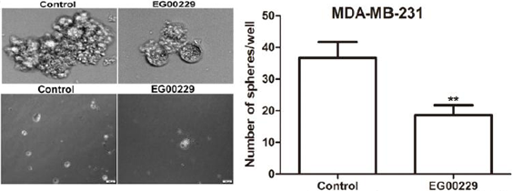
EG00229 purchased from MedChemExpress. Usage Cited in: Cell Physiol Biochem. 2017;44(3):1251-1262. [Abstract]
The inhibitor of NRP-1 called EG00229 decreases sphere and clone formation in MDA-MB-231 cells.
-
bioRxiv
2024 Oct 21:2023.12.06.570398. PMID: 38106002

Protocol
-
Kinase activity and phosphorylation assays
Kinase activity assays measure the ability of kinases to transfer phosphate groups from ATP to specific substrates, while phosphorylation assays detect the presence and levels of phosphorylated proteins. Common methods include radiolabeled ATP incorporation (e. g. ,), ADP release detection via bioluminescence (e. g. ,[3]), enzyme-linked immunosorbent assays (ELISA) for phospho-specific epitopes (e. g. ,[6]), and microtiter-based formats for high-throughput screening (e. g. ,[8]). The ADP-Glo assay quantifies kinase activity by measuring ADP produced during phosphorylation using a luciferase-based system. Radiometric assays involve autoradiography or scintillation counting after incorporation of 32P-labeled ATP into substrate proteins. ELISA-based approaches rely on phospho-specific antibodies to detect activated kinases in cell lysates or purified samples.
-
Western Blot
Western blotting (WB) is a commonly used experimental method in molecular biology, biochemistry, and immunogenetics for identifying and quantifying target proteins. It combines gel electrophoresis with immunoassay, enabling researchers to analyze protein expression, post-translational modifications, and molecular weight.
-
Cell Cytotoxicity Assay
Cytotoxicity assays are usually based on the assessment of cell membrane damage, which can also be indirectly detected by measuring cell viability. Detection methods include MTT assay, CKK-8 assay, LDH assay and ATP assay, etc.
-
Mammalian live/dead viability and cytotoxicity staining
Live/dead viability and cytotoxicity staining assays are based on the simultaneous detection of intracellular esterase activity in metabolically active (viable) cells and membrane integrity loss in non-viable cells. In commonly used dual-staining approaches, membrane-permeant fluorogenic substrates are converted by intracellular esterases into fluorescent products in live cells, while impermeant DNA-binding dyes selectively enter cells with compromised plasma membranes and label nucleic acids in dead or dying cells, enabling discrimination between viable and non-viable populations by fluorescence microscopy or flow cytometry.
-
Cell migration
Cell migration is a method that plays an important role in wound healing, cell differentiation, embryonic development, etc.
-
Endothelial Tube Formation Assay
Endothelial tube formation assay evaluates the ability of endothelial cells to attach, migrate, align, and organize into capillary-like networks when cultured on gelled basement membrane extract or Matrigel; the readout is the morphology and quantity of tube-like networks, which reflects an in vitro endothelial morphogenesis step related to angiogenesis. Basement membrane extract/Matrigel provides laminin-rich extracellular matrix cues that support endothelial differentiation into capillary-like structures, but it can contain biologically active growth factors, so growth-factor-reduced matrix is preferred when testing defined angiogenic stimulators or inhibitors.
-
Protocol for Kinase activity and phosphorylation assays
Kinase activity assays measure transfer of phosphate from ATP to a protein or peptide substrate, generating phosphorylated substrate, ADP, or incorporated radiolabeled phosphate as the readout; phosphorylation assays measure site-specific phosphorylation in cells or tissues as a proxy for kinase-pathway activation, inhibition, or substrate regulation. Phosphorylation can be detected by phospho-specific Western blot, immunoprecipitation kinase assay, phospho-immunofluorescence, phospho-flow cytometry, luminescent ADP detection, radiolabeled ATP incorporation, or reporter-based pathway assays, and these readouts can be applied to cancer cells, primary neurons, mouse tumors, organoids, inflammatory macrophages, ferroptosis studies, and mitophagy studies when the kinase target is biologically relevant.
-
Cell Viability Determination by MTT Colorimetric Assay
The following protocol uses the MTT colorimetric assay as a classic literature-established method for assessing cell viability/metabolic activity in cultured mammalian cells. MTT[3-(4,5-dimethylthiazol-2-yl)-2,5-diphenyltetrazolium bromide] is reduced by metabolically active cells to a colored formazan product; the amount of formazan is quantified spectrophotometrically and provides an indirect measure of metabolically active viable cells. Importantly, MTT reduction reflects cellular oxidoreductase/metabolic activity rather than an absolute direct count of living cells, so changes in cellular metabolism can alter the signal independently of cell number.


순도&문서
References

Calculators
Concentration (start) × Volume (start) = Concentration (final) × Volume (final)


EG00229 Related Classifications
HY-18333 Related Classifications
 Powered by Bioz