



EN106
Based on 1 publication(s) in Google Scholar
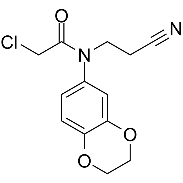
EN106 is a potent inhibitor of FEMIB. EN106 is a cysteine-reactive covalent ligand. EN106 disrupts recognition of the key reductive stress substrate of FEM1B, FNIP1. EN106 reduces oxidative stress and rescues high glucose-induced impaired angiogenesis in HUVECs.
For research use only. We do not sell to patients.
- Purity : 99.92%
- CAS No.: 757192-67-9
- Formula: C13H13ClN2O3
- Molecular Weight:280.71
-
Storage:Powder -20°C, 3 years , 4°C, 2 years ; In solvent -80°C, 6 months , -20°C, 1 month

To place orders, for customer services and technical support, please contact: MedChemExpress USA
Tel: 609-228-6898 E-mail: [email protected] [email protected]


-
 Biological Activity
-
Chemical Information
-
Solvent & Solubility
- Protocols
- Purity & Documentation
- References
-
 Help & FAQs


Publications Citing Use of MedChemExpress (MCE) EN106
More

Biological Activity
Description
In Vitro
EN106 (1 μM, 24 h) reduces the MDA content in HUVECs, inhibits the ROS generation and the activation of NF-κB signaling pathway, increases SOD activity, exhibiting antioxidant effects[2].
EN106 (1 μM, 24 h) increases the expression of cell cycle-related proteins (Cyclin D1, Cyclin D3) and VEGFA, promotes the cell proliferation, migration and angiogenesis in HUVEC, rescuing high glucose-impaired angiogenesis of HUVECs[2].
MedChemExpress (MCE) has not independently confirmed the accuracy of these methods. They are for reference only.
In Vivo
MedChemExpress (MCE) has not independently confirmed the accuracy of these methods. They are for reference only.
-
Animal Model:Mouse Streptozotocin (HY-13753)-induced type 2 diabetes models[2]
-
Dosage:
-
Administration:HA-PBA-FA/EN106 hydrogel, topical application, administration on days 0, 3, 5, 7, 10, and 14 after wound establishment.
-
Result:Accelerated the wound healing, promoted collagen deposition, upregulated the expression of FNIP1 and VEGFA proteins, increased the number of CD31(+) cells, and decreased the ROS level.


Chemical Information
-
CAS No. 757192-67-9
-
Appearance Solid
-
Molecular Weight 280.71
-
Formula C13H13ClN2O3
-
Color White to off-white
-
SMILES
O=C(N(CCC#N)C1=CC=C(OCCO2)C2=C1)CCl
-
Shipping
Room temperature in continental US; may vary elsewhere.
-
Storage
Powder -20°C 3 years 4°C 2 years In solvent -80°C 6 months -20°C 1 month


Publications (1)
-
Journal Impact Factor
-
Most Recent
-
bioRxiv
2026 May 8:2026.05.07.723605. PMID: 42146656


Solvent & Solubility
In Vitro:
DMSO : 250 mg/mL (890.60 mM; Need ultrasonic; Hygroscopic DMSO has a significant impact on the solubility of product, please use newly opened DMSO)
Please refer to the solubility information to select the appropriate solvent. Once prepared, please aliquot and store the solution to prevent product inactivation from repeated freeze-thaw cycles.
Storage method and period of stock solution: -80°C, 6 months; -20°C, 1 month. When stored at -80°C, please use it within 6 months. When stored at -20°C, please use it within 1 month.
Please refer to the solubility information to select the appropriate solvent. Once prepared, please aliquot and store the solution to prevent product inactivation from repeated freeze-thaw cycles.
Storage method and period of stock solution: -80°C, 6 months; -20°C, 1 month. When stored at -80°C, please use it within 6 months. When stored at -20°C, please use it within 1 month.
Concentration (start) × Volume (start) = Concentration (final) × Volume (final)


Purity & Documentation
-
Data Sheet (277 KB)
-
SDS (393 KB)
- English - EN (393 KB)
- Français - FR (393 KB)
- Deutsch - DE (393 KB)
- Norwegian - NO (393 KB)
- Español - ES (393 KB)
- Swedish - SV (393 KB)
- Italian - IT (393 KB)
- Korean - KR (393 KB)
- Portuguese - PT (393 KB)
-
Handling Instructions (2659 KB)
References
[1]. Henning NJ, et al. Discovery of a Covalent FEM1B Recruiter for Targeted Protein Degradation Applications. J Am Chem Soc. 2022;144(2):701-708. [Content Brief]
[2]. Zhang W, et al., Glucose-responsive, antioxidative HA-PBA-FA/EN106 hydrogel enhanced diabetic wound healing through modulation of FEM1b-FNIP1 axis and promoting angiogenesis. Bioact Mater. 2023 Jul 22;30:29-45. [Content Brief]


Complete Stock Solution Preparation Table
Please refer to the solubility information to select the appropriate solvent. Once prepared, please aliquot and store the solution to prevent product inactivation from repeated freeze-thaw cycles.
Storage method and period of stock solution: -80°C, 6 months; -20°C, 1 month. When stored at -80°C, please use it within 6 months. When stored at -20°C, please use it within 1 month.
| Optional Solvent | Concentration Solvent Mass | 1 mg | 5 mg | 10 mg | 25 mg |
|---|---|---|---|---|---|
| DMSO | 1 mM | 3.5624 mL | 17.8119 mL | 35.6238 mL | 89.0596 mL |
| 5 mM | 0.7125 mL | 3.5624 mL | 7.1248 mL | 17.8119 mL | |
| 10 mM | 0.3562 mL | 1.7812 mL | 3.5624 mL | 8.9060 mL | |
| 15 mM | 0.2375 mL | 1.1875 mL | 2.3749 mL | 5.9373 mL | |
| 20 mM | 0.1781 mL | 0.8906 mL | 1.7812 mL | 4.4530 mL | |
| 25 mM | 0.1425 mL | 0.7125 mL | 1.4250 mL | 3.5624 mL | |
| 30 mM | 0.1187 mL | 0.5937 mL | 1.1875 mL | 2.9687 mL | |
| 40 mM | 0.0891 mL | 0.4453 mL | 0.8906 mL | 2.2265 mL | |
| 50 mM | 0.0712 mL | 0.3562 mL | 0.7125 mL | 1.7812 mL | |
| 60 mM | 0.0594 mL | 0.2969 mL | 0.5937 mL | 1.4843 mL | |
| 80 mM | 0.0445 mL | 0.2226 mL | 0.4453 mL | 1.1132 mL | |
| 100 mM | 0.0356 mL | 0.1781 mL | 0.3562 mL | 0.8906 mL |


EN106 Related Classifications
HY-W229874 Related Classifications
 Powered by Bioz