



Enterolactone-d6

Based on 1 Customer Validation
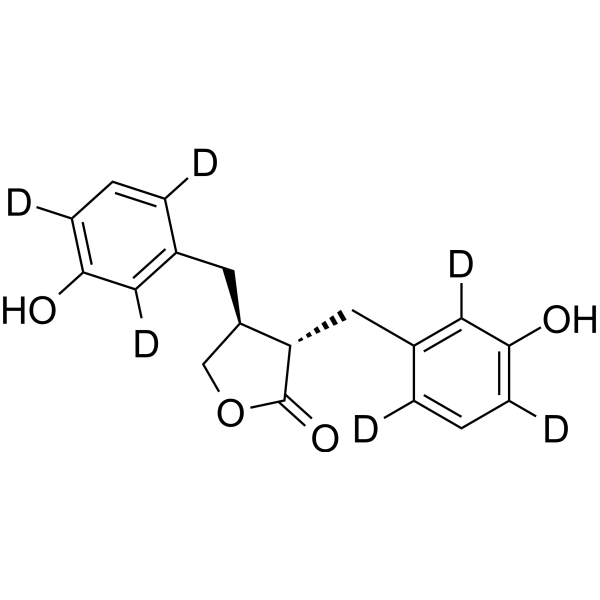
Enterolactone-d6 is the deuterium labeled Enterolactone. Enterolactone is a bioactive phenolic metabolite known as a mammalian lignan derived from dietary lignans. Enterolactone has estrogenic properties and anti-breast cancer activity. Enterolactone is a radiosensitizer for human breast cancer cell lines through impaired DNA repair and increased apoptosis.
Nur für Forschungszwecke. Wir verkaufen nicht an Patienten.
- Reinheit : 95.0%
- CAS. Nr.: 104411-11-2
- Formel: C18H12D6O4
- Molecular Weight:304.37
-
Speicherung:Powder -20°C, 3 years , 4°C, 2 years ; In solvent -80°C, 6 months , -20°C, 1 month

To place orders, for customer services and technical support, please contact: MedChemExpress USA
Tel: 609-228-6898 E-mail: [email protected] [email protected]


-
 Biologische Aktivität
-
Chemical Information
-
Lösungsmittel & Löslichkeit
- Protokoll
- Reinheit & Dokumentation
- Verweise
-
 Help & FAQs


Alle Endogenous Metabolite Isoform-spezifische Produkte anzeigen
More

Biologische Aktivität
Beschreibung
In Vitro
Stable heavy isotopes of hydrogen, carbon, and other elements have been incorporated into drug molecules, largely as tracers for quantitation during the drug development process. Deuteration has gained attention because of its potential to affect the pharmacokinetic and metabolic profiles of drugs[1].
MedChemExpress (MCE) has not independently confirmed the accuracy of these methods. They are for reference only.
Anwendung
1. This compound can be used as a tracer
2. This compound can be used as an internal standard for quantitative analysis by NMR, GC-MS, or LC-MS.


Chemical Information
-
CAS. Nr. 104411-11-2
-
Unlabeled CAS 78473-71-9
-
Appearance Solid
-
Molecular Weight 304.37
-
Formel C18H12D6O4
-
Color White to light yellow
-
SMILES
C([C@H]1[C@H](CC2=C(C(O)=C(C=C2[2H])[2H])[2H])C(=O)OC1)C3=C(C(O)=C(C=C3[2H])[2H])[2H]
-
Versand
Room temperature in continental US; may vary elsewhere.
-
Speicherung
Powder -20°C 3 years 4°C 2 years In solvent -80°C 6 months -20°C 1 month


Lösungsmittel & Löslichkeit
In Vitro:
DMSO : 30 mg/mL (98.56 mM; Need ultrasonic and warming; Hygroscopic DMSO has a significant impact on the solubility of product, please use newly opened DMSO)
Please refer to the solubility information to select the appropriate solvent. Once prepared, please aliquot and store the solution to prevent product inactivation from repeated freeze-thaw cycles.
Storage method and period of stock solution: -80°C, 6 months; -20°C, 1 month. When stored at -80°C, please use it within 6 months. When stored at -20°C, please use it within 1 month.
Please refer to the solubility information to select the appropriate solvent. Once prepared, please aliquot and store the solution to prevent product inactivation from repeated freeze-thaw cycles.
Storage method and period of stock solution: -80°C, 6 months; -20°C, 1 month. When stored at -80°C, please use it within 6 months. When stored at -20°C, please use it within 1 month.
Konzentration (Stammlösung) × Volumen (Stammlösung) = Konzentration (Ziellösung) × Volumen (Ziellösung)


Reinheit & Dokumentation
-
Data Sheet (275 KB)
-
SDS (251 KB)
- English - EN (251 KB)
- Français - FR (251 KB)
- Deutsch - DE (251 KB)
- Norwegian - NO (251 KB)
- Español - ES (251 KB)
- Swedish - SV (251 KB)
- Italian - IT (251 KB)
- Korean - KR (251 KB)
- Portuguese - PT (251 KB)
-
Handling Instructions (2659 KB)
Verweise
[1]. Russak EM, et al. Impact of Deuterium Substitution on the Pharmacokinetics of Pharmaceuticals. Ann Pharmacother. 2019;53(2):211-216. [Content Brief]
[2]. Bigdeli B, et al. Enterolactone: A novel radiosensitizer for human breast cancer cell lines through impaired DNA repair and increased apoptosis. Toxicol Appl Pharmacol. 2016;313:180-194. [Content Brief]
[3]. Mali AV, et al. Enterolactone modulates the ERK/NF-κB/Snail signaling pathway in triple-negative breast cancer cell line MDA-MB-231 to revert the TGF-β-induced epithelial-mesenchymal transition. Cancer Biol Med. 2018;15(2):137-156. [Content Brief]


Complete Stock Solution Preparation Table
Please refer to the solubility information to select the appropriate solvent. Once prepared, please aliquot and store the solution to prevent product inactivation from repeated freeze-thaw cycles.
Storage method and period of stock solution: -80°C, 6 months; -20°C, 1 month. When stored at -80°C, please use it within 6 months. When stored at -20°C, please use it within 1 month.
| Optional Solvent | Concentration Solvent Mass | 1 mg | 5 mg | 10 mg | 25 mg |
|---|---|---|---|---|---|
| DMSO | 1 mM | 3.2855 mL | 16.4274 mL | 32.8547 mL | 82.1369 mL |
| 5 mM | 0.6571 mL | 3.2855 mL | 6.5709 mL | 16.4274 mL | |
| 10 mM | 0.3285 mL | 1.6427 mL | 3.2855 mL | 8.2137 mL | |
| 15 mM | 0.2190 mL | 1.0952 mL | 2.1903 mL | 5.4758 mL | |
| 20 mM | 0.1643 mL | 0.8214 mL | 1.6427 mL | 4.1068 mL | |
| 25 mM | 0.1314 mL | 0.6571 mL | 1.3142 mL | 3.2855 mL | |
| 30 mM | 0.1095 mL | 0.5476 mL | 1.0952 mL | 2.7379 mL | |
| 40 mM | 0.0821 mL | 0.4107 mL | 0.8214 mL | 2.0534 mL | |
| 50 mM | 0.0657 mL | 0.3285 mL | 0.6571 mL | 1.6427 mL | |
| 60 mM | 0.0548 mL | 0.2738 mL | 0.5476 mL | 1.3689 mL | |
| 80 mM | 0.0411 mL | 0.2053 mL | 0.4107 mL | 1.0267 mL |


Enterolactone-d6 Related Classifications
HY-108692S Related Classifications
 Powered by Bioz