



Entrectinib-d8

Based on 1 Customer Validation
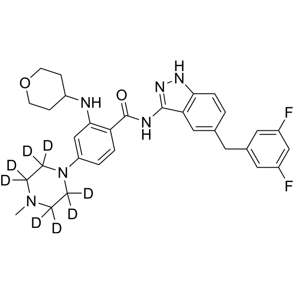
Entrectinib-d8 (NMS-E628-d8; RXDX-101-d8) is a deuterated version of Entrectinib (HY-12678). Entrectinib (NMS-E628) is an orally available, blood-brain barrier permeable, central nervous system active TrkA/B/C, ROS1 and ALK inhibitor with IC50 values of 1, 3, 5, 12 and 12, respectively. 7 nM. Entrectinib induces apoptosis and cycle arrest in cancer cells, has anti-tumor activity, and also alleviates bleomycin-induced pulmonary fibrosis in mice.
For research use only. We do not sell to patients.
- Purity : 99.82%
- CAS No.: 2251773-94-9
- Formula: C31H26D8F2N6O2
- Molecular Weight:568.69
-
Storage:Powder -20°C, 3 years , 4°C, 2 years ; In solvent -80°C, 6 months , -20°C, 1 month

To place orders, for customer services and technical support, please contact: MedChemExpress USA
Tel: 609-228-6898 E-mail: [email protected] [email protected]


-
 Biological Activity
-
Chemical Information
-
Solvent & Solubility
- Protocols
- Purity & Documentation
- References
-
 Help & FAQs




Biological Activity
Description
In Vitro
Stable heavy isotopes of hydrogen,carbon,and other elements have been incorporated into drug molecules,largely as tracers for quantitation during the drug development process. Deuteration has gained attention because of its potential to affect the pharmacokinetic and metabolic profiles of drugs[1].
MedChemExpress (MCE) has not independently confirmed the accuracy of these methods. They are for reference only.
Application
1. This compound can be used as a tracer
2. This compound can be used as an internal standard for quantitative analysis by NMR, GC-MS, or LC-MS.


Chemical Information
-
CAS No. 2251773-94-9
-
Unlabeled CAS 1108743-60-7
-
Appearance Solid
-
Molecular Weight 568.69
-
Formula C31H26D8F2N6O2
-
Color White to off-white
-
SMILES
O=C(NC1=NNC2=C1C=C(CC3=CC(F)=CC(F)=C3)C=C2)C4=C(NC5CCOCC5)C=C(N6C([2H])([2H])C([2H])([2H])N(C)C([2H])([2H])C6([2H])[2H])C=C4
-
Synonyms
NMS-E628-d8; RXDX-101-d8
-
Shipping
Room temperature in continental US; may vary elsewhere.
-
Storage
Powder -20°C 3 years 4°C 2 years In solvent -80°C 6 months -20°C 1 month


Solvent & Solubility
In Vitro:
DMSO : 100 mg/mL (175.84 mM; Need ultrasonic; Hygroscopic DMSO has a significant impact on the solubility of product, please use newly opened DMSO)
Please refer to the solubility information to select the appropriate solvent. Once prepared, please aliquot and store the solution to prevent product inactivation from repeated freeze-thaw cycles.
Storage method and period of stock solution: -80°C, 6 months; -20°C, 1 month. When stored at -80°C, please use it within 6 months. When stored at -20°C, please use it within 1 month.
Please refer to the solubility information to select the appropriate solvent. Once prepared, please aliquot and store the solution to prevent product inactivation from repeated freeze-thaw cycles.
Storage method and period of stock solution: -80°C, 6 months; -20°C, 1 month. When stored at -80°C, please use it within 6 months. When stored at -20°C, please use it within 1 month.
Concentration (start) × Volume (start) = Concentration (final) × Volume (final)


Purity & Documentation
-
Data Sheet (277 KB)
-
SDS (252 KB)
- English - EN (252 KB)
- Français - FR (252 KB)
- Deutsch - DE (252 KB)
- Norwegian - NO (252 KB)
- Español - ES (252 KB)
- Swedish - SV (252 KB)
- Italian - IT (252 KB)
- Korean - KR (252 KB)
- Portuguese - PT (252 KB)
-
Handling Instructions (2659 KB)
References
[1]. Russak EM, et al. Impact of Deuterium Substitution on the Pharmacokinetics of Pharmaceuticals. Ann Pharmacother. 2019;53(2):211-216. [Content Brief]
[2]. Miao Y, et al. Entrectinib ameliorates bleomycin-induced pulmonary fibrosis in mice by inhibiting TGF-β1 signaling pathway. Int Immunopharmacol. 2022 Dec;113(Pt B):109427. [Content Brief]
[3]. Ardini E, et al. Entrectinib, a Pan-TRK, ROS1, and ALK Inhibitor with Activity in Multiple Molecularly Defined Cancer Indications. Mol Cancer Ther. 2016 Apr;15(4):628-39. [Content Brief]
[4]. Iyer R, et al. Entrectinib is a potent inhibitor of Trk-driven neuroblastomas in a xenograft mouse model. Cancer Lett. 2016 Mar 28;372(2):179-86. [Content Brief]


Complete Stock Solution Preparation Table
Please refer to the solubility information to select the appropriate solvent. Once prepared, please aliquot and store the solution to prevent product inactivation from repeated freeze-thaw cycles.
Storage method and period of stock solution: -80°C, 6 months; -20°C, 1 month. When stored at -80°C, please use it within 6 months. When stored at -20°C, please use it within 1 month.
| Optional Solvent | Concentration Solvent Mass | 1 mg | 5 mg | 10 mg | 25 mg |
|---|---|---|---|---|---|
| DMSO | 1 mM | 1.7584 mL | 8.7921 mL | 17.5843 mL | 43.9607 mL |
| 5 mM | 0.3517 mL | 1.7584 mL | 3.5169 mL | 8.7921 mL | |
| 10 mM | 0.1758 mL | 0.8792 mL | 1.7584 mL | 4.3961 mL | |
| 15 mM | 0.1172 mL | 0.5861 mL | 1.1723 mL | 2.9307 mL | |
| 20 mM | 0.0879 mL | 0.4396 mL | 0.8792 mL | 2.1980 mL | |
| 25 mM | 0.0703 mL | 0.3517 mL | 0.7034 mL | 1.7584 mL | |
| 30 mM | 0.0586 mL | 0.2931 mL | 0.5861 mL | 1.4654 mL | |
| 40 mM | 0.0440 mL | 0.2198 mL | 0.4396 mL | 1.0990 mL | |
| 50 mM | 0.0352 mL | 0.1758 mL | 0.3517 mL | 0.8792 mL | |
| 60 mM | 0.0293 mL | 0.1465 mL | 0.2931 mL | 0.7327 mL | |
| 80 mM | 0.0220 mL | 0.1099 mL | 0.2198 mL | 0.5495 mL | |
| 100 mM | 0.0176 mL | 0.0879 mL | 0.1758 mL | 0.4396 mL |


Entrectinib-d8 Related Classifications
HY-12678S1 Related Classifications
 Powered by Bioz